 Movie
Movie Controller

[English] 日本語
 Yorodumi
- EMDB-4887: Cryo-EM reconstruction of Thermus thermophilus bactofilin double ... -
+ Open data

- Basic information
| Entry | Database: EMDB / ID: EMD-4887 | |||||||||
|---|---|---|---|---|---|---|---|---|---|---|
| Title | Cryo-EM reconstruction of Thermus thermophilus bactofilin double helical filaments | |||||||||
 Map data Map data | Thermus thermophilus bactofilin double filament | |||||||||
 Sample Sample |
| |||||||||
 Keywords Keywords | Prokaryotic cytoskeletons / PROTEIN FIBRIL | |||||||||
| Function / homology | Bactofilin A/B / Polymer-forming cytoskeletal / Polymer-forming cytoskeletal protein Function and homology information Function and homology information | |||||||||
| Biological species |   Thermus thermophilus (strain HB8 / ATCC 27634 / DSM 579) (bacteria) Thermus thermophilus (strain HB8 / ATCC 27634 / DSM 579) (bacteria) | |||||||||
| Method | helical reconstruction / cryo EM / Resolution: 4.2 Å | |||||||||
 Authors Authors | Deng X / Lowe J | |||||||||
| Funding support |  United Kingdom, 2 items United Kingdom, 2 items
| |||||||||
 Citation Citation | Journal: Nat Microbiol / Year: 2019 Title: The structure of bactofilin filaments reveals their mode of membrane binding and lack of polarity. Authors: Xian Deng / Andres Gonzalez Llamazares / James M Wagstaff / Victoria L Hale / Giuseppe Cannone / Stephen H McLaughlin / Danguole Kureisaite-Ciziene / Jan Löwe / Abstract: Bactofilins are small β-helical proteins that form cytoskeletal filaments in a range of bacteria. Bactofilins have diverse functions, from cell stalk formation in Caulobacter crescentus to ...Bactofilins are small β-helical proteins that form cytoskeletal filaments in a range of bacteria. Bactofilins have diverse functions, from cell stalk formation in Caulobacter crescentus to chromosome segregation and motility in Myxococcus xanthus. However, the precise molecular architecture of bactofilin filaments has remained unclear. Here, sequence analysis and electron microscopy results reveal that, in addition to being widely distributed across bacteria and archaea, bactofilins are also present in a few eukaryotic lineages such as the Oomycetes. Electron cryomicroscopy analysis demonstrated that the sole bactofilin from Thermus thermophilus (TtBac) forms constitutive filaments that polymerize through end-to-end association of the β-helical domains. Using a nanobody, we determined the near-atomic filament structure, showing that the filaments are non-polar. A polymerization-impairing mutation enabled crystallization and structure determination, while reaffirming the lack of polarity and the strength of the β-stacking interface. To confirm the generality of the lack of polarity, we performed coevolutionary analysis on a large set of sequences. Finally, we determined that the widely conserved N-terminal disordered tail of TtBac is responsible for direct binding to lipid membranes, both on liposomes and in Escherichia coli cells. Membrane binding is probably a common feature of these widespread but only recently discovered filaments of the prokaryotic cytoskeleton. | |||||||||
| History |
|
- Structure visualization
| Movie |
Movie viewer |
|---|---|
| Structure viewer | EM map: SurfViewMolmilJmol/JSmol |
| Supplemental images |

- Downloads & links
-EMDB archive
| Map data | emd_4887.map.gz | 96.2 MB | EMDB map data format | |
|---|---|---|---|---|
| Header (meta data) | emd-4887-v30.xmlemd-4887.xml | 9.8 KB 9.8 KB | Display Display | EMDB header |
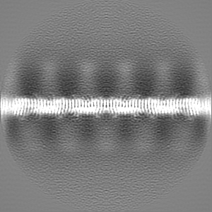
| Images | 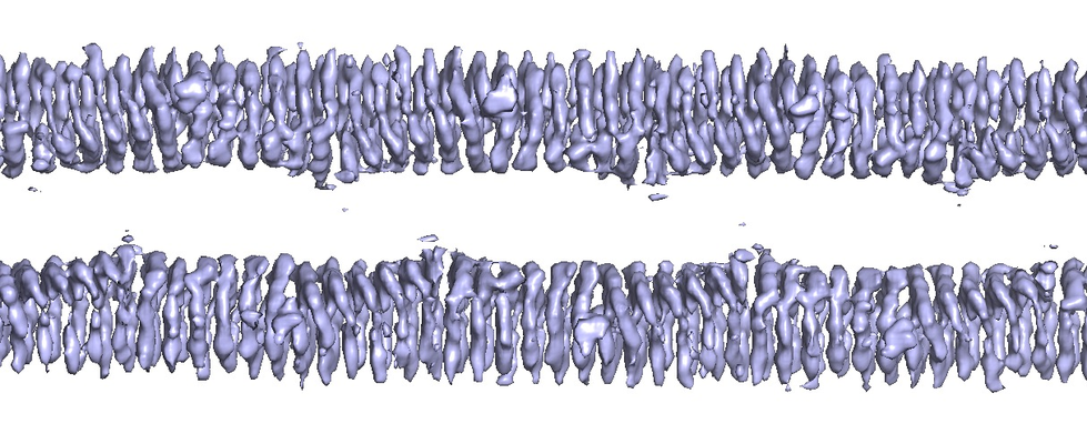 emd_4887.png emd_4887.png | 528.9 KB | ||
| Filedesc metadata | emd-4887.cif.gz | 5.2 KB | ||
| Archive directory |  http://ftp.pdbj.org/pub/emdb/structures/EMD-4887ftp://ftp.pdbj.org/pub/emdb/structures/EMD-4887 http://ftp.pdbj.org/pub/emdb/structures/EMD-4887ftp://ftp.pdbj.org/pub/emdb/structures/EMD-4887 | HTTPS FTP |
-Related structure data
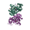
| Related structure data |  6ribMC  6riaC M: atomic model generated by this map C: citing same article ( |
|---|---|
| Similar structure data |
-Links
| EMDB pages | EMDB (EBI/PDBe) / EMDataResource |
|---|
-Map
| File | Download / File: emd_4887.map.gz / Format: CCP4 / Size: 103 MB / Type: IMAGE STORED AS FLOATING POINT NUMBER (4 BYTES) | ||||||||||||||||||||||||||||||||||||||||||||||||||||||||||||||||||||
|---|---|---|---|---|---|---|---|---|---|---|---|---|---|---|---|---|---|---|---|---|---|---|---|---|---|---|---|---|---|---|---|---|---|---|---|---|---|---|---|---|---|---|---|---|---|---|---|---|---|---|---|---|---|---|---|---|---|---|---|---|---|---|---|---|---|---|---|---|---|
| Annotation | Thermus thermophilus bactofilin double filament | ||||||||||||||||||||||||||||||||||||||||||||||||||||||||||||||||||||
| Projections & slices | Image control
Images are generated by Spider. | ||||||||||||||||||||||||||||||||||||||||||||||||||||||||||||||||||||
| Voxel size | X=Y=Z: 1.07 Å | ||||||||||||||||||||||||||||||||||||||||||||||||||||||||||||||||||||
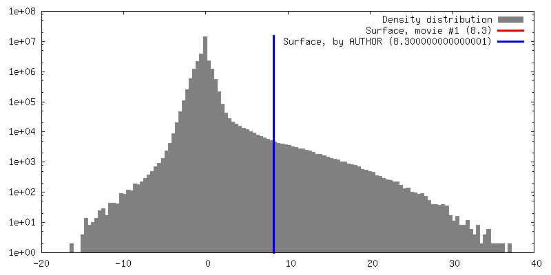
| Density |
| ||||||||||||||||||||||||||||||||||||||||||||||||||||||||||||||||||||
| Symmetry | Space group: 1 | ||||||||||||||||||||||||||||||||||||||||||||||||||||||||||||||||||||
| Details | EMDB XML:
CCP4 map header:
| ||||||||||||||||||||||||||||||||||||||||||||||||||||||||||||||||||||
 X (Sec.)
 Y (Row.)
 Z (Col.)


-Supplemental data
- Sample components
-Entire : bactofilin
| Entire | Name: bactofilin |
|---|---|
| Components |
|
-Supramolecule #1: bactofilin
| Supramolecule | Name: bactofilin / type: complex / ID: 1 / Parent: 0 / Macromolecule list: all |
|---|---|
| Source (natural) | Organism: Thermus thermophilus (strain HB8 / ATCC 27634 / DSM 579) (bacteria) |
-Macromolecule #1: bactofilin
| Macromolecule | Name: bactofilin / type: protein_or_peptide / ID: 1 / Number of copies: 22 / Enantiomer: LEVO |
|---|---|
| Source (natural) | Organism: Thermus thermophilus (strain HB8 / ATCC 27634 / DSM 579) (bacteria) |
| Molecular weight | Theoretical: 13.142075 KDa |
| Recombinant expression | Organism: |
| Sequence | String: MGRMLGRKER TLTYLGPDTE VLGDMRAKGQ VRIDGLVRGS VLVEGELEVG PTGRVEGERV EARSVLIHGE VKAELTAEKV VLSKTARFT GQLKAQALEV EAGAVFVGQS VAGEHKALEA PKEA UniProtKB: Polymer-forming cytoskeletal protein |
-Experimental details
-Structure determination
| Method | cryo EM |
|---|---|
 Processing Processing | helical reconstruction |
| Aggregation state | helical array |
-Sample preparation
| Buffer | pH: 11 |
|---|---|
| Vitrification | Cryogen name: ETHANE |
- Electron microscopy
| Microscope | FEI TITAN KRIOS |
|---|---|
| Image recording | Film or detector model: FEI FALCON III (4k x 4k) / Detector mode: COUNTING / Number real images: 2130 / Average electron dose: 40.0 e/Å2 |
| Electron beam | Acceleration voltage: 300 kV / Electron source:  FIELD EMISSION GUN FIELD EMISSION GUN |
| Electron optics | Illumination mode: FLOOD BEAM / Imaging mode: BRIGHT FIELD |
| Experimental equipment |  Model: Titan Krios / Image courtesy: FEI Company |
-Image processing
| Final reconstruction | Applied symmetry - Helical parameters - Δz: 57.46 Å Applied symmetry - Helical parameters - Δ&Phi: 4.89 ° Applied symmetry - Helical parameters - Axial symmetry: C1 (asymmetric) Resolution.type: BY AUTHOR / Resolution: 4.2 Å / Resolution method: OTHER / Details: FSC: map versus refined atomic model / Number images used: 346000 |
|---|---|
| Startup model | Type of model: OTHER / Details: RELION 3 |
| Final angle assignment | Type: NOT APPLICABLE |
-Atomic model buiding 1
| Refinement | Protocol: AB INITIO MODEL |
|---|---|
| Output model | PDB-6rib: |
