 Movie
Movie Controller

[English] 日本語
 Yorodumi
- EMDB-48467: Structure of native murine cardiac thin filament at pCa=5.8 in Ca... -
+ Open data

- Basic information
| Entry |  | |||||||||
|---|---|---|---|---|---|---|---|---|---|---|
| Title | Structure of native murine cardiac thin filament at pCa=5.8 in Ca2+-free rotated state (lower strand) | |||||||||
 Map data Map data | ||||||||||
 Sample Sample |
| |||||||||
 Keywords Keywords | thin filament / cryo-EM / troponin / tropomyosin / muscle structure / motor protein | |||||||||
| Function / homology |  Function and homology information Function and homology informationatrial cardiac muscle tissue morphogenesis / positive regulation of heart rate by epinephrine / muscle thin filament tropomyosin / RHOB GTPase cycle / regulation of systemic arterial blood pressure by ischemic conditions / Formation of the dystrophin-glycoprotein complex (DGC) / troponin C binding / Striated Muscle Contraction / Regulation of CDH1 Function / RHOA GTPase cycle ...atrial cardiac muscle tissue morphogenesis / positive regulation of heart rate by epinephrine / muscle thin filament tropomyosin / RHOB GTPase cycle / regulation of systemic arterial blood pressure by ischemic conditions / Formation of the dystrophin-glycoprotein complex (DGC) / troponin C binding / Striated Muscle Contraction / Regulation of CDH1 Function / RHOA GTPase cycle / contractile muscle fiber / diaphragm contraction / regulation of muscle filament sliding speed / troponin T binding / cytoplasmic actin-based contraction involved in cell motility / cardiac Troponin complex / cardiac myofibril / actin crosslink formation / Smooth Muscle Contraction / negative regulation of ATP-dependent activity / troponin complex / actin-myosin filament sliding / cardiac myofibril assembly / regulation of muscle contraction / regulation of smooth muscle contraction / positive regulation of ATP-dependent activity / actin filament-based movement / ruffle organization / bleb / transition between fast and slow fiber / Ion homeostasis / cardiac muscle tissue morphogenesis / actomyosin structure organization / actin filament capping / muscle filament sliding / regulation of cardiac muscle contraction by calcium ion signaling / response to metal ion / I band / cardiac muscle cell contraction / sarcomere organization / ventricular cardiac muscle tissue morphogenesis / microfilament motor activity / myosin binding / heart contraction / tropomyosin binding / regulation of heart contraction / negative regulation of vascular associated smooth muscle cell migration / troponin I binding / mesenchyme migration / myofibril / striated muscle thin filament / negative regulation of vascular associated smooth muscle cell proliferation / skeletal muscle thin filament assembly / vasculogenesis / calcium channel inhibitor activity / striated muscle contraction / skeletal muscle contraction / cardiac muscle contraction / cytoskeletal protein binding / positive regulation of stress fiber assembly / stress fiber / positive regulation of cell adhesion / muscle contraction / negative regulation of cell migration / actin filament organization / sarcomere / response to bacterium / filopodium / actin filament / wound healing / response to calcium ion / structural constituent of cytoskeleton / Hydrolases; Acting on acid anhydrides; Acting on acid anhydrides to facilitate cellular and subcellular movement / ruffle membrane / intracellular calcium ion homeostasis / disordered domain specific binding / calcium-dependent protein binding / actin filament binding / regulation of cell shape / lamellipodium / actin cytoskeleton / heart development / actin binding / cell body / in utero embryonic development / response to ethanol / protein-macromolecule adaptor activity / response to xenobiotic stimulus / protein heterodimerization activity / protein domain specific binding / hydrolase activity / calcium ion binding / positive regulation of gene expression / synapse / protein kinase binding / negative regulation of apoptotic process / protein-containing complex binding / glutamatergic synapse / protein homodimerization activity / protein-containing complex Similarity search - Function | |||||||||
| Biological species |  | |||||||||
| Method | single particle reconstruction / cryo EM / Resolution: 5.4 Å | |||||||||
 Authors Authors | Galkin VE / Risi CM | |||||||||
| Funding support |  United States, 1 items United States, 1 items
| |||||||||
 Citation Citation | Journal: J Mol Cell Cardiol / Year: 2025 Title: The role of the troponin T interactions with actin in regulation of cardiac thin filament revealed by the troponin T pathogenic variant Ile79Asn. Authors: Cristina M Risi / Maicon Landim-Vieira / Betty Belknap / P Bryant Chase / Jose R Pinto / Vitold E Galkin / Abstract: Cardiac muscle contraction/relaxation cycle depends on the rising and falling Ca levels in sarcomeres that control the extent of interactions between myosin-based thick and actin-based thin filaments. ...Cardiac muscle contraction/relaxation cycle depends on the rising and falling Ca levels in sarcomeres that control the extent of interactions between myosin-based thick and actin-based thin filaments. Cardiac thin filament (cTF) consists of actin, tropomyosin (Tm) that regulates myosin binding to actin, and troponin complex that governs Tm position upon Ca-binding. Troponin has three subunits - Ca-binding troponin C (TnC), Tm stabilizing troponin T (TnT), and inhibitory troponin I (TnI). TnT N-terminus (TnT1) interactions with actin stabilize the inhibited state of cTF. TnC, TnI, and Tm work in concert to control actomyosin interactions. Cryo-electron microscopy (cryo-EM) provided factual structures of healthy cTF, but structures of cTF carrying missense mutations linked to human cardiomyopathy are unknown. Variant Ile79Asn in human cardiac TnT (TnT-I79N) increases myofilament Ca sensitivity and slows cross-bridge kinetics, leading to severe hypertrophic/restrictive cardiomyopathy. Here, we used TnT-I79N mutation as a tool to examine the role of TnT1 in the complex mechanism of cTF regulation. Comparison of the cryo-EM structures of murine wild type and TnT-I79N native cTFs at systolic Ca levels (pCa = 5.8) demonstrates that TnT-I79N causes 1) dissociation of the TnT1 loop from its actin interface that results in Tm release to a more activated position, 2) reduced interaction of TnI C-terminus with actin-Tm, and 3) increased frequency of Ca-bound regulatory units. Our data indicate that the TnT1 loop is a crucial element of the allosteric regulatory network that couples Tn subunits and Tm to maintain adequate cTF response to physiological Ca levels during a heartbeat. | |||||||||
| History |
|
- Structure visualization
| Supplemental images |
|---|

- Downloads & links
-EMDB archive
| Map data | emd_48467.map.gz | 8.4 MB | EMDB map data format | |
|---|---|---|---|---|
| Header (meta data) | emd-48467-v30.xmlemd-48467.xml | 24.7 KB 24.7 KB | Display Display | EMDB header |
| FSC (resolution estimation) | emd_48467_fsc.xml | 11.5 KB | Display | FSC data file |
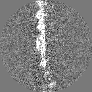
| Images |  emd_48467.png emd_48467.png | 29.3 KB | ||
| Filedesc metadata | emd-48467.cif.gz | 7.2 KB | ||
| Others | emd_48467_half_map_1.map.gzemd_48467_half_map_2.map.gz | 102.2 MB 102.2 MB | ||
| Archive directory |  http://ftp.pdbj.org/pub/emdb/structures/EMD-48467ftp://ftp.pdbj.org/pub/emdb/structures/EMD-48467 http://ftp.pdbj.org/pub/emdb/structures/EMD-48467ftp://ftp.pdbj.org/pub/emdb/structures/EMD-48467 | HTTPS FTP |
-Related structure data
| Related structure data |  9moiMC  9e2eC  9mo4C  9mo5C  9mo6C 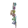 9mo7C  9mo8C  9mo9C  9moaC 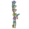 9mobC  9mocC  9modC  9mokC  9molC  9momC  9monC  9mooC  9mopC  9mouC  9mowC  9moxC M: atomic model generated by this map C: citing same article ( |
|---|---|
| Similar structure data |


-Links
| EMDB pages | EMDB (EBI/PDBe) / EMDataResource |
|---|---|
| Related items in Molecule of the Month |


-Map
| File | Download / File: emd_48467.map.gz / Format: CCP4 / Size: 129.7 MB / Type: IMAGE STORED AS FLOATING POINT NUMBER (4 BYTES) | ||||||||||||||||||||||||||||||||||||
|---|---|---|---|---|---|---|---|---|---|---|---|---|---|---|---|---|---|---|---|---|---|---|---|---|---|---|---|---|---|---|---|---|---|---|---|---|---|
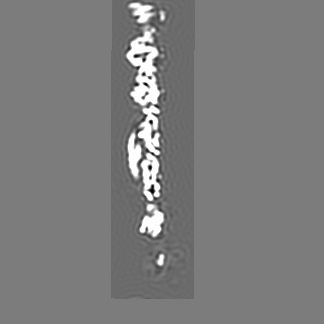
| Projections & slices | Image control
Images are generated by Spider. | ||||||||||||||||||||||||||||||||||||
| Voxel size | X=Y=Z: 1.356 Å | ||||||||||||||||||||||||||||||||||||
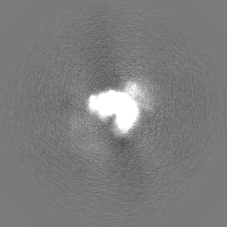
| Density |
| ||||||||||||||||||||||||||||||||||||
| Symmetry | Space group: 1 | ||||||||||||||||||||||||||||||||||||
| Details | EMDB XML:
|
 Z (Sec.)
 Y (Row.)
 X (Col.)


-Supplemental data
-Half map: #2
| File | emd_48467_half_map_1.map | ||||||||||||
|---|---|---|---|---|---|---|---|---|---|---|---|---|---|
| Projections & Slices |
| ||||||||||||
| Density Histograms |


-Half map: #1
| File | emd_48467_half_map_2.map | ||||||||||||
|---|---|---|---|---|---|---|---|---|---|---|---|---|---|
| Projections & Slices |
| ||||||||||||
| Density Histograms |


- Sample components
-Entire : Native murine cardiac thin filament at pCa=5.8
| Entire | Name: Native murine cardiac thin filament at pCa=5.8 |
|---|---|
| Components |
|
-Supramolecule #1: Native murine cardiac thin filament at pCa=5.8
| Supramolecule | Name: Native murine cardiac thin filament at pCa=5.8 / type: complex / ID: 1 / Parent: 0 / Macromolecule list: #1-#5 |
|---|---|
| Source (natural) | Organism: |
-Macromolecule #1: Actin, alpha cardiac muscle 1
| Macromolecule | Name: Actin, alpha cardiac muscle 1 / type: protein_or_peptide / ID: 1 / Number of copies: 6 / Enantiomer: LEVO EC number: Hydrolases; Acting on acid anhydrides; Acting on acid anhydrides to facilitate cellular and subcellular movement |
|---|---|
| Source (natural) | Organism: |
| Molecular weight | Theoretical: 42.064891 KDa |
| Sequence | String: MCDDEETTAL VCDNGSGLVK AGFAGDDAPR AVFPSIVGRP RHQGVMVGMG QKDSYVGDEA QSKRGILTLK YPIEHGIITN WDDMEKIWH HTFYNELRVA PEEHPTLLTE APLNPKANRE KMTQIMFETF NVPAMYVAIQ AVLSLYASGR TTGIVLDSGD G VTHNVPIY ...String: MCDDEETTAL VCDNGSGLVK AGFAGDDAPR AVFPSIVGRP RHQGVMVGMG QKDSYVGDEA QSKRGILTLK YPIEHGIITN WDDMEKIWH HTFYNELRVA PEEHPTLLTE APLNPKANRE KMTQIMFETF NVPAMYVAIQ AVLSLYASGR TTGIVLDSGD G VTHNVPIY EGYALPHAIM RLDLAGRDLT DYLMKILTER GYSFVTTAER EIVRDIKEKL CYVALDFENE MATAASSSSL EK SYELPDG QVITIGNERF RCPETLFQPS FIGMESAGIH ETTYNSIMKC DIDIRKDLYA NNVLSGGTTM YPGIADRMQK EIT ALAPST MKIKIIAPPE RKYSVWIGGS ILASLSTFQQ MWISKQEYDE AGPSIVHRKC F UniProtKB: Actin, alpha cardiac muscle 1 |
-Macromolecule #2: Troponin C, slow skeletal and cardiac muscles
| Macromolecule | Name: Troponin C, slow skeletal and cardiac muscles / type: protein_or_peptide / ID: 2 / Number of copies: 1 / Enantiomer: LEVO |
|---|---|
| Source (natural) | Organism: |
| Molecular weight | Theoretical: 18.43752 KDa |
| Sequence | String: MDDIYKAAVE QLTEEQKNEF KAAFDIFVLG AEDGCISTKE LGKVMRMLGQ NPTPEELQEM IDEVDEDGSG TVDFDEFLVM MVRCMKDDS KGKSEEELSD LFRMFDKNAD GYIDLDELKM MLQATGETIT EDDIEELMKD GDKNNDGRID YDEFLEFMKG V E UniProtKB: Troponin C, slow skeletal and cardiac muscles |
-Macromolecule #3: Troponin I, cardiac muscle
| Macromolecule | Name: Troponin I, cardiac muscle / type: protein_or_peptide / ID: 3 / Number of copies: 1 / Enantiomer: LEVO |
|---|---|
| Source (natural) | Organism: |
| Molecular weight | Theoretical: 24.308908 KDa |
| Sequence | String: MADESSDAAG EPQPAPAPVR RRSSANYRAY ATEPHAKKKS KISASRKLQL KTLMLQIAKQ EMEREAEERR GEKGRVLRTR CQPLELDGL GFEELQDLCR QLHARVDKVD EERYDVEAKV TKNITEIADL TQKIYDLRGK FKRPTLRRVR ISADAMMQAL L GTRAKESL ...String: MADESSDAAG EPQPAPAPVR RRSSANYRAY ATEPHAKKKS KISASRKLQL KTLMLQIAKQ EMEREAEERR GEKGRVLRTR CQPLELDGL GFEELQDLCR QLHARVDKVD EERYDVEAKV TKNITEIADL TQKIYDLRGK FKRPTLRRVR ISADAMMQAL L GTRAKESL DLRAHLKQVK KEDIEKENRE VGDWRKNIDA LSGMEGRKKK FEG UniProtKB: Troponin I, cardiac muscle |
-Macromolecule #4: Isoform A2 of Troponin T, cardiac muscle
| Macromolecule | Name: Isoform A2 of Troponin T, cardiac muscle / type: protein_or_peptide / ID: 4 / Number of copies: 2 / Enantiomer: LEVO |
|---|---|
| Source (natural) | Organism: |
| Molecular weight | Theoretical: 34.616367 KDa |
| Sequence | String: MSDAEEVVEE YEEEQEEQEE AVEEEEAGGA EPEPEGEAET EEANVEEVGP DEEAKDAEEG PVEDTKPKPS RLFMPNLVPP KIPDGERVD FDDIHRKRVE KDLNELQTLI EAHFENRKKE EEELISLKDR IEKRRAERAE QQRIRNEREK ERQNRLAEER A RREEEENR ...String: MSDAEEVVEE YEEEQEEQEE AVEEEEAGGA EPEPEGEAET EEANVEEVGP DEEAKDAEEG PVEDTKPKPS RLFMPNLVPP KIPDGERVD FDDIHRKRVE KDLNELQTLI EAHFENRKKE EEELISLKDR IEKRRAERAE QQRIRNEREK ERQNRLAEER A RREEEENR RKAEDEARKK KALSNMMHFG GYIQKQAQTE RKSGKRQTER EKKKKILAER RKALAIDHLN EDQLREKAKE LW QSIHNLE AEKFDLQEKF KQQKYEINVL RNRINDNQKV SKTRGKAKVT GRWK UniProtKB: Troponin T, cardiac muscle |
-Macromolecule #5: Tropomyosin alpha-1 chain
| Macromolecule | Name: Tropomyosin alpha-1 chain / type: protein_or_peptide / ID: 5 / Number of copies: 4 / Enantiomer: LEVO |
|---|---|
| Source (natural) | Organism: |
| Molecular weight | Theoretical: 32.735609 KDa |
| Sequence | String: MDAIKKKMQM LKLDKENALD RAEQAEADKK AAEDRSKQLE DELVSLQKKL KGTEDELDKY SEALKDAQEK LELAEKKATD AEADVASLN RRIQLVEEEL DRAQERLATA LQKLEEAEKA ADESERGMKV IESRAQKDEE KMEIQEIQLK EAKHIAEDAD R KYEEVARK ...String: MDAIKKKMQM LKLDKENALD RAEQAEADKK AAEDRSKQLE DELVSLQKKL KGTEDELDKY SEALKDAQEK LELAEKKATD AEADVASLN RRIQLVEEEL DRAQERLATA LQKLEEAEKA ADESERGMKV IESRAQKDEE KMEIQEIQLK EAKHIAEDAD R KYEEVARK LVIIESDLER AEERAELSEG KCAELEEELK TVTNNLKSLE AQAEKYSQKE DKYEEEIKVL SDKLKEAETR AE FAERSVT KLEKSIDDLE DELYAQKLKY KAISEELDHA LNDMTSI UniProtKB: Tropomyosin alpha-1 chain |
-Macromolecule #6: ADENOSINE-5'-DIPHOSPHATE
| Macromolecule | Name: ADENOSINE-5'-DIPHOSPHATE / type: ligand / ID: 6 / Number of copies: 6 / Formula: ADP |
|---|---|
| Molecular weight | Theoretical: 427.201 Da |
| Chemical component information |  ChemComp-ADP: |
-Macromolecule #7: MAGNESIUM ION
| Macromolecule | Name: MAGNESIUM ION / type: ligand / ID: 7 / Number of copies: 6 / Formula: MG |
|---|---|
| Molecular weight | Theoretical: 24.305 Da |
-Experimental details
-Structure determination
| Method | cryo EM |
|---|---|
 Processing Processing | single particle reconstruction |
| Aggregation state | filament |
-Sample preparation
| Buffer | pH: 7 |
|---|---|
| Grid | Model: EMS Lacey Carbon / Material: COPPER / Mesh: 300 / Pretreatment - Type: GLOW DISCHARGE / Pretreatment - Time: 45 sec. |
| Vitrification | Cryogen name: ETHANE |
- Electron microscopy
| Microscope | TFS KRIOS |
|---|---|
| Image recording | Film or detector model: GATAN K3 (6k x 4k) / Number real images: 21091 / Average electron dose: 34.0 e/Å2 |
| Electron beam | Acceleration voltage: 300 kV / Electron source:  FIELD EMISSION GUN FIELD EMISSION GUN |
| Electron optics | Illumination mode: FLOOD BEAM / Imaging mode: BRIGHT FIELD / Nominal defocus max: 3.5 µm / Nominal defocus min: 0.5 µm |
| Sample stage | Specimen holder model: FEI TITAN KRIOS AUTOGRID HOLDER / Cooling holder cryogen: NITROGEN |
| Experimental equipment |  Model: Titan Krios / Image courtesy: FEI Company |
+Image processing

-Atomic model buiding 1
| Initial model |
| ||||||||||
|---|---|---|---|---|---|---|---|---|---|---|---|
| Refinement | Space: REAL / Protocol: FLEXIBLE FIT | ||||||||||
| Output model | PDB-9moi: |


