 Movie
Movie Controller

[English] 日本語
 Yorodumi
- EMDB-46577: Structure of Citrobacter multi-ubiquitin protein, local refinemen... -
+ Open data

- Basic information
| Entry |  | |||||||||
|---|---|---|---|---|---|---|---|---|---|---|
| Title | Structure of Citrobacter multi-ubiquitin protein, local refinement of one full-length protomer | |||||||||
 Map data Map data | ||||||||||
 Sample Sample |
| |||||||||
 Keywords Keywords | filament / beta-grasp / PROTEIN BINDING | |||||||||
| Biological species |  Citrobacter sp. RHBSTW-00271 (bacteria) Citrobacter sp. RHBSTW-00271 (bacteria) | |||||||||
| Method | single particle reconstruction / cryo EM / Resolution: 2.73 Å | |||||||||
 Authors Authors | Gong M / Gu Y / Corbett KD | |||||||||
| Funding support |  United States, 1 items United States, 1 items
| |||||||||
 Citation Citation | Journal: Structure / Year: 2025 Title: Structural diversity and oligomerization of bacterial ubiquitin-like proteins. Authors: Minheng Gong / Qiaozhen Ye / Yajie Gu / Lydia R Chambers / Andrey A Bobkov / Neal K Arakawa / Mariusz Matyszewski / Kevin D Corbett / Abstract: Bacteria possess a variety of operons with homology to eukaryotic ubiquitination pathways that encode predicted E1, E2, E3, deubiquitinase, and ubiquitin-like proteins. Some of these pathways have ...Bacteria possess a variety of operons with homology to eukaryotic ubiquitination pathways that encode predicted E1, E2, E3, deubiquitinase, and ubiquitin-like proteins. Some of these pathways have recently been shown to function in anti-bacteriophage immunity, but the biological functions of others remain unknown. Here, we show that ubiquitin-like proteins in two bacterial operon families show surprising architectural diversity, possessing one to three β-grasp domains preceded by diverse N-terminal domains. We find that a large group of bacterial ubiquitin-like proteins possess three β-grasp domains and form homodimers and helical filaments mediated by conserved Ca ion binding sites. Our findings highlight a distinctive mode of self-assembly for ubiquitin-like proteins and suggest that Ca-mediated ubiquitin-like protein filament assembly and/or disassembly enables cells to sense and respond to stress conditions that alter intracellular metal ion concentration. | |||||||||
| History |
|
- Structure visualization
| Supplemental images |
|---|

- Downloads & links
-EMDB archive
| Map data | emd_46577.map.gz | 62.8 MB |  EMDB map data format EMDB map data format | |
|---|---|---|---|---|
| Header (meta data) | emd-46577-v30.xmlemd-46577.xml | 19.8 KB 19.8 KB | Display Display | EMDB header |
| FSC (resolution estimation) | emd_46577_fsc.xml | 10.5 KB | Display | FSC data file |
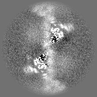
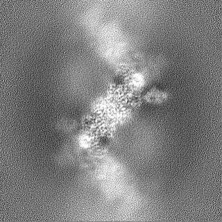
| Images |  emd_46577.png emd_46577.png | 108.4 KB | ||
| Filedesc metadata | emd-46577.cif.gz | 6.1 KB | ||
| Others | emd_46577_half_map_1.map.gzemd_46577_half_map_2.map.gz | 116.1 MB 116.1 MB | ||
| Archive directory |  http://ftp.pdbj.org/pub/emdb/structures/EMD-46577ftp://ftp.pdbj.org/pub/emdb/structures/EMD-46577 http://ftp.pdbj.org/pub/emdb/structures/EMD-46577ftp://ftp.pdbj.org/pub/emdb/structures/EMD-46577 | HTTPS FTP |
-Related structure data
| Related structure data |  9d5aMC  8u38C  9cd2C  9d59C  9d5bC M: atomic model generated by this map C: citing same article ( |
|---|


-Links
| EMDB pages | EMDB (EBI/PDBe) / EMDataResource |
|---|
-Map
| File | Download / File: emd_46577.map.gz / Format: CCP4 / Size: 125 MB / Type: IMAGE STORED AS FLOATING POINT NUMBER (4 BYTES) | ||||||||||||||||||||||||||||||||||||
|---|---|---|---|---|---|---|---|---|---|---|---|---|---|---|---|---|---|---|---|---|---|---|---|---|---|---|---|---|---|---|---|---|---|---|---|---|---|
| Projections & slices | Image control
Images are generated by Spider. | ||||||||||||||||||||||||||||||||||||
| Voxel size | X=Y=Z: 0.935 Å | ||||||||||||||||||||||||||||||||||||
| Density |
| ||||||||||||||||||||||||||||||||||||
| Symmetry | Space group: 1 | ||||||||||||||||||||||||||||||||||||
| Details | EMDB XML:
|
 Z (Sec.)
 Y (Row.)
 X (Col.)


-Supplemental data
-Half map: #1
| File | emd_46577_half_map_1.map | ||||||||||||
|---|---|---|---|---|---|---|---|---|---|---|---|---|---|
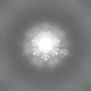
| Projections & Slices |
| ||||||||||||
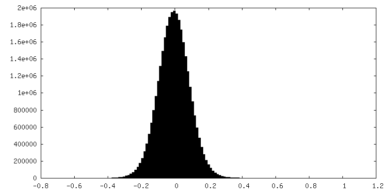
| Density Histograms |


-Half map: #2
| File | emd_46577_half_map_2.map | ||||||||||||
|---|---|---|---|---|---|---|---|---|---|---|---|---|---|
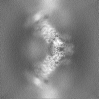
| Projections & Slices |
| ||||||||||||
| Density Histograms |


- Sample components
-Entire : Filament assembly of Citrobacter sp. ubiquitin-like (Ubl) protein...
| Entire | Name: Filament assembly of Citrobacter sp. ubiquitin-like (Ubl) protein with Ca ion |
|---|---|
| Components |
|
-Supramolecule #1: Filament assembly of Citrobacter sp. ubiquitin-like (Ubl) protein...
| Supramolecule | Name: Filament assembly of Citrobacter sp. ubiquitin-like (Ubl) protein with Ca ion type: complex / ID: 1 / Parent: 0 / Macromolecule list: #1 |
|---|---|
| Source (natural) | Organism: Citrobacter sp. RHBSTW-00271 (bacteria) |
| Molecular weight | Theoretical: 20 kDa/nm |
-Macromolecule #1: Multi-ubiquitin domain-containing protein
| Macromolecule | Name: Multi-ubiquitin domain-containing protein / type: protein_or_peptide / ID: 1 / Number of copies: 1 / Enantiomer: LEVO |
|---|---|
| Source (natural) | Organism: Citrobacter sp. RHBSTW-00271 (bacteria) |
| Molecular weight | Theoretical: 28.376852 KDa |
| Recombinant expression | Organism: |
| Sequence | String: MKSSHHHHHH ENLYFQSNAQ DIQSQHHHRR FIEVADETLS FRQVVMEDST PNGSQISAAS GFKPDQMPVV LMLLPNGSLE DIRPDEVVD LSSEVRRFIV VESDRTYFFT IDGARLEWPC RFITGYSIRQ LGDIGDNKKL LLEREDEADL EVQNDQIIDL D GDGIERFI ...String: MKSSHHHHHH ENLYFQSNAQ DIQSQHHHRR FIEVADETLS FRQVVMEDST PNGSQISAAS GFKPDQMPVV LMLLPNGSLE DIRPDEVVD LSSEVRRFIV VESDRTYFFT IDGARLEWPC RFITGYSIRQ LGDIGDNKKL LLEREDEADL EVQNDQIIDL D GDGIERFI SRKATWKLNI QGKEFTFDTP TVVIRDAVIR AGLNPNQAWH IFLKVEGQPK VEKNIDDVID LRTPGIEKLR LT PKDVNNG |
-Macromolecule #2: CALCIUM ION
| Macromolecule | Name: CALCIUM ION / type: ligand / ID: 2 / Number of copies: 5 / Formula: CA |
|---|---|
| Molecular weight | Theoretical: 40.078 Da |
-Experimental details
-Structure determination
| Method | cryo EM |
|---|---|
 Processing Processing | single particle reconstruction |
| Aggregation state | filament |
-Sample preparation
| Buffer | pH: 8.5 Component:
| ||||||||||||||||||
|---|---|---|---|---|---|---|---|---|---|---|---|---|---|---|---|---|---|---|---|
| Grid | Model: Quantifoil R1.2/1.3 / Material: COPPER / Support film - Material: CARBON / Support film - topology: HOLEY / Pretreatment - Type: PLASMA CLEANING / Pretreatment - Time: 10 sec. | ||||||||||||||||||
| Vitrification | Cryogen name: ETHANE / Chamber humidity: 100 % / Chamber temperature: 298 K / Instrument: FEI VITROBOT MARK III |
- Electron microscopy
| Microscope | FEI TITAN KRIOS |
|---|---|
| Image recording | Film or detector model: FEI FALCON IV (4k x 4k) / Average electron dose: 45.0 e/Å2 |
| Electron beam | Acceleration voltage: 300 kV / Electron source:  FIELD EMISSION GUN FIELD EMISSION GUN |
| Electron optics | C2 aperture diameter: 100.0 µm / Illumination mode: FLOOD BEAM / Imaging mode: BRIGHT FIELD / Cs: 2.7 mm / Nominal defocus max: 2.2 µm / Nominal defocus min: 0.5 µm / Nominal magnification: 130000 |
| Sample stage | Cooling holder cryogen: NITROGEN |
| Experimental equipment |  Model: Titan Krios / Image courtesy: FEI Company |

