National Institutes of Health/Eunice Kennedy Shriver National Institute of Child Health & Human Development (NIH/NICHD)
1DP2HL163810-01
United States
American Heart Association
847236
United States
Citation
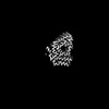
Journal: Structure / Year: 2024 Title: ATTRv-V30M amyloid fibrils from heart and nerves exhibit structural homogeneity. Authors: Binh An Nguyen / Shumaila Afrin / Anna Yakubovska / Virender Singh / Rose Pedretti / Parker Bassett / Maja Pekala / Jaime Vaquer Alicea / Peter Kunach / Lanie Wang / Andrew Lemoff / Barbara ...Authors: Binh An Nguyen / Shumaila Afrin / Anna Yakubovska / Virender Singh / Rose Pedretti / Parker Bassett / Maja Pekala / Jaime Vaquer Alicea / Peter Kunach / Lanie Wang / Andrew Lemoff / Barbara Kluve-Beckerman / Lorena Saelices / Abstract: Amyloidogenic transthyretin (ATTR) amyloidosis is a systemic disease characterized by the deposition of amyloid fibrils made of transthyretin. Transthyretin is primarily produced in tetrameric form ...Amyloidogenic transthyretin (ATTR) amyloidosis is a systemic disease characterized by the deposition of amyloid fibrils made of transthyretin. Transthyretin is primarily produced in tetrameric form by the liver, but also by retinal epithelium and choroid plexus. The deposition of these fibrils in the myocardium and peripheral nerves causes cardiomyopathies and neuropathies, respectively. Using cryoelectron microscopy (cryo-EM), we investigated fibrils extracted from cardiac and nerve tissues of an ATTRv-V30M patient. We found consistent fibril structures from both tissues, similar to cardiac fibrils previously described, but different from vitreous humor fibrils of the same genotype. Our findings, along with previous ATTR fibrils structural studies, suggest a uniform fibrillar architecture across different tissues when transthyretin originates from the liver. This study advances our understanding of how deposition and production sites influence fibril structure in ATTRv-V30M amyloidosis.
Model: Quantifoil R1.2/1.3 / Material: COPPER / Mesh: 300 / Support film - Material: CARBON / Support film - topology: HOLEY / Pretreatment - Type: GLOW DISCHARGE / Pretreatment - Time: 30 sec. / Pretreatment - Atmosphere: AIR
Vitrification
Cryogen name: ETHANE / Chamber humidity: 100 % / Chamber temperature: 298 K / Instrument: FEI VITROBOT MARK IV / Details: Blot-force 0, blotting time 3 sec.
Details
Sample was extracted using water-based extraction method.
-
Electron microscopy
Microscope
FEI TITAN KRIOS
Image recording
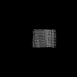
Film or detector model: FEI FALCON IV (4k x 4k) / Number grids imaged: 1 / Number real images: 8957 / Average exposure time: 4.57 sec. / Average electron dose: 40.0 e/Å2
Electron beam
Acceleration voltage: 300 kV / Electron source: FIELD EMISSION GUN
In the structure databanks used in Yorodumi, some data are registered as the other names, "COVID-19 virus" and "2019-nCoV". Here are the details of the virus and the list of structure data.
Jan 31, 2019. EMDB accession codes are about to change! (news from PDBe EMDB page)
EMDB accession codes are about to change! (news from PDBe EMDB page)
The allocation of 4 digits for EMDB accession codes will soon come to an end. Whilst these codes will remain in use, new EMDB accession codes will include an additional digit and will expand incrementally as the available range of codes is exhausted. The current 4-digit format prefixed with “EMD-” (i.e. EMD-XXXX) will advance to a 5-digit format (i.e. EMD-XXXXX), and so on. It is currently estimated that the 4-digit codes will be depleted around Spring 2019, at which point the 5-digit format will come into force.
The EM Navigator/Yorodumi systems omit the EMD- prefix.
Related info.:Q: What is EMD? / ID/Accession-code notation in Yorodumi/EM Navigator
Yorodumi is a browser for structure data from EMDB, PDB, SASBDB, etc.
This page is also the successor to EM Navigator detail page, and also detail information page/front-end page for Omokage search.
The word "yorodu" (or yorozu) is an old Japanese word meaning "ten thousand". "mi" (miru) is to see.
Related info.:EMDB / PDB / SASBDB / Comparison of 3 databanks / Yorodumi Search / Aug 31, 2016. New EM Navigator & Yorodumi / Yorodumi Papers / Jmol/JSmol / Function and homology information / Changes in new EM Navigator and Yorodumi
 Movie
Movie Controller

 Yorodumi
 Open data
 Basic information
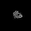
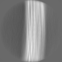
 Map data
 Sample
 Keywords
 Function and homology information
 Homo sapiens (human)
 Authors
 United States, 2 items
  Citation
 Structure visualization
 Downloads & links
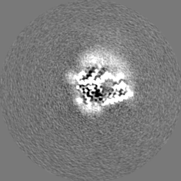
 emd_45039.png
 http://ftp.pdbj.org/pub/emdb/structures/EMD-45039


 Z (Sec.)
 Y (Row.)
 X (Col.)


 Sample components
 Processing
 Electron microscopy
 FIELD EMISSION GUN


