Movie
Movie Controller


+ Open data

- Basic information
| Entry |  | |||||||||
|---|---|---|---|---|---|---|---|---|---|---|
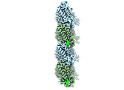
| Title | Structure of alpha1B and betaI/IVb microtubule bound to GMPCPP | |||||||||
 Map data Map data | Full B-factor sharpened map of a1B/bI bIVb microtubule | |||||||||
 Sample Sample |
| |||||||||
 Keywords Keywords | human microtubule / tubulin isoforms / cytoskeleton / STRUCTURAL PROTEIN | |||||||||
| Function / homology |  Function and homology information Function and homology informationodontoblast differentiation / Post-chaperonin tubulin folding pathway / Carboxyterminal post-translational modifications of tubulin / Microtubule-dependent trafficking of connexons from Golgi to the plasma membrane / Cilium Assembly / Sealing of the nuclear envelope (NE) by ESCRT-III / Intraflagellar transport / cytoskeleton-dependent intracellular transport / Formation of tubulin folding intermediates by CCT/TriC / Gap junction assembly ...odontoblast differentiation / Post-chaperonin tubulin folding pathway / Carboxyterminal post-translational modifications of tubulin / Microtubule-dependent trafficking of connexons from Golgi to the plasma membrane / Cilium Assembly / Sealing of the nuclear envelope (NE) by ESCRT-III / Intraflagellar transport / cytoskeleton-dependent intracellular transport / Formation of tubulin folding intermediates by CCT/TriC / Gap junction assembly / COPI-independent Golgi-to-ER retrograde traffic / natural killer cell mediated cytotoxicity / Assembly and cell surface presentation of NMDA receptors / Kinesins / GTPase activating protein binding / COPI-dependent Golgi-to-ER retrograde traffic / intercellular bridge / regulation of synapse organization / nuclear envelope lumen / MHC class I protein binding / Recycling pathway of L1 / cytoplasmic microtubule / microtubule-based process / RHOH GTPase cycle / spindle assembly / RHO GTPases activate IQGAPs / cellular response to interleukin-4 / Hedgehog 'off' state / COPI-mediated anterograde transport / Activation of AMPK downstream of NMDARs / Mitotic Prometaphase / EML4 and NUDC in mitotic spindle formation / Loss of Nlp from mitotic centrosomes / Loss of proteins required for interphase microtubule organization from the centrosome / Recruitment of mitotic centrosome proteins and complexes / Resolution of Sister Chromatid Cohesion / Recruitment of NuMA to mitotic centrosomes / Anchoring of the basal body to the plasma membrane / HSP90 chaperone cycle for steroid hormone receptors (SHR) in the presence of ligand / MHC class II antigen presentation / AURKA Activation by TPX2 / Translocation of SLC2A4 (GLUT4) to the plasma membrane / RHO GTPases Activate Formins / Hydrolases; Acting on acid anhydrides; Acting on GTP to facilitate cellular and subcellular movement / PKR-mediated signaling / structural constituent of cytoskeleton / mitotic spindle / cytoplasmic ribonucleoprotein granule / Aggrephagy / microtubule cytoskeleton organization / HCMV Early Events / Separation of Sister Chromatids / The role of GTSE1 in G2/M progression after G2 checkpoint / microtubule cytoskeleton / Regulation of PLK1 Activity at G2/M Transition / azurophil granule lumen / double-stranded RNA binding / mitotic cell cycle / cell body / microtubule / Potential therapeutics for SARS / cytoskeleton / membrane raft / protein domain specific binding / cell division / GTPase activity / ubiquitin protein ligase binding / Neutrophil degranulation / protein-containing complex binding / GTP binding / structural molecule activity / protein-containing complex / extracellular exosome / extracellular region / nucleus / metal ion binding / cytoplasm / cytosol Similarity search - Function | |||||||||
| Biological species |  Homo sapiens (human) Homo sapiens (human) | |||||||||
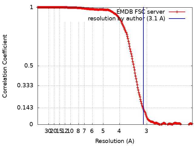
| Method | single particle reconstruction / cryo EM / Resolution: 3.1 Å | |||||||||
 Authors Authors | Zehr EA / Roll-Mecak A | |||||||||
| Funding support |  United States, 1 items United States, 1 items
| |||||||||
 Citation Citation | Journal: Biorxiv / Year: 2023 Title: Cryo-EM structures of human alpha 1B/ beta I+ beta IVb microtubules shed light on isoform specific assembly Authors: Zehr EA / Roll-Mecak A | |||||||||
| History |
|
- Structure visualization
| Supplemental images |
|---|
- Downloads & links
-EMDB archive
| Map data | emd_44762.map.gz | 30 MB | EMDB map data format | |
|---|---|---|---|---|
| Header (meta data) | emd-44762-v30.xmlemd-44762.xml | 25.8 KB 25.8 KB | Display Display | EMDB header |
| FSC (resolution estimation) | emd_44762_fsc.xml | 25.8 KB | Display | FSC data file |
| Images |  emd_44762.png emd_44762.png | 46.3 KB | ||
| Masks | emd_44762_msk_1.map | 699 MB | Mask map | |
| Filedesc metadata | emd-44762.cif.gz | 6.9 KB | ||
| Others | emd_44762_additional_1.map.gzemd_44762_additional_2.map.gzemd_44762_half_map_1.map.gzemd_44762_half_map_2.map.gz | 560.5 MB 8.8 MB 563.1 MB 562.4 MB | ||
| Archive directory |  http://ftp.pdbj.org/pub/emdb/structures/EMD-44762ftp://ftp.pdbj.org/pub/emdb/structures/EMD-44762 http://ftp.pdbj.org/pub/emdb/structures/EMD-44762ftp://ftp.pdbj.org/pub/emdb/structures/EMD-44762 | HTTPS FTP |
-Validation report
| Summary document | emd_44762_validation.pdf.gz | 664.1 KB | Display | EMDB validaton report |
|---|---|---|---|---|
| Full document | emd_44762_full_validation.pdf.gz | 663.7 KB | Display | |
| Data in XML | emd_44762_validation.xml.gz | 27 KB | Display | |
| Data in CIF | emd_44762_validation.cif.gz | 36.1 KB | Display | |
| Arichive directory | https://ftp.pdbj.org/pub/emdb/validation_reports/EMD-44762ftp://ftp.pdbj.org/pub/emdb/validation_reports/EMD-44762 | HTTPS FTP |
-Related structure data
| Related structure data |  9bp6MC  42916 M: atomic model generated by this map C: citing same article ( |
|---|---|
| Similar structure data |
-Links
| EMDB pages | EMDB (EBI/PDBe) / EMDataResource |
|---|---|
| Related items in Molecule of the Month |


-Map
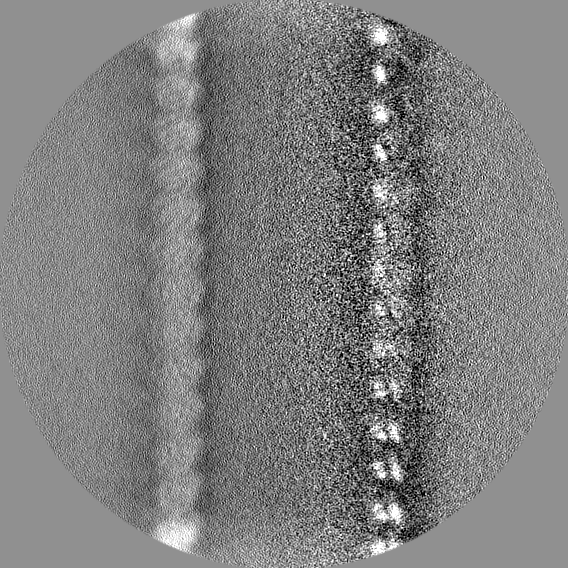
| File | Download / File: emd_44762.map.gz / Format: CCP4 / Size: 699 MB / Type: IMAGE STORED AS FLOATING POINT NUMBER (4 BYTES) | ||||||||||||||||||||||||||||||||||||
|---|---|---|---|---|---|---|---|---|---|---|---|---|---|---|---|---|---|---|---|---|---|---|---|---|---|---|---|---|---|---|---|---|---|---|---|---|---|
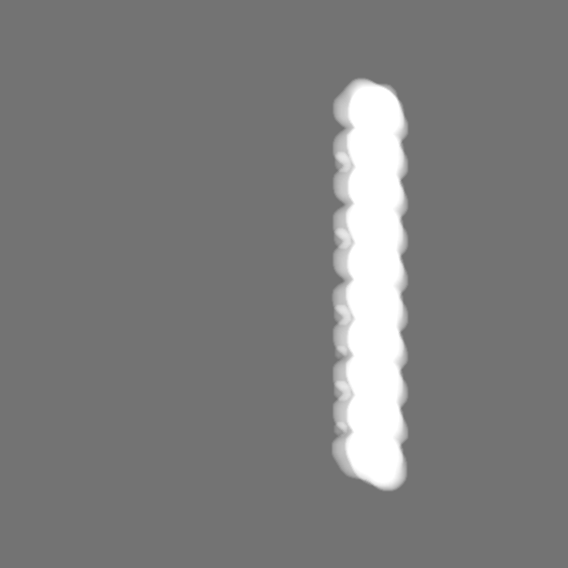
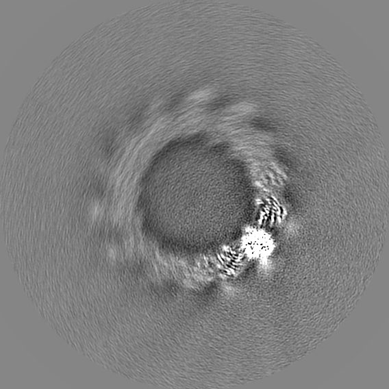
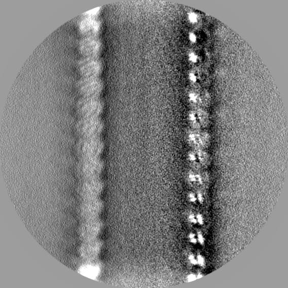
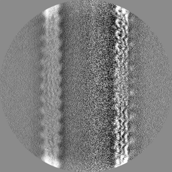
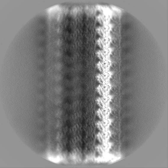
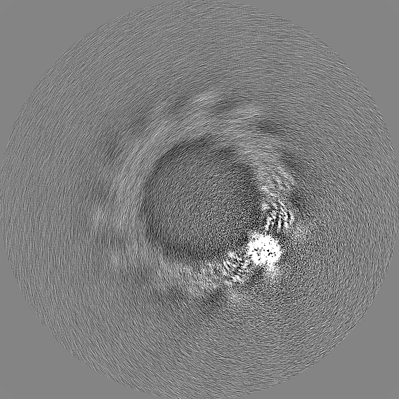
| Annotation | Full B-factor sharpened map of a1B/bI bIVb microtubule | ||||||||||||||||||||||||||||||||||||
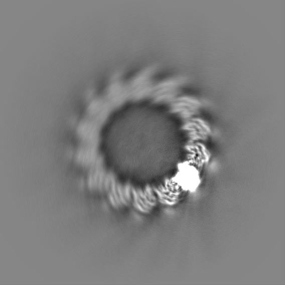
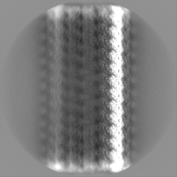
| Projections & slices | Image control
Images are generated by Spider. | ||||||||||||||||||||||||||||||||||||
| Voxel size | X=Y=Z: 1.06 Å | ||||||||||||||||||||||||||||||||||||
| Density |
| ||||||||||||||||||||||||||||||||||||
| Symmetry | Space group: 1 | ||||||||||||||||||||||||||||||||||||
| Details | EMDB XML:
|
 Z (Sec.)
 Y (Row.)
 X (Col.)


-Supplemental data
-Mask #1
| File | emd_44762_msk_1.map | ||||||||||||
|---|---|---|---|---|---|---|---|---|---|---|---|---|---|
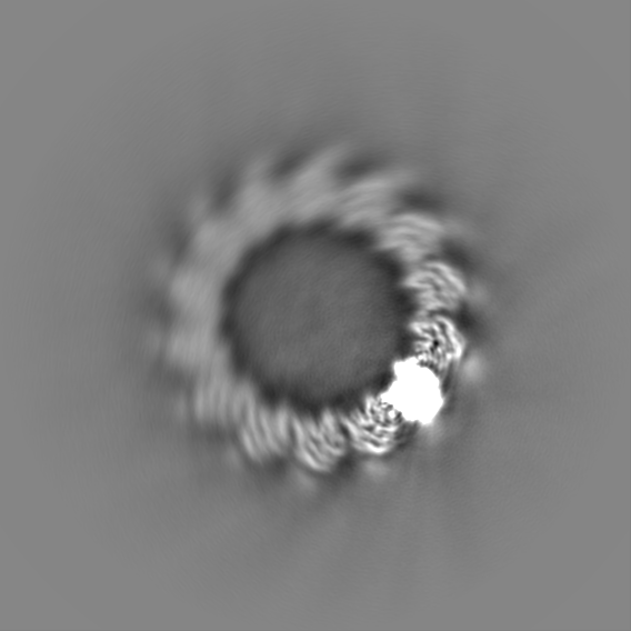
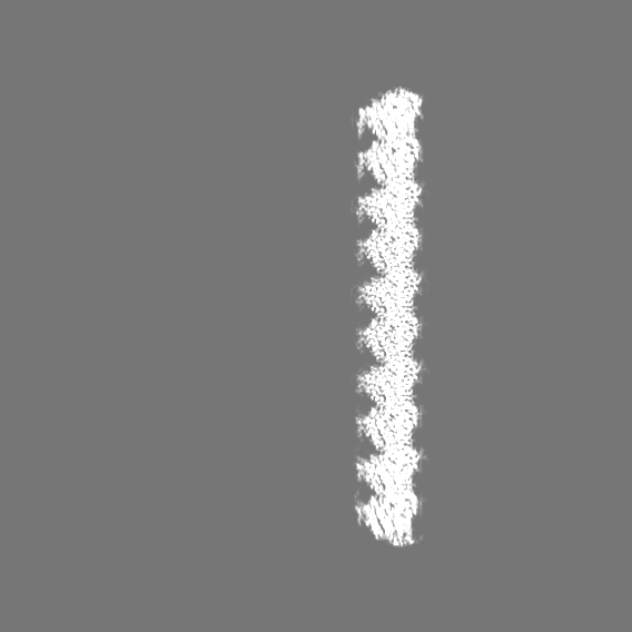
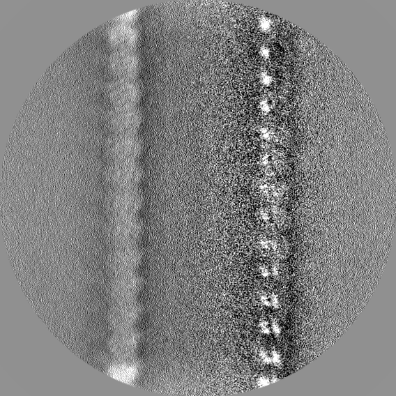
| Projections & Slices |
| ||||||||||||
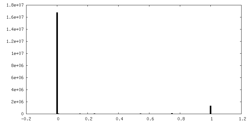
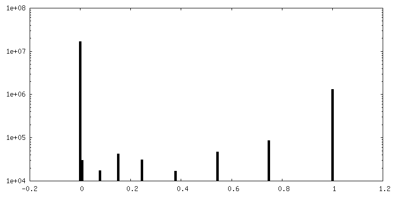
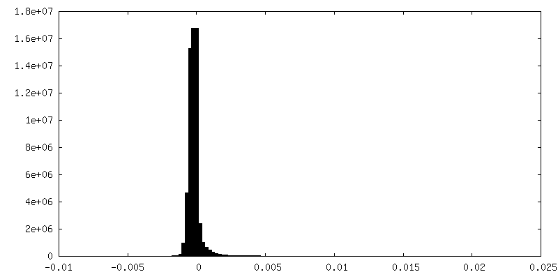
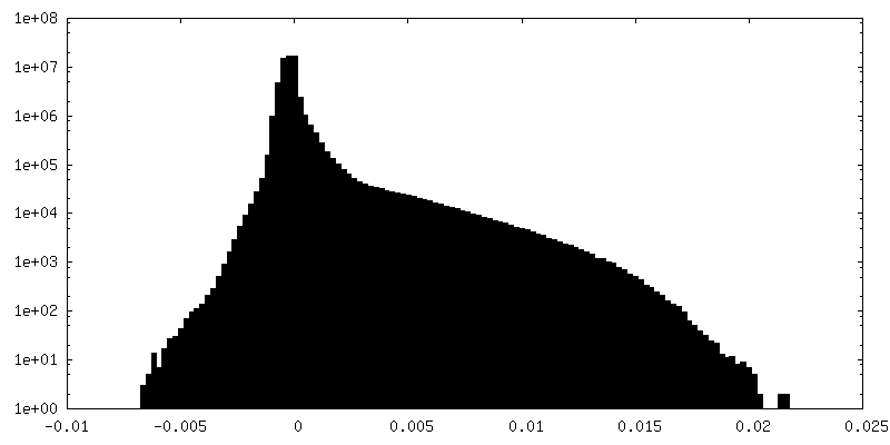
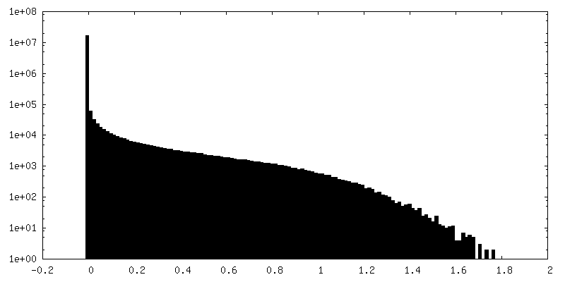
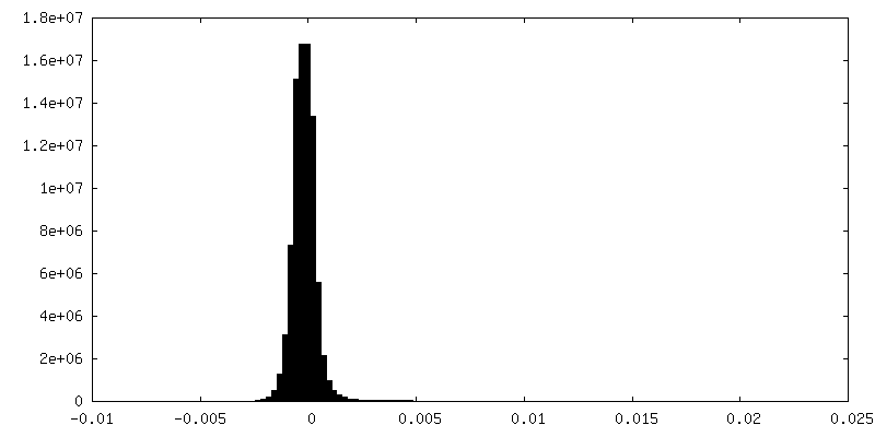
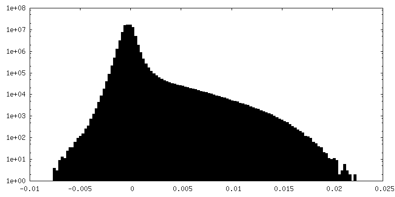
| Density Histograms |


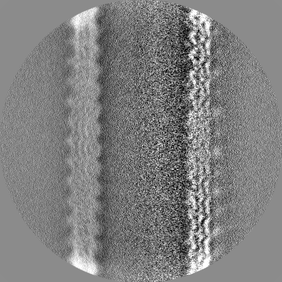
-Additional map: Full raw map of a1B/bI bIVb microtubule
| File | emd_44762_additional_1.map | ||||||||||||
|---|---|---|---|---|---|---|---|---|---|---|---|---|---|
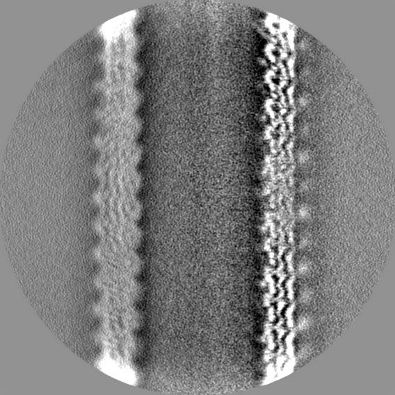
| Annotation | Full raw map of a1B/bI bIVb microtubule | ||||||||||||
| Projections & Slices |
| ||||||||||||
| Density Histograms |


-Additional map: Full map of a1B/bI bIVb microtubule modified with DeepEMhancer
| File | emd_44762_additional_2.map | ||||||||||||
|---|---|---|---|---|---|---|---|---|---|---|---|---|---|
| Annotation | Full map of a1B/bI bIVb microtubule modified with DeepEMhancer | ||||||||||||
| Projections & Slices |
| ||||||||||||
| Density Histograms |


-Half map: First half-map of a1B/bI bIVb microtubule
| File | emd_44762_half_map_1.map | ||||||||||||
|---|---|---|---|---|---|---|---|---|---|---|---|---|---|
| Annotation | First half-map of a1B/bI bIVb microtubule | ||||||||||||
| Projections & Slices |
| ||||||||||||
| Density Histograms |


-Half map: Second half-map of a1B/bI bIVb microtubule
| File | emd_44762_half_map_2.map | ||||||||||||
|---|---|---|---|---|---|---|---|---|---|---|---|---|---|
| Annotation | Second half-map of a1B/bI bIVb microtubule | ||||||||||||
| Projections & Slices |
| ||||||||||||
| Density Histograms |


- Sample components
-Entire : alpha1B and betaI/IVb microtubule
| Entire | Name: alpha1B and betaI/IVb microtubule |
|---|---|
| Components |
|
-Supramolecule #1: alpha1B and betaI/IVb microtubule
| Supramolecule | Name: alpha1B and betaI/IVb microtubule / type: complex / ID: 1 / Parent: 0 / Macromolecule list: #1-#2 / Details: Microtubules were double-cycled with GMPCPP |
|---|---|
| Source (natural) | Organism: Homo sapiens (human) / Organ: kidney / Location in cell: cytoplasm |
| Molecular weight | Theoretical: 300 KDa |
-Macromolecule #1: Tubulin alpha-1B chain
| Macromolecule | Name: Tubulin alpha-1B chain / type: protein_or_peptide / ID: 1 / Number of copies: 2 / Enantiomer: LEVO |
|---|---|
| Source (natural) | Organism: Homo sapiens (human) / Organ: kidney / Location in cell: cytoplasm |
| Molecular weight | Theoretical: 50.204445 KDa |
| Sequence | String: MRECISIHVG QAGVQIGNAC WELYCLEHGI QPDGQMPSDK TIGGGDDSFN TFFSETGAGK HVPRAVFVDL EPTVIDEVRT GTYRQLFHP EQLITGKEDA ANNYARGHYT IGKEIIDLVL DRIRKLADQC TGLQGFLVFH SFGGGTGSGF TSLLMERLSV D YGKKSKLE ...String: MRECISIHVG QAGVQIGNAC WELYCLEHGI QPDGQMPSDK TIGGGDDSFN TFFSETGAGK HVPRAVFVDL EPTVIDEVRT GTYRQLFHP EQLITGKEDA ANNYARGHYT IGKEIIDLVL DRIRKLADQC TGLQGFLVFH SFGGGTGSGF TSLLMERLSV D YGKKSKLE FSIYPAPQVS TAVVEPYNSI LTTHTTLEHS DCAFMVDNEA IYDICRRNLD IERPTYTNLN RLISQIVSSI TA SLRFDGA LNVDLTEFQT NLVPYPRIHF PLATYAPVIS AEKAYHEQLS VAEITNACFE PANQMVKCDP RHGKYMACCL LYR GDVVPK DVNAAIATIK TKRSIQFVDW CPTGFKVGIN YQPPTVVPGG DLAKVQRAVC MLSNTTAIAE AWARLDHKFD LMYA KRAFV HWYVGEGMEE GEFSEAREDM AALEKDYEEV GVDSVEGEGE EEGEEY UniProtKB: Tubulin alpha-1B chain |
-Macromolecule #2: Tubulin beta chain
| Macromolecule | Name: Tubulin beta chain / type: protein_or_peptide / ID: 2 / Number of copies: 2 / Enantiomer: LEVO |
|---|---|
| Source (natural) | Organism: Homo sapiens (human) / Organ: kidney / Location in cell: cytoplasm |
| Molecular weight | Theoretical: 49.717629 KDa |
| Sequence | String: MREIVHIQAG QCGNQIGAKF WEVISDEHGI DPTGTYHGDS DLQLDRISVY YNEATGGKYV PRAILVDLEP GTMDSVRSGP FGQIFRPDN FVFGQSGAGN NWAKGHYTEG AELVDSVLDV VRKEAESCDC LQGFQLTHSL GGGTGSGMGT LLISKIREEY P DRIMNTFS ...String: MREIVHIQAG QCGNQIGAKF WEVISDEHGI DPTGTYHGDS DLQLDRISVY YNEATGGKYV PRAILVDLEP GTMDSVRSGP FGQIFRPDN FVFGQSGAGN NWAKGHYTEG AELVDSVLDV VRKEAESCDC LQGFQLTHSL GGGTGSGMGT LLISKIREEY P DRIMNTFS VVPSPKVSDT VVEPYNATLS VHQLVENTDE TYCIDNEALY DICFRTLKLT TPTYGDLNHL VSATMSGVTT CL RFPGQLN ADLRKLAVNM VPFPRLHFFM PGFAPLTSRG SQQYRALTVP ELTQQVFDAK NMMAACDPRH GRYLTVAAVF RGR MSMKEV DEQMLNVQNK NSSYFVEWIP NNVKTAVCDI PPRGLKMAVT FIGNSTAIQE LFKRISEQFT AMFRRKAFLH WYTG EGMDE MEFTEAESNM NDLVSEYQQY QDATAEEEED FGEEAEEEA UniProtKB: Tubulin beta chain |
-Macromolecule #3: GUANOSINE-5'-TRIPHOSPHATE
| Macromolecule | Name: GUANOSINE-5'-TRIPHOSPHATE / type: ligand / ID: 3 / Number of copies: 2 / Formula: GTP |
|---|---|
| Molecular weight | Theoretical: 523.18 Da |
| Chemical component information |  ChemComp-GTP: |
-Macromolecule #4: MAGNESIUM ION
| Macromolecule | Name: MAGNESIUM ION / type: ligand / ID: 4 / Number of copies: 4 / Formula: MG |
|---|---|
| Molecular weight | Theoretical: 24.305 Da |
-Macromolecule #5: PHOSPHOMETHYLPHOSPHONIC ACID GUANYLATE ESTER
| Macromolecule | Name: PHOSPHOMETHYLPHOSPHONIC ACID GUANYLATE ESTER / type: ligand / ID: 5 / Number of copies: 2 / Formula: G2P |
|---|---|
| Molecular weight | Theoretical: 521.208 Da |
| Chemical component information |  ChemComp-G2P: |
-Experimental details
-Structure determination
| Method | cryo EM |
|---|---|
 Processing Processing | single particle reconstruction |
| Aggregation state | filament |
-Sample preparation
| Concentration | 0.1 mg/mL | |||||||||||||||
|---|---|---|---|---|---|---|---|---|---|---|---|---|---|---|---|---|
| Buffer | pH: 7 Component:
| |||||||||||||||
| Grid | Model: C-flat-1.2/1.3 / Material: GOLD / Mesh: 400 / Support film - Material: CARBON / Support film - topology: HOLEY / Support film - Film thickness: 100 / Pretreatment - Type: GLOW DISCHARGE / Pretreatment - Time: 30 sec. | |||||||||||||||
| Vitrification | Cryogen name: ETHANE / Chamber humidity: 100 % / Chamber temperature: 303 K / Instrument: LEICA EM GP Details: Double-cycled GMPCPP microtubules were diluted to 2.5uM in Brb80 buffer. 5 ul of microtubules were applied to glow-discharged cryo-EM grid and allowed to absorb for 30 sec.. | |||||||||||||||
| Details | Microtubules were double-cycled with GMPCPP |
- Electron microscopy
| Microscope | FEI TITAN KRIOS |
|---|---|
| Specialist optics | Energy filter - Name: GIF Bioquantum / Energy filter - Slit width: 20 eV |
| Software | Name: SerialEM |
| Image recording | Film or detector model: GATAN K2 SUMMIT (4k x 4k) / Detector mode: COUNTING / Digitization - Frames/image: 1-40 / Number real images: 6930 / Average exposure time: 7.8 sec. / Average electron dose: 56.0 e/Å2 |
| Electron beam | Acceleration voltage: 300 kV / Electron source:  FIELD EMISSION GUN FIELD EMISSION GUN |
| Electron optics | Illumination mode: FLOOD BEAM / Imaging mode: BRIGHT FIELD / Cs: 2.7 mm / Nominal defocus max: 2.5 µm / Nominal defocus min: 0.5 µm / Nominal magnification: 130000 |
| Experimental equipment |  Model: Titan Krios / Image courtesy: FEI Company |
+Image processing
-Atomic model buiding 1
| Initial model | PDB ID: Chain - Residue range: 1-437 / Chain - Source name: PDB / Chain - Initial model type: experimental model |
|---|---|
| Software | Name: Coot |
| Refinement | Space: REAL / Protocol: FLEXIBLE FIT / Target criteria: Cross-correlation |
| Output model | PDB-9bp6: |