[English] 日本語
 Yorodumi
- EMDB-44539: Cryo-EM structure of human monoclonal antibody C74 targeting PFD1... -
+ Open data
- Basic information
| Entry |  | |||||||||
|---|---|---|---|---|---|---|---|---|---|---|
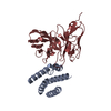
| Title | Cryo-EM structure of human monoclonal antibody C74 targeting PFD1235w (CIDRa1.6) PfEMP1 | |||||||||
 Map data Map data | ||||||||||
 Sample Sample |
| |||||||||
 Keywords Keywords | Antibody / Plasmodium / Monoclonal / Neutralizing / IMMUNE SYSTEM | |||||||||
| Function / homology |  Function and homology information Function and homology informationsymbiont-mediated perturbation of host erythrocyte aggregation / infected host cell surface knob / antigenic variation / adhesion of symbiont to microvasculature / cell adhesion molecule binding / cell-cell adhesion / host cell surface receptor binding / host cell plasma membrane / membrane Similarity search - Function | |||||||||
| Biological species |   Homo sapiens (human) Homo sapiens (human) | |||||||||
| Method | single particle reconstruction / cryo EM / Resolution: 3.1 Å | |||||||||
 Authors Authors | Raghavan SSR / Ward AB | |||||||||
| Funding support |  United States, United States,  Denmark, 2 items Denmark, 2 items
| |||||||||
 Citation Citation |  Journal: To Be Published Journal: To Be PublishedTitle: Cryo-EM structure of human monoclonal C74 targeting CIDRa1.6 PfEMP1 malarial protein Authors: Raghavan SSR / Ward AB | |||||||||
| History |
|
- Structure visualization
| Supplemental images |
|---|
- Downloads & links
-EMDB archive
| Map data |  emd_44539.map.gz emd_44539.map.gz | 168 MB |  EMDB map data format EMDB map data format | |
|---|---|---|---|---|
| Header (meta data) |  emd-44539-v30.xml emd-44539-v30.xml emd-44539.xml emd-44539.xml | 15.4 KB 15.4 KB | Display Display |  EMDB header EMDB header |
| Images |  emd_44539.png emd_44539.png | 37.9 KB | ||
| Filedesc metadata |  emd-44539.cif.gz emd-44539.cif.gz | 5.8 KB | ||
| Others |  emd_44539_half_map_1.map.gz emd_44539_half_map_1.map.gz emd_44539_half_map_2.map.gz emd_44539_half_map_2.map.gz | 165.4 MB 165.4 MB | ||
| Archive directory |  http://ftp.pdbj.org/pub/emdb/structures/EMD-44539 http://ftp.pdbj.org/pub/emdb/structures/EMD-44539 ftp://ftp.pdbj.org/pub/emdb/structures/EMD-44539 ftp://ftp.pdbj.org/pub/emdb/structures/EMD-44539 | HTTPS FTP |
-Validation report
| Summary document |  emd_44539_validation.pdf.gz emd_44539_validation.pdf.gz | 903.4 KB | Display |  EMDB validaton report EMDB validaton report |
|---|---|---|---|---|
| Full document |  emd_44539_full_validation.pdf.gz emd_44539_full_validation.pdf.gz | 902.9 KB | Display | |
| Data in XML |  emd_44539_validation.xml.gz emd_44539_validation.xml.gz | 14.7 KB | Display | |
| Data in CIF |  emd_44539_validation.cif.gz emd_44539_validation.cif.gz | 17.6 KB | Display | |
| Arichive directory |  https://ftp.pdbj.org/pub/emdb/validation_reports/EMD-44539 https://ftp.pdbj.org/pub/emdb/validation_reports/EMD-44539 ftp://ftp.pdbj.org/pub/emdb/validation_reports/EMD-44539 ftp://ftp.pdbj.org/pub/emdb/validation_reports/EMD-44539 | HTTPS FTP |
-Related structure data
| Related structure data |  9bhbMC M: atomic model generated by this map C: citing same article ( |
|---|---|
| Similar structure data | Similarity search - Function & homology  F&H Search F&H Search |
- Links
Links
| EMDB pages |  EMDB (EBI/PDBe) / EMDB (EBI/PDBe) /  EMDataResource EMDataResource |
|---|
- Map
Map
| File |  Download / File: emd_44539.map.gz / Format: CCP4 / Size: 178 MB / Type: IMAGE STORED AS FLOATING POINT NUMBER (4 BYTES) Download / File: emd_44539.map.gz / Format: CCP4 / Size: 178 MB / Type: IMAGE STORED AS FLOATING POINT NUMBER (4 BYTES) | ||||||||||||||||||||||||||||||||||||
|---|---|---|---|---|---|---|---|---|---|---|---|---|---|---|---|---|---|---|---|---|---|---|---|---|---|---|---|---|---|---|---|---|---|---|---|---|---|
| Projections & slices | Image control
Images are generated by Spider. | ||||||||||||||||||||||||||||||||||||
| Voxel size | X=Y=Z: 0.725 Å | ||||||||||||||||||||||||||||||||||||
| Density |
| ||||||||||||||||||||||||||||||||||||
| Symmetry | Space group: 1 | ||||||||||||||||||||||||||||||||||||
| Details | EMDB XML:
|
-Supplemental data
-Half map: #2
| File | emd_44539_half_map_1.map | ||||||||||||
|---|---|---|---|---|---|---|---|---|---|---|---|---|---|
| Projections & Slices |
| ||||||||||||
| Density Histograms |
-Half map: #1
| File | emd_44539_half_map_2.map | ||||||||||||
|---|---|---|---|---|---|---|---|---|---|---|---|---|---|
| Projections & Slices |
| ||||||||||||
| Density Histograms |
- Sample components
-Entire : Complex of human monoclonal C74 with PfEMP1 PFD1235w N-terminal d...
| Entire | Name: Complex of human monoclonal C74 with PfEMP1 PFD1235w N-terminal domain complex |
|---|---|
| Components |
|
-Supramolecule #1: Complex of human monoclonal C74 with PfEMP1 PFD1235w N-terminal d...
| Supramolecule | Name: Complex of human monoclonal C74 with PfEMP1 PFD1235w N-terminal domain complex type: cell / ID: 1 / Parent: 0 / Macromolecule list: all |
|---|---|
| Source (natural) | Organism:  |
-Macromolecule #1: C74 heavy chain
| Macromolecule | Name: C74 heavy chain / type: protein_or_peptide / ID: 1 / Number of copies: 1 / Enantiomer: LEVO |
|---|---|
| Source (natural) | Organism:  Homo sapiens (human) Homo sapiens (human) |
| Molecular weight | Theoretical: 13.441013 KDa |
| Recombinant expression | Organism:  |
| Sequence | String: EVQLVQSGGA LVRPGGSLRL SCAASGFDFS DFEMNWVRQA PGKGLEWISY ISKISAASFY ADSVEGRFTI SRDNTKNLLW LEMTSLRDE DTAVYYCARD LPGYLERVFD LWGQGTLVSV SS |
-Macromolecule #2: C74 kappa chain
| Macromolecule | Name: C74 kappa chain / type: protein_or_peptide / ID: 2 / Number of copies: 1 / Enantiomer: LEVO |
|---|---|
| Source (natural) | Organism:  Homo sapiens (human) Homo sapiens (human) |
| Molecular weight | Theoretical: 12.178488 KDa |
| Recombinant expression | Organism:  |
| Sequence | String: EIVLTQSPAT LSLSPGEDAT LSCRASQSVG SALAWYQHRP GQSPRLLIYD ASTRATGIPA RFSGSGSGTE FTLTVSSLTS EDFAVYYCQ EYKNSVPPTW TFGQGTKVEI KRTV |
-Macromolecule #3: Erythrocyte membrane protein 1, PfEMP1
| Macromolecule | Name: Erythrocyte membrane protein 1, PfEMP1 / type: protein_or_peptide / ID: 3 / Number of copies: 1 / Enantiomer: LEVO |
|---|---|
| Source (natural) | Organism:  |
| Molecular weight | Theoretical: 85.069266 KDa |
| Recombinant expression | Organism:  |
| Sequence | String: MGNASSSEGE AKTPSLTESH NSARNILEGY AESIKEQASK DAKIHGHHLK GDLAKAVFRH PFSAYRPNYG NPCELDYRFH TNVWHRNAE DRNPCLFSRA KRFSNEGEAE CNGGIITGNK GECGACAPYR RRHICDYNLH HINENNIRNT HDLLGNLLVM A RSEGESIV ...String: MGNASSSEGE AKTPSLTESH NSARNILEGY AESIKEQASK DAKIHGHHLK GDLAKAVFRH PFSAYRPNYG NPCELDYRFH TNVWHRNAE DRNPCLFSRA KRFSNEGEAE CNGGIITGNK GECGACAPYR RRHICDYNLH HINENNIRNT HDLLGNLLVM A RSEGESIV KSHEYTGYGI YKSGICTSLA RSFADIGDII RGKDLYRRDS RTDKLEENLR KIFANIYKEL KNGKKWAEAK EY YQDDGTG NYYKLREAWW ALNRKDVWKA LTCSAPRDAQ YFIKSSVRDQ TFSNDYCGHG EHEVLTNLDY VPQFLRWFEE WAE EFCRIK KIKLGKVKEA CRDDSKKLYC SHNGYDCTKT IRNKDILSDN PKCTGCSVKC KVYELWLRNQ RNEFEKQKKK YYKE IQTYT SKDAKTDSNI NNEYYKEFYD KLKNEGYETL NKFIKLLNEG RYCKEKISGE RNIDFTMTGD KDAFYRSDYC QICPE CGVQ CSGTTCTPKK VIHPNCKDKE TYEPGDAKTT DITVLYSGDE EGDIAQKLQD FCNDKNKEND ENYEKWQCYY KSSEIN KCQ MTPSSHKVPK HGYIMSFYAF FDLWVKNLLI DSINWKNDLT NCINNTNVTD CKNDCNTNCK CFENWAKTKE NEWKKVK TI YKNENGNTNN YYKKLNNHFQ GYFFHVMKEL NKEEKWYKLM EDLKEKIDSS NLKNGTKDSE GAIKVLFDHL KDIAERCI D NNSKDSC UniProtKB: Erythrocyte membrane protein 1, PfEMP1 |
-Experimental details
-Structure determination
| Method | cryo EM |
|---|---|
 Processing Processing | single particle reconstruction |
| Aggregation state | particle |
- Sample preparation
| Buffer | pH: 7.5 |
|---|---|
| Grid | Material: GOLD / Mesh: 300 |
| Vitrification | Cryogen name: ETHANE / Chamber humidity: 95 % / Instrument: FEI VITROBOT MARK IV |
- Electron microscopy
| Microscope | TFS GLACIOS |
|---|---|
| Image recording | Film or detector model: FEI FALCON IV (4k x 4k) / Average electron dose: 50.0 e/Å2 |
| Electron beam | Acceleration voltage: 200 kV / Electron source:  FIELD EMISSION GUN FIELD EMISSION GUN |
| Electron optics | Illumination mode: FLOOD BEAM / Imaging mode: BRIGHT FIELD / Nominal defocus max: 2.0 µm / Nominal defocus min: 0.8 µm |
- Image processing
| Startup model | Type of model: INSILICO MODEL |
|---|---|
| Final reconstruction | Resolution.type: BY AUTHOR / Resolution: 3.1 Å / Resolution method: FSC 0.143 CUT-OFF / Number images used: 130000 |
| Initial angle assignment | Type: ANGULAR RECONSTITUTION |
| Final angle assignment | Type: ANGULAR RECONSTITUTION |
 Movie
Movie Controller


 Z (Sec.)
 Y (Row.)
 X (Col.)


