 Movie
Movie Controller

[English] 日本語
 Yorodumi
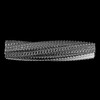
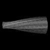
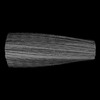
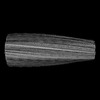
- EMDB-44183: Filament of D-TLKIVWR, a D-peptide that disaggregates Alzheimer's... -
+ Open data

- Basic information
| Entry | 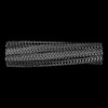 | ||||||||||||
|---|---|---|---|---|---|---|---|---|---|---|---|---|---|
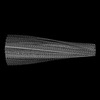
| Title | Filament of D-TLKIVWR, a D-peptide that disaggregates Alzheimer's Paired Helical Filaments, determined by Cryo-EM | ||||||||||||
 Map data Map data | |||||||||||||
 Sample Sample |
| ||||||||||||
 Keywords Keywords | Alzheimer's disease / Tau / fibril / cryo-EM / helix / UNKNOWN FUNCTION | ||||||||||||
| Biological species | synthetic construct (others) | ||||||||||||
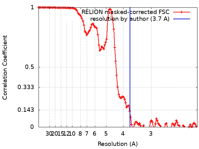
| Method | helical reconstruction / cryo EM / Resolution: 3.7 Å | ||||||||||||
 Authors Authors | Hou K / Ge P / Sawaya MR / Eisenberg DS | ||||||||||||
| Funding support |  United States, 3 items United States, 3 items
| ||||||||||||
 Citation Citation | Journal: Nature / Year: 2025 Title: How short peptides disassemble tau fibrils in Alzheimer's disease. Authors: Ke Hou / Peng Ge / Michael R Sawaya / Liisa Lutter / Joshua L Dolinsky / Yuan Yang / Yi Xiao Jiang / David R Boyer / Xinyi Cheng / Justin Pi / Jeffrey Zhang / Jiahui Lu / Romany Abskharon / ...Authors: Ke Hou / Peng Ge / Michael R Sawaya / Liisa Lutter / Joshua L Dolinsky / Yuan Yang / Yi Xiao Jiang / David R Boyer / Xinyi Cheng / Justin Pi / Jeffrey Zhang / Jiahui Lu / Romany Abskharon / Shixin Yang / Zhiheng Yu / Juli Feigon / David S Eisenberg / Abstract: Reducing fibrous aggregates of the protein tau is a possible strategy for halting the progression of Alzheimer's disease (AD). Previously, we found that in vitro, the D-enantiomeric peptide (D- ...Reducing fibrous aggregates of the protein tau is a possible strategy for halting the progression of Alzheimer's disease (AD). Previously, we found that in vitro, the D-enantiomeric peptide (D-peptide) D-TLKIVWC disassembles ultra-stable tau fibrils extracted from the autopsied brains of individuals with AD (hereafter, these tau fibrils are referred to as AD-tau) into benign segments, with no energy source other than ambient thermal agitation. To consider D-peptide-mediated disassembly as a potential route to therapeutics for AD, it is essential to understand the mechanism and energy source of the disassembly action. Here, we show that the assembly of D-peptides into amyloid-like ('mock-amyloid') fibrils is essential for AD-tau disassembly. These mock-amyloid fibrils have a right-handed twist but are constrained to adopt a left-handed twist when templated in complex with AD-tau. The release of strain that accompanies the conversion of left-twisted to right-twisted, relaxed mock-amyloid produces a torque that is sufficient to break the local hydrogen bonding between tau molecules, and leads to the fragmentation of AD-tau. This strain-relief mechanism seems to operate in other examples of amyloid fibril disassembly, and could inform the development of first-in-class therapeutics for amyloid diseases. | ||||||||||||
| History |
|
- Structure visualization
| Supplemental images |
|---|

- Downloads & links
-EMDB archive
| Map data | emd_44183.map.gz | 91.1 MB |  EMDB map data format EMDB map data format | |
|---|---|---|---|---|
| Header (meta data) | emd-44183-v30.xmlemd-44183.xml | 20.9 KB 20.9 KB | Display Display | EMDB header |
| FSC (resolution estimation) | emd_44183_fsc.xml | 11.4 KB | Display | FSC data file |
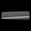
| Images |  emd_44183.png emd_44183.png | 76.6 KB | ||
| Masks | emd_44183_msk_1.map | 125 MB | Mask map | |
| Filedesc metadata | emd-44183.cif.gz | 5.6 KB | ||
| Others | emd_44183_half_map_1.map.gzemd_44183_half_map_2.map.gz | 9 MB 9 MB | ||
| Archive directory |  http://ftp.pdbj.org/pub/emdb/structures/EMD-44183ftp://ftp.pdbj.org/pub/emdb/structures/EMD-44183 http://ftp.pdbj.org/pub/emdb/structures/EMD-44183ftp://ftp.pdbj.org/pub/emdb/structures/EMD-44183 | HTTPS FTP |
-Related structure data
| Related structure data |  9b4kMC  9b4iC  9b4jC  9b4lC  9b4mC  9b4nC  9b4oC M: atomic model generated by this map C: citing same article ( |
|---|
-Links
| EMDB pages | EMDB (EBI/PDBe) / EMDataResource |
|---|
-Map
| File | Download / File: emd_44183.map.gz / Format: CCP4 / Size: 125 MB / Type: IMAGE STORED AS FLOATING POINT NUMBER (4 BYTES) | ||||||||||||||||||||||||||||||||||||
|---|---|---|---|---|---|---|---|---|---|---|---|---|---|---|---|---|---|---|---|---|---|---|---|---|---|---|---|---|---|---|---|---|---|---|---|---|---|
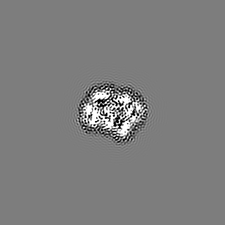
| Projections & slices | Image control
Images are generated by Spider. | ||||||||||||||||||||||||||||||||||||
| Voxel size | X=Y=Z: 1.067 Å | ||||||||||||||||||||||||||||||||||||
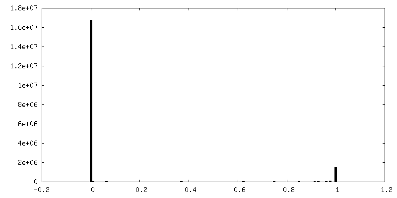
| Density |
| ||||||||||||||||||||||||||||||||||||
| Symmetry | Space group: 1 | ||||||||||||||||||||||||||||||||||||
| Details | EMDB XML:
|
 X (Sec.)
 Y (Row.)
 Z (Col.)


-Supplemental data
-Mask #1
| File | emd_44183_msk_1.map | ||||||||||||
|---|---|---|---|---|---|---|---|---|---|---|---|---|---|
| Projections & Slices |
| ||||||||||||
| Density Histograms |


-Half map: #2
| File | emd_44183_half_map_1.map | ||||||||||||
|---|---|---|---|---|---|---|---|---|---|---|---|---|---|
| Projections & Slices |
| ||||||||||||
| Density Histograms |


-Half map: #1
| File | emd_44183_half_map_2.map | ||||||||||||
|---|---|---|---|---|---|---|---|---|---|---|---|---|---|
| Projections & Slices |
| ||||||||||||
| Density Histograms |


- Sample components
-Entire : D-peptide TLKIVWR
| Entire | Name: D-peptide TLKIVWR |
|---|---|
| Components |
|
-Supramolecule #1: D-peptide TLKIVWR
| Supramolecule | Name: D-peptide TLKIVWR / type: complex / ID: 1 / Parent: 0 / Macromolecule list: all |
|---|---|
| Source (natural) | Organism: synthetic construct (others) / Synthetically produced: Yes |
-Macromolecule #1: DTH-DLE-DLY-DIL-DVA-DTR-DAR
| Macromolecule | Name: DTH-DLE-DLY-DIL-DVA-DTR-DAR / type: protein_or_peptide / ID: 1 / Number of copies: 52 / Enantiomer: DEXTRO |
|---|---|
| Source (natural) | Organism: synthetic construct (others) |
| Molecular weight | Theoretical: 916.142 Da |
| Sequence | String: (DTH)(DLE)(DLY)(DIL)(DVA)(DTR)(DAR) |
-Experimental details
-Structure determination
| Method | cryo EM |
|---|---|
 Processing Processing | helical reconstruction |
| Aggregation state | filament |
-Sample preparation
| Concentration | 10 mg/mL |
|---|---|
| Buffer | pH: 7 / Details: Dissolved in de-ionized water |
| Grid | Model: Quantifoil R1.2/1.3 / Material: COPPER / Mesh: 300 / Pretreatment - Type: GLOW DISCHARGE |
| Vitrification | Cryogen name: ETHANE / Chamber humidity: 100 % / Chamber temperature: 295 K / Instrument: FEI VITROBOT MARK IV |
- Electron microscopy
| Microscope | FEI TITAN KRIOS |
|---|---|
| Image recording | Film or detector model: GATAN K3 BIOQUANTUM (6k x 4k) / Average electron dose: 50.0 e/Å2 |
| Electron beam | Acceleration voltage: 300 kV / Electron source:  FIELD EMISSION GUN FIELD EMISSION GUN |
| Electron optics | Illumination mode: FLOOD BEAM / Imaging mode: BRIGHT FIELD / Cs: 2.7 mm / Nominal defocus max: 2.5 µm / Nominal defocus min: 1.8 µm / Nominal magnification: 81000 |
| Sample stage | Specimen holder model: FEI TITAN KRIOS AUTOGRID HOLDER / Cooling holder cryogen: NITROGEN |
| Experimental equipment |  Model: Titan Krios / Image courtesy: FEI Company |
+Image processing
-Atomic model buiding 1
| Refinement | Space: REAL / Protocol: AB INITIO MODEL |
|---|---|
| Output model | PDB-9b4k: |
