National Institutes of Health/National Institute of General Medical Sciences (NIH/NIGMS)
R35GM148412
United States
National Institutes of Health/National Institute of General Medical Sciences (NIH/NIGMS)
F32GM142266
United States
Citation
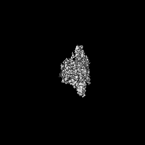
Journal: Nat Commun / Year: 2025 Title: Structural basis for regulation of CELSR1 by a compact module in its extracellular region. Authors: Sumit J Bandekar / Krassimira Garbett / Szymon P Kordon / Ethan E Dintzner / Jingxian Li / Tanner Shearer / Richard C Sando / Demet Araç / Abstract: The Cadherin EGF Laminin G seven-pass G-type receptor subfamily (CELSR/ADGRC) is one of the most conserved among adhesion G protein-coupled receptors and is essential for animal development. The ...The Cadherin EGF Laminin G seven-pass G-type receptor subfamily (CELSR/ADGRC) is one of the most conserved among adhesion G protein-coupled receptors and is essential for animal development. The extracellular regions (ECRs) of CELSRs are large with 23 adhesion domains. However, molecular insight into CELSR function is sparsely available. Here, we report the 3.8 Å cryo-EM reconstruction of the mouse CELSR1 ECR and reveal that 14 domains form a compact module mediated by conserved interactions majorly between the CADH9 and C-terminal GAIN domains. In the presence of Ca, the CELSR1 ECR forms a dimer species mediated by the cadherin repeats putatively in an antiparallel fashion. Cell-based assays reveal the N-terminal CADH1-8 repeat is required for cell-cell adhesion and the C-terminal CADH9-GAIN compact module can regulate cellular adhesion. Our work provides molecular insight into how one of the largest GPCRs uses defined structural modules to regulate receptor function.
In the structure databanks used in Yorodumi, some data are registered as the other names, "COVID-19 virus" and "2019-nCoV". Here are the details of the virus and the list of structure data.
Jan 31, 2019. EMDB accession codes are about to change! (news from PDBe EMDB page)
EMDB accession codes are about to change! (news from PDBe EMDB page)
The allocation of 4 digits for EMDB accession codes will soon come to an end. Whilst these codes will remain in use, new EMDB accession codes will include an additional digit and will expand incrementally as the available range of codes is exhausted. The current 4-digit format prefixed with “EMD-” (i.e. EMD-XXXX) will advance to a 5-digit format (i.e. EMD-XXXXX), and so on. It is currently estimated that the 4-digit codes will be depleted around Spring 2019, at which point the 5-digit format will come into force.
The EM Navigator/Yorodumi systems omit the EMD- prefix.
Related info.:Q: What is EMD? / ID/Accession-code notation in Yorodumi/EM Navigator
Yorodumi is a browser for structure data from EMDB, PDB, SASBDB, etc.
This page is also the successor to EM Navigator detail page, and also detail information page/front-end page for Omokage search.
The word "yorodu" (or yorozu) is an old Japanese word meaning "ten thousand". "mi" (miru) is to see.
Related info.:EMDB / PDB / SASBDB / Comparison of 3 databanks / Yorodumi Search / Aug 31, 2016. New EM Navigator & Yorodumi / Yorodumi Papers / Jmol/JSmol / Function and homology information / Changes in new EM Navigator and Yorodumi
 Movie
Movie Controller

 Yorodumi
 Open data
 Basic information
 Map data
 Sample
 Keywords
 Function and homology information
 Authors
 United States, 2 items
  Citation
 Structure visualization
 Downloads & links
 emd_43644.png
 http://ftp.pdbj.org/pub/emdb/structures/EMD-43644
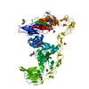


 Z (Sec.)
 Y (Row.)
 X (Col.)


 Sample components
 Trichoplusia ni (cabbage looper)
 Processing
 Electron microscopy
 FIELD EMISSION GUN

