+Open data
-Basic information
Entry | Database: EMDB / ID: EMD-43224 | |||||||||
---|---|---|---|---|---|---|---|---|---|---|
Title | Structure of the HKU1 RBD bound to the human TMPRSS2 receptor | |||||||||
Map data | ||||||||||
Sample |
| |||||||||
Keywords | Spike glycoprotein / fusion protein / Structural Genomics / Seattle Structural Genomics Center for Infectious Disease / SSGCID / inhibitor / VIRAL PROTEIN | |||||||||
Function / homology | Function and homology information transmembrane protease serine 2 / protein autoprocessing / Attachment and Entry / serine-type peptidase activity / endocytosis involved in viral entry into host cell / viral translation / Induction of Cell-Cell Fusion / host cell endoplasmic reticulum-Golgi intermediate compartment membrane / Attachment and Entry / positive regulation of viral entry into host cell ...transmembrane protease serine 2 / protein autoprocessing / Attachment and Entry / serine-type peptidase activity / endocytosis involved in viral entry into host cell / viral translation / Induction of Cell-Cell Fusion / host cell endoplasmic reticulum-Golgi intermediate compartment membrane / Attachment and Entry / positive regulation of viral entry into host cell / receptor-mediated virion attachment to host cell / fusion of virus membrane with host plasma membrane / serine-type endopeptidase activity / fusion of virus membrane with host endosome membrane / viral envelope / host cell plasma membrane / virion membrane / proteolysis / extracellular exosome / extracellular region / nucleoplasm / membrane / plasma membrane Similarity search - Function | |||||||||
Biological species | Homo sapiens (human) / Human coronavirus HKU1 (isolate N1) | |||||||||
Method | single particle reconstruction / cryo EM / Resolution: 2.9 Å | |||||||||
Authors | Park YJ / Seattle Structural Genomics Center for Infectious Disease (SSGCID) / Veesler D | |||||||||
Funding support | United States, 1 items
| |||||||||
Citation | 
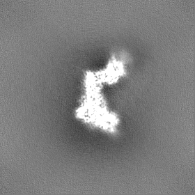
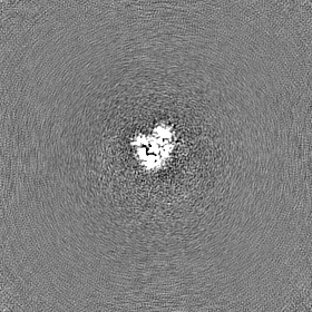
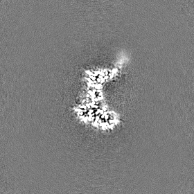
Journal: Cell / Year: 2024 Title: Human coronavirus HKU1 recognition of the TMPRSS2 host receptor. Authors: Matthew McCallum / Young-Jun Park / Cameron Stewart / Kaitlin R Sprouse / Amin Addetia / Jack Brown / M Alejandra Tortorici / Cecily Gibson / Emily Wong / Margareta Ieven / Amalio Telenti / David Veesler / Abstract: The human coronavirus HKU1 spike (S) glycoprotein engages host cell surface sialoglycans and transmembrane protease serine 2 (TMPRSS2) to initiate infection. The molecular basis of HKU1 binding to ...The human coronavirus HKU1 spike (S) glycoprotein engages host cell surface sialoglycans and transmembrane protease serine 2 (TMPRSS2) to initiate infection. The molecular basis of HKU1 binding to TMPRSS2 and determinants of host receptor tropism remain elusive. We designed an active human TMPRSS2 construct enabling high-yield recombinant production in human cells of this key therapeutic target. We determined a cryo-electron microscopy structure of the HKU1 RBD bound to human TMPRSS2, providing a blueprint of the interactions supporting viral entry and explaining the specificity for TMPRSS2 among orthologous proteases. We identified TMPRSS2 orthologs from five mammalian orders promoting HKU1 S-mediated entry into cells along with key residues governing host receptor usage. Our data show that the TMPRSS2 binding motif is a site of vulnerability to neutralizing antibodies and suggest that HKU1 uses S conformational masking and glycan shielding to balance immune evasion and receptor engagement. | |||||||||
History |
|
-Structure visualization
Supplemental images |
---|
-Downloads & links
-EMDB archive
Map data | emd_43224.map.gz | 78.9 MB | EMDB map data format | |
---|---|---|---|---|
Header (meta data) | emd-43224-v30.xml emd-43224.xml | 17.5 KB 17.5 KB | Display Display | EMDB header |
Images | emd_43224.png | 75.8 KB | ||
Filedesc metadata | emd-43224.cif.gz | 6 KB | ||
Others | emd_43224_additional_1.map.gz emd_43224_half_map_1.map.gz emd_43224_half_map_2.map.gz | 42.1 MB 77.7 MB 77.7 MB | ||
Archive directory | http://ftp.pdbj.org/pub/emdb/structures/EMD-43224 ftp://ftp.pdbj.org/pub/emdb/structures/EMD-43224 | HTTPS FTP |
-Validation report
Summary document | emd_43224_validation.pdf.gz | 708.2 KB | Display | EMDB validaton report |
---|---|---|---|---|
Full document | emd_43224_full_validation.pdf.gz | 707.8 KB | Display | |
Data in XML | emd_43224_validation.xml.gz | 11.7 KB | Display | |
Data in CIF | emd_43224_validation.cif.gz | 13.8 KB | Display | |
Arichive directory | https://ftp.pdbj.org/pub/emdb/validation_reports/EMD-43224 ftp://ftp.pdbj.org/pub/emdb/validation_reports/EMD-43224 | HTTPS FTP |
-Related structure data
Related structure data | 8vgtMC M: atomic model generated by this map C: citing same article (ref.) |
---|---|
Similar structure data | Similarity search - Function & homologyF&H Search |
-Links
EMDB pages | EMDB (EBI/PDBe) / EMDataResource |
---|---|
Related items in Molecule of the Month |
-Map
File | Download / File: emd_43224.map.gz / Format: CCP4 / Size: 83.7 MB / Type: IMAGE STORED AS FLOATING POINT NUMBER (4 BYTES) | ||||||||||||||||||||
---|---|---|---|---|---|---|---|---|---|---|---|---|---|---|---|---|---|---|---|---|---|
Voxel size | X=Y=Z: 1 Å | ||||||||||||||||||||
Density |
| ||||||||||||||||||||
Symmetry | Space group: 1 | ||||||||||||||||||||
Details | EMDB XML:
|
-Supplemental data
-Additional map: #1
File | emd_43224_additional_1.map | ||||||||||||
---|---|---|---|---|---|---|---|---|---|---|---|---|---|
Projections & Slices |
| ||||||||||||
Density Histograms |
-Half map: #2
File | emd_43224_half_map_1.map | ||||||||||||
---|---|---|---|---|---|---|---|---|---|---|---|---|---|
Projections & Slices |
| ||||||||||||
Density Histograms |
-Half map: #1
File | emd_43224_half_map_2.map | ||||||||||||
---|---|---|---|---|---|---|---|---|---|---|---|---|---|
Projections & Slices |
| ||||||||||||
Density Histograms |
-Sample components
-Entire : HKU1 RBD bound to the human TMPRSS2 receptor
Entire | Name: HKU1 RBD bound to the human TMPRSS2 receptor |
---|---|
Components |
|
-Supramolecule #1: HKU1 RBD bound to the human TMPRSS2 receptor
Supramolecule | Name: HKU1 RBD bound to the human TMPRSS2 receptor / type: complex / ID: 1 / Parent: 0 / Macromolecule list: #1-#2 |
---|---|
Source (natural) | Organism: Homo sapiens (human) |
-Macromolecule #1: Spike protein S1
Macromolecule | Name: Spike protein S1 / type: protein_or_peptide / ID: 1 / Number of copies: 1 / Enantiomer: LEVO |
---|---|
Source (natural) | Organism: Human coronavirus HKU1 (isolate N1) |
Molecular weight | Theoretical: 37.321785 KDa |
Recombinant expression | Organism: Homo sapiens (human) |
Sequence | String: MGILPSPGMP ALLSLVSLLS VLLMGCVAET GTRIPDLPDC DIDKWLNNFN VPSPLNWERK IFSNCNFNLS TLLRLVHTDS FSCNNFDES KIYGSCFKSI VLDKFAIPNS RRSDLQLGSS GFLQSSNYKI DTTSSSCQLY YSLPAINVTI NNYNPSSWNR R YGFNNFNL ...String: MGILPSPGMP ALLSLVSLLS VLLMGCVAET GTRIPDLPDC DIDKWLNNFN VPSPLNWERK IFSNCNFNLS TLLRLVHTDS FSCNNFDES KIYGSCFKSI VLDKFAIPNS RRSDLQLGSS GFLQSSNYKI DTTSSSCQLY YSLPAINVTI NNYNPSSWNR R YGFNNFNL SSHSVVYSRY CFSVNNTFCP CAKPSFASSC KSHKPPSASC PIGTNYRSCE STTVLDHTDW CRCSCLPDPI TA YDPRSCS QKKSLVGVGE HCAGFGVDEE KCGVLDGSYN VSCLCSTDAF LGWSYDTCVS NNRCNIFSNF ILNGINSGTT CSN DLLQPN TGGSHHHHHH HH UniProtKB: Spike glycoprotein |
-Macromolecule #2: Transmembrane protease serine 2
Macromolecule | Name: Transmembrane protease serine 2 / type: protein_or_peptide / ID: 2 / Number of copies: 1 / Enantiomer: LEVO / EC number: transmembrane protease serine 2 |
---|---|
Source (natural) | Organism: Homo sapiens (human) |
Molecular weight | Theoretical: 46.232004 KDa |
Recombinant expression | Organism: Homo sapiens (human) |
Sequence | String: MKWVTFISLL FLFSSAYSMG SKCSNSGIEC DSSGTCINPS NWCDGVSHCP GGEDENRCVR LYGPNFILQV YSSQRKSWHP VCQDDWNEN YGRAACRDMG YKNNFYSSQG IVDDSGSTSF MKLNTSAGNV DIYKKLYHSD ACSSKAVVSL RCIACGVNLN D DDDKIVGG ...String: MKWVTFISLL FLFSSAYSMG SKCSNSGIEC DSSGTCINPS NWCDGVSHCP GGEDENRCVR LYGPNFILQV YSSQRKSWHP VCQDDWNEN YGRAACRDMG YKNNFYSSQG IVDDSGSTSF MKLNTSAGNV DIYKKLYHSD ACSSKAVVSL RCIACGVNLN D DDDKIVGG ESALPGAWPW QVSLHVQNVH VCGGSIITPE WIVTAAHCVE KPLNNPWHWT AFAGILRQSF MFYGAGYQVE KV ISHPNYD SKTKNNDIAL MKLQKPLTFN DLVKPVCLPN PGMMLQPEQL CWISGWGATE EKGKTSEVLN AAKVLLIETQ RCN SRYVYD NLITPAMICA GFLQGNVDSC QGDAGGPLVC SKNNIWWLIG DTSWGSGCAK AYRPGVYGNV MVFTDWIYRQ MRAD GDDDD KSGHHHHHHH H UniProtKB: Transmembrane protease serine 2 |
-Macromolecule #4: 2-acetamido-2-deoxy-beta-D-glucopyranose
Macromolecule | Name: 2-acetamido-2-deoxy-beta-D-glucopyranose / type: ligand / ID: 4 / Number of copies: 3 / Formula: NAG |
---|---|
Molecular weight | Theoretical: 221.208 Da |
Chemical component information | ChemComp-NAG: |
-Experimental details
-Structure determination
Method | cryo EM |
---|---|
Processing | single particle reconstruction |
Aggregation state | particle |
-Sample preparation
Buffer | pH: 8 |
---|---|
Vitrification | Cryogen name: ETHANE |
-Electron microscopy
Microscope | FEI TITAN KRIOS |
---|---|
Image recording | Film or detector model: GATAN K3 (6k x 4k) / Average electron dose: 60.0 e/Å2 |
Electron beam | Acceleration voltage: 300 kV / Electron source: FIELD EMISSION GUN |
Electron optics | Illumination mode: FLOOD BEAM / Imaging mode: BRIGHT FIELD / Nominal defocus max: 3.5 µm / Nominal defocus min: 0.5 µm |
Experimental equipment | Model: Titan Krios / Image courtesy: FEI Company |
-Image processing
Startup model | Type of model: NONE |
---|---|
Final reconstruction | Resolution.type: BY AUTHOR / Resolution: 2.9 Å / Resolution method: FSC 0.143 CUT-OFF / Software - Name: cryoSPARC / Number images used: 810357 |
Initial angle assignment | Type: PROJECTION MATCHING |
Final angle assignment | Type: PROJECTION MATCHING |