 Movie
Movie Controller

[English] 日本語
 Yorodumi
- EMDB-42851: 5x5 tiled montage tomogram of a holey carbon grid with apoferriti... -
+ Open data

- Basic information
| Entry | 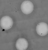 | |||||||||
|---|---|---|---|---|---|---|---|---|---|---|
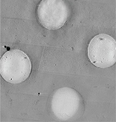
| Title | 5x5 tiled montage tomogram of a holey carbon grid with apoferritin imaged with a square electron beam | |||||||||
 Map data Map data | 5x5 tiled montage tomogram of a holey carbon grid with apoferritin imaged with a square electron beam | |||||||||
 Sample Sample |
| |||||||||
 Keywords Keywords | Iron binding / storage / METAL BINDING PROTEIN | |||||||||
| Biological species |  | |||||||||
| Method | electron tomography / cryo EM | |||||||||
 Authors Authors | Chua EYD / Alink LM / Kopylov M / Johnston J / Einsenstein F / de Marco A | |||||||||
| Funding support |  United States, 2 items United States, 2 items
| |||||||||
 Citation Citation | Journal: Nat Methods / Year: 2024 Title: Square beams for optimal tiling in transmission electron microscopy. Authors: Eugene Y D Chua / Lambertus M Alink / Mykhailo Kopylov / Jake D Johnston / Fabian Eisenstein / Alex de Marco /  Abstract: Imaging large fields of view at a high magnification requires tiling. Transmission electron microscopes typically have round beam profiles; therefore, tiling across a large area is either imperfect ...Imaging large fields of view at a high magnification requires tiling. Transmission electron microscopes typically have round beam profiles; therefore, tiling across a large area is either imperfect or results in uneven exposures, a problem for dose-sensitive samples. Here, we introduce a square electron beam that can easily be retrofitted in existing microscopes, and demonstrate its application, showing that it can tile nearly perfectly and deliver cryo-electron microscopy imaging with a resolution comparable to conventional set-ups. | |||||||||
| History |
|
- Structure visualization
| Supplemental images |
|---|
- Downloads & links
-EMDB archive
| Map data | emd_42851.map.gz | 9.6 GB |  EMDB map data format EMDB map data format | |
|---|---|---|---|---|
| Header (meta data) | emd-42851-v30.xmlemd-42851.xml | 8.2 KB 8.2 KB | Display Display | EMDB header |
| Images |  emd_42851.png emd_42851.png | 243.3 KB | ||
| Filedesc metadata | emd-42851.cif.gz | 3.8 KB | ||
| Archive directory |  http://ftp.pdbj.org/pub/emdb/structures/EMD-42851ftp://ftp.pdbj.org/pub/emdb/structures/EMD-42851 http://ftp.pdbj.org/pub/emdb/structures/EMD-42851ftp://ftp.pdbj.org/pub/emdb/structures/EMD-42851 | HTTPS FTP |
-Validation report
| Summary document | emd_42851_validation.pdf.gz | 369.1 KB | Display | EMDB validaton report |
|---|---|---|---|---|
| Full document | emd_42851_full_validation.pdf.gz | 368.7 KB | Display | |
| Data in XML | emd_42851_validation.xml.gz | 4.9 KB | Display | |
| Data in CIF | emd_42851_validation.cif.gz | 5.4 KB | Display | |
| Arichive directory | https://ftp.pdbj.org/pub/emdb/validation_reports/EMD-42851ftp://ftp.pdbj.org/pub/emdb/validation_reports/EMD-42851 | HTTPS FTP |
-Related structure data


-Links
| EMDB pages | EMDB (EBI/PDBe) / EMDataResource |
|---|
-Map
| File | Download / File: emd_42851.map.gz / Format: CCP4 / Size: 11.4 GB / Type: IMAGE STORED AS FLOATING POINT NUMBER (4 BYTES) | ||||||||||||||||||||||||||||||||
|---|---|---|---|---|---|---|---|---|---|---|---|---|---|---|---|---|---|---|---|---|---|---|---|---|---|---|---|---|---|---|---|---|---|
| Annotation | 5x5 tiled montage tomogram of a holey carbon grid with apoferritin imaged with a square electron beam | ||||||||||||||||||||||||||||||||
| Projections & slices | Image control
Images are generated by Spider. generated in cubic-lattice coordinate | ||||||||||||||||||||||||||||||||
| Voxel size | X=Y=Z: 14.04 Å | ||||||||||||||||||||||||||||||||
| Density |
| ||||||||||||||||||||||||||||||||
| Symmetry | Space group: 1 | ||||||||||||||||||||||||||||||||
| Details | EMDB XML:
|
 Z (Sec.)
 Y (Row.)
 X (Col.)


-Supplemental data
- Sample components
-Entire : Mouse apoferritin at 8 mg/ml, on a carbon foil support
| Entire | Name: Mouse apoferritin at 8 mg/ml, on a carbon foil support |
|---|---|
| Components |
|
-Supramolecule #1: Mouse apoferritin at 8 mg/ml, on a carbon foil support
| Supramolecule | Name: Mouse apoferritin at 8 mg/ml, on a carbon foil support type: complex / ID: 1 / Parent: 0 |
|---|---|
| Source (natural) | Organism: |
| Molecular weight | Theoretical: 460 KDa |
-Experimental details
-Structure determination
| Method | cryo EM |
|---|---|
 Processing Processing | electron tomography |
| Aggregation state | particle |
-Sample preparation
| Concentration | 8 mg/mL |
|---|---|
| Buffer | pH: 7.5 |
| Grid | Model: Quantifoil R1.2/1.3 / Support film - Material: CARBON / Support film - topology: HOLEY |
| Vitrification | Cryogen name: ETHANE |
| Sectioning | Other: NO SECTIONING |
- Electron microscopy
| Microscope | FEI TITAN KRIOS |
|---|---|
| Image recording | Film or detector model: GATAN K3 BIOQUANTUM (6k x 4k) / Average electron dose: 2.55 e/Å2 |
| Electron beam | Acceleration voltage: 300 kV / Electron source:  FIELD EMISSION GUN FIELD EMISSION GUN |
| Electron optics | C2 aperture diameter: 50.0 µm / Illumination mode: FLOOD BEAM / Imaging mode: BRIGHT FIELD / Cs: 2.7 mm / Nominal defocus max: 5.0 µm / Nominal defocus min: 2.5 µm |
| Experimental equipment |  Model: Titan Krios / Image courtesy: FEI Company |
-Image processing
| Final reconstruction | Number images used: 875 |
|---|
