[English] 日本語
 Yorodumi
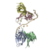
- EMDB-42842: A Mitochondrial Replication Complex: PolG/PolG2 Bound to DNA in c... -
+ Open data
- Basic information
| Entry |  | |||||||||
|---|---|---|---|---|---|---|---|---|---|---|
| Title | A Mitochondrial Replication Complex: PolG/PolG2 Bound to DNA in complex with the Single-Stranded Binding Protein (mtSSB) | |||||||||
 Map data Map data | ||||||||||
 Sample Sample |
| |||||||||
 Keywords Keywords | DNA BINDING PROTEIN / DNA polymerase / mitochondrial DNA replication / DNA polymerase gamma / mitochondrial single-stranded binding protein / SSB / SSBP1 / DNA BINDING PROTEIN-DNA complex | |||||||||
| Biological species |  Homo sapiens (human) / synthetic construct (others) Homo sapiens (human) / synthetic construct (others) | |||||||||
| Method | single particle reconstruction / cryo EM / Resolution: 3.5 Å | |||||||||
 Authors Authors | Riccio AA / Bouvette J / Borgnia MJ / Copeland WC | |||||||||
| Funding support |  United States, 2 items United States, 2 items
| |||||||||
 Citation Citation |  Journal: Nucleic Acids Res / Year: 2024 Journal: Nucleic Acids Res / Year: 2024Title: Structures of the mitochondrial single-stranded DNA binding protein with DNA and DNA polymerase γ. Authors: Amanda A Riccio / Jonathan Bouvette / Lars C Pedersen / Shruti Somai / Robert C Dutcher / Mario J Borgnia / William C Copeland /  Abstract: The mitochondrial single-stranded DNA (ssDNA) binding protein, mtSSB or SSBP1, binds to ssDNA to prevent secondary structures of DNA that could impede downstream replication or repair processes. ...The mitochondrial single-stranded DNA (ssDNA) binding protein, mtSSB or SSBP1, binds to ssDNA to prevent secondary structures of DNA that could impede downstream replication or repair processes. Clinical mutations in the SSBP1 gene have been linked to a range of mitochondrial disorders affecting nearly all organs and systems. Yet, the molecular determinants governing the interaction between mtSSB and ssDNA have remained elusive. Similarly, the structural interaction between mtSSB and other replisome components, such as the mitochondrial DNA polymerase, Polγ, has been minimally explored. Here, we determined a 1.9-Å X-ray crystallography structure of the human mtSSB bound to ssDNA. This structure uncovered two distinct DNA binding sites, a low-affinity site and a high-affinity site, confirmed through site-directed mutagenesis. The high-affinity binding site encompasses a clinically relevant residue, R38, and a highly conserved DNA base stacking residue, W84. Employing cryo-electron microscopy, we confirmed the tetrameric assembly in solution and capture its interaction with Polγ. Finally, we derived a model depicting modes of ssDNA wrapping around mtSSB and a region within Polγ that mtSSB binds. | |||||||||
| History |
|
- Structure visualization
| Supplemental images |
|---|
- Downloads & links
-EMDB archive
| Map data |  emd_42842.map.gz emd_42842.map.gz | 348.4 MB |  EMDB map data format EMDB map data format | |
|---|---|---|---|---|
| Header (meta data) |  emd-42842-v30.xml emd-42842-v30.xml emd-42842.xml emd-42842.xml | 18.4 KB 18.4 KB | Display Display |  EMDB header EMDB header |
| FSC (resolution estimation) |  emd_42842_fsc.xml emd_42842_fsc.xml | 21.2 KB | Display |  FSC data file FSC data file |
| Images |  emd_42842.png emd_42842.png | 58.2 KB | ||
| Filedesc metadata |  emd-42842.cif.gz emd-42842.cif.gz | 6.3 KB | ||
| Others |  emd_42842_half_map_1.map.gz emd_42842_half_map_1.map.gz emd_42842_half_map_2.map.gz emd_42842_half_map_2.map.gz | 647.8 MB 647.8 MB | ||
| Archive directory |  http://ftp.pdbj.org/pub/emdb/structures/EMD-42842 http://ftp.pdbj.org/pub/emdb/structures/EMD-42842 ftp://ftp.pdbj.org/pub/emdb/structures/EMD-42842 ftp://ftp.pdbj.org/pub/emdb/structures/EMD-42842 | HTTPS FTP |
-Validation report
| Summary document |  emd_42842_validation.pdf.gz emd_42842_validation.pdf.gz | 956.6 KB | Display |  EMDB validaton report EMDB validaton report |
|---|---|---|---|---|
| Full document |  emd_42842_full_validation.pdf.gz emd_42842_full_validation.pdf.gz | 956.2 KB | Display | |
| Data in XML |  emd_42842_validation.xml.gz emd_42842_validation.xml.gz | 28.4 KB | Display | |
| Data in CIF |  emd_42842_validation.cif.gz emd_42842_validation.cif.gz | 37.6 KB | Display | |
| Arichive directory |  https://ftp.pdbj.org/pub/emdb/validation_reports/EMD-42842 https://ftp.pdbj.org/pub/emdb/validation_reports/EMD-42842 ftp://ftp.pdbj.org/pub/emdb/validation_reports/EMD-42842 ftp://ftp.pdbj.org/pub/emdb/validation_reports/EMD-42842 | HTTPS FTP |
-Related structure data
- Links
Links
| EMDB pages |  EMDB (EBI/PDBe) / EMDB (EBI/PDBe) /  EMDataResource EMDataResource |
|---|
- Map
Map
| File |  Download / File: emd_42842.map.gz / Format: CCP4 / Size: 699 MB / Type: IMAGE STORED AS FLOATING POINT NUMBER (4 BYTES) Download / File: emd_42842.map.gz / Format: CCP4 / Size: 699 MB / Type: IMAGE STORED AS FLOATING POINT NUMBER (4 BYTES) | ||||||||||||||||||||||||||||||||||||
|---|---|---|---|---|---|---|---|---|---|---|---|---|---|---|---|---|---|---|---|---|---|---|---|---|---|---|---|---|---|---|---|---|---|---|---|---|---|
| Projections & slices | Image control
Images are generated by Spider. | ||||||||||||||||||||||||||||||||||||
| Voxel size | X=Y=Z: 0.85 Å | ||||||||||||||||||||||||||||||||||||
| Density |
| ||||||||||||||||||||||||||||||||||||
| Symmetry | Space group: 1 | ||||||||||||||||||||||||||||||||||||
| Details | EMDB XML:
|
-Supplemental data
-Half map: #2
| File | emd_42842_half_map_1.map | ||||||||||||
|---|---|---|---|---|---|---|---|---|---|---|---|---|---|
| Projections & Slices |
| ||||||||||||
| Density Histograms |
-Half map: #1
| File | emd_42842_half_map_2.map | ||||||||||||
|---|---|---|---|---|---|---|---|---|---|---|---|---|---|
| Projections & Slices |
| ||||||||||||
| Density Histograms |
- Sample components
-Entire : The Mitchondrial DNA polymerase, catalytic (PolG) and accessory s...
| Entire | Name: The Mitchondrial DNA polymerase, catalytic (PolG) and accessory subunits (PolG2), bound to DNA in complex with the mitochondrial DNA single-stranded binding protein (mtSSB) |
|---|---|
| Components |
|
-Supramolecule #1: The Mitchondrial DNA polymerase, catalytic (PolG) and accessory s...
| Supramolecule | Name: The Mitchondrial DNA polymerase, catalytic (PolG) and accessory subunits (PolG2), bound to DNA in complex with the mitochondrial DNA single-stranded binding protein (mtSSB) type: complex / ID: 1 / Parent: 0 / Macromolecule list: all |
|---|---|
| Source (natural) | Organism:  Homo sapiens (human) Homo sapiens (human) |
| Molecular weight | Theoretical: 230 KDa |
-Macromolecule #1: PolG, mitochondrial DNA polymerase catalytic subunit
| Macromolecule | Name: PolG, mitochondrial DNA polymerase catalytic subunit / type: protein_or_peptide / ID: 1 / Enantiomer: LEVO |
|---|---|
| Source (natural) | Organism:  Homo sapiens (human) Homo sapiens (human) |
| Recombinant expression | Organism:  |
| Sequence | String: MGGSHHHHHH GSRFMVSSSV PASDPSDGQR RRQQQQQQQQ QQQQQPQQPQ VLSSEGGQL RHNPLDIQML SRGLHEQIFG QGGEMPGEAA VRRSVEHLQK H GLWGQPAV PLPDVELRLP PLYGDNLDQH FRLLAQKQSL PYLEAANLLL QA QLPPKPP AWAWAEGWTR ...String: MGGSHHHHHH GSRFMVSSSV PASDPSDGQR RRQQQQQQQQ QQQQQPQQPQ VLSSEGGQL RHNPLDIQML SRGLHEQIFG QGGEMPGEAA VRRSVEHLQK H GLWGQPAV PLPDVELRLP PLYGDNLDQH FRLLAQKQSL PYLEAANLLL QA QLPPKPP AWAWAEGWTR YGPEGEAVPV AIPEERALVF AVAVCLAEGT CPT LAVAIS PSAWYSWCSQ RLVEERYSWT SQLSPADLIP LEVPTGASSP TQRD WQEQL VVGHNVSFDR AHIREQYLIQ GSRMRFLDTM SMHMAISGLS SFQRS LWIA AKQGKHKVQP PTKQGQKSQR KARRGPAISS WDWLDISSVN SLAEVH RLY VGGPPLEKEP RELFVKGTMK DIRENFQDLM QYCAQDVWAT HEVFQQQ LP LFLERCPHPV TLAGMLEMGV SYLPVNQNWE RYLAEAQGTY EELQREMK K SLMDLANDAC QLLSGERYKE DPWLWDLEWD LQEFKQKKAK KVKKEPATA SKLPIEGAGA PGDPMDQEDL GPCSEEEEFQ QDVMARACLQ KLKGTTELLP KRPQHLPGH PGWYRKLCPR LDDPAWTPGP SLLSLQMRVT PKLMALTWDG F PLHYSERH GWGYLVPGRR DNLAKLPTGT TLESAGVVCP YRAIESLYRK HC LEQGKQQ LMPQEAGLAE EFLLTDNSAI WQTVEELDYL EVEAEAKMEN LRA AVPGQP LALTARGGPK DTQPSYHHGN GPYNDVDIPG CWFFKLPHKD GNSC NVGSP FAKDFLPKME DGTLQAGPGG ASGPRALEIN KMISFWRNAH KRISS QMVV WLPRSALPRA VIRHPDYDEE GLYGAILPQV VTAGTITRRA VEPTWL TAS NARPDRVGSE LKAMVQAPPG YTLVGADVDS QELWIAAVLG DAHFAGM HG CTAFGWMTLQ GRKSRGTDLH SKTATTVGIS REHAKIFNYG RIYGAGQP F AERLLMQFNH RLTQQEAAEK AQQMYAATKG LRWYRLSDEG EWLVRELNL PVDRTEGGWI SLQDLRKVQR ETARKSQWKK WEVVAERAWK GGTESEMFNK LESIATSDI PRTPVLGCCI SRALEPSAVQ EEFMTSRVNW VVQSSAVDYL H LMLVAMKW LFEEFAIDGR FCISIHDEVR YLVREEDRYR AALALQITNL LT RCMFAYK LGLNDLPQSV AFFSAVDIDR CLRKEVTMDC KTPSNPTGME RRY GIPQGE ALDIYQIIEL TKGSLEKRSQ PGP |
-Macromolecule #2: PolG2, mitochondrial DNA polymerase gamma accessory subunit
| Macromolecule | Name: PolG2, mitochondrial DNA polymerase gamma accessory subunit type: protein_or_peptide / ID: 2 / Enantiomer: LEVO |
|---|---|
| Source (natural) | Organism:  Homo sapiens (human) Homo sapiens (human) |
| Sequence | String: M A S R G S H H H H H H G A DA GQPELLTERS SPKGGHVKSH AELEGNGEHP EAP GSGEGS EALLEICQRR HFLSGSKQQL SRDSLLSGCH PGFGPLGVEL RKNLAAEWWT SVVV FREQV FPVDALHHKP GPLLPGDSAF RLVSAETLRE ILQDKELSKE ...String: M A S R G S H H H H H H G A DA GQPELLTERS SPKGGHVKSH AELEGNGEHP EAP GSGEGS EALLEICQRR HFLSGSKQQL SRDSLLSGCH PGFGPLGVEL RKNLAAEWWT SVVV FREQV FPVDALHHKP GPLLPGDSAF RLVSAETLRE ILQDKELSKE QLVAFLENVL KTSGK LREN LLHGALEHYV NCLDLVNKRL PYGLAQIGVC FHPVFDTKQI RNGVKSIGEK TEASLV WFT PPRTSNQWLD FWLRHRLQWW RKFAMSPSNF SSSDCQDEEG RKGNKLYYNF PWGKELI ET LWNLGDHELL HMYPGNVSKL HGRDGRKNVV PCVLSVNGDL DRGMLAYLYD SFQLTENS F TRKKNLHRKV LKLHPCLAPI KVALDVGRGP TLELRQVCQG LFNELLENGI SVWPGYLET MQSSLEQLYS KYDEMSILFT VLVTETTLEN GLIHLRSRDT TMKEMMHISK LKDFLIKYIS SAKNV |
-Macromolecule #3: Single-stranded DNA-binding protein, mitochondrial
| Macromolecule | Name: Single-stranded DNA-binding protein, mitochondrial / type: protein_or_peptide / ID: 3 / Enantiomer: LEVO |
|---|---|
| Source (natural) | Organism:  Homo sapiens (human) Homo sapiens (human) |
| Sequence | String: MGSSHHHHHH SSGENLYFQG SMESETTTSL VLERSLNRVH LLGRVGQDPV LRQVEGKNP VTIFSLATNE MWRSGDSEVY QLGDVSQKTT WHRISVFRPG L RDVAYQYV KKGSRIYLEG KIDYGEYMDK NNVRRQATTI IADNIIFLSD QT KEKE |
-Macromolecule #4: DNA1
| Macromolecule | Name: DNA1 / type: dna / ID: 4 / Classification: DNA |
|---|---|
| Source (natural) | Organism: synthetic construct (others) |
| Sequence | String: ATTTCGTACA TTACTGCCAG CCACCATGA ATATTGTACG GTACCATAAA TACTTGACCA CCTGTAGTAC ATAAAAACCC A |
-Macromolecule #5: DNA2
| Macromolecule | Name: DNA2 / type: dna / ID: 5 / Classification: DNA |
|---|---|
| Source (natural) | Organism: synthetic construct (others) |
| Sequence | String: TGGGTTTTTA TGTACTACAG GTGGTCAAGT ATTTATGGTA CCGTACAATA |
-Macromolecule #6: DNA3
| Macromolecule | Name: DNA3 / type: dna / ID: 6 / Classification: DNA |
|---|---|
| Source (natural) | Organism: synthetic construct (others) |
| Sequence | String: AGACACAGCA CTACATTGAG GTTGGTACGA GCAGTTAGGG AGAATTGAGC TCTAACGGAT CTGGCAGTA ATGTACGAAA T |
-Experimental details
-Structure determination
| Method | cryo EM |
|---|---|
 Processing Processing | single particle reconstruction |
| Aggregation state | particle |
- Sample preparation
| Concentration | 1.2 mg/mL |
|---|---|
| Buffer | pH: 8 |
| Vitrification | Cryogen name: ETHANE |
- Electron microscopy
| Microscope | TFS KRIOS |
|---|---|
| Image recording | Film or detector model: GATAN K3 BIOQUANTUM (6k x 4k) / Average electron dose: 60.0 e/Å2 |
| Electron beam | Acceleration voltage: 300 kV / Electron source:  FIELD EMISSION GUN FIELD EMISSION GUN |
| Electron optics | Illumination mode: FLOOD BEAM / Imaging mode: BRIGHT FIELD / Nominal defocus max: 2.5 µm / Nominal defocus min: 1.0 µm |
| Experimental equipment |  Model: Titan Krios / Image courtesy: FEI Company |
 Movie
Movie Controller


 Z (Sec.)
 Y (Row.)
 X (Col.)


