autophagy of mitochondrion / positive regulation of mitochondrial fission / regulation of apoptotic process / mitochondrial outer membrane / protein kinase activity / non-specific serine/threonine protein kinase / mitochondrial inner membrane / protein serine/threonine kinase activity / ATP binding / metal ion binding / cytosol 類似検索 - 分子機能
: / Serine/threonine-protein kinase, active site / Serine/Threonine protein kinases active-site signature. / Protein kinase domain / Serine/Threonine protein kinases, catalytic domain / Protein kinase domain profile. / Protein kinase domain / Protein kinase-like domain superfamily 類似検索 - ドメイン・相同性
National Health and Medical Research Council (NHMRC, Australia)
オーストラリア
Michael J. Fox Foundation
米国
引用
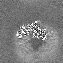
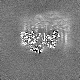
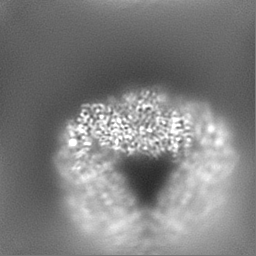
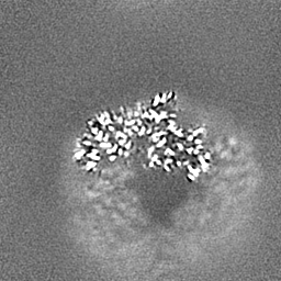
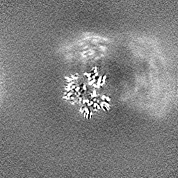
ジャーナル: Sci Adv / 年: 2024 タイトル: Interaction of PINK1 with nucleotides and kinetin. 著者: Zhong Yan Gan / Sylvie Callegari / Thanh N Nguyen / Nicholas S Kirk / Andrew Leis / Michael Lazarou / Grant Dewson / David Komander / 要旨: The ubiquitin kinase PINK1 accumulates on damaged mitochondria to trigger mitophagy, and PINK1 loss-of-function mutations cause early onset Parkinson's disease. Nucleotide analogs such as kinetin ...The ubiquitin kinase PINK1 accumulates on damaged mitochondria to trigger mitophagy, and PINK1 loss-of-function mutations cause early onset Parkinson's disease. Nucleotide analogs such as kinetin triphosphate (KTP) were reported to enhance PINK1 activity and may represent a therapeutic strategy for the treatment of Parkinson's disease. Here, we investigate the interaction of PINK1 with nucleotides, including KTP. We establish a cryo-EM platform exploiting the dodecamer assembly of () PINK1 and determine PINK1 structures bound to AMP-PNP and ADP, revealing conformational changes in the kinase N-lobe that help establish PINK1's ubiquitin binding site. Notably, we find that KTP is unable to bind PINK1 or human () PINK1 due to a steric clash with the kinase "gatekeeper" methionine residue, and mutation to Ala or Gly is required for PINK1 to bind and use KTP as a phosphate donor in ubiquitin phosphorylation and mitophagy. PINK1 M318G can be used to conditionally uncouple PINK1 stabilization and activity on mitochondria.
 ムービー
ムービー コントローラー


 データを開く
 基本情報
基本情報
 マップデータ
 試料
試料 キーワード
キーワード 機能・相同性情報
 Pediculus humanus corporis (キモノジラミ)
 データ登録者
 オーストラリア,
  米国, 2件
  引用
引用 構造の表示
構造の表示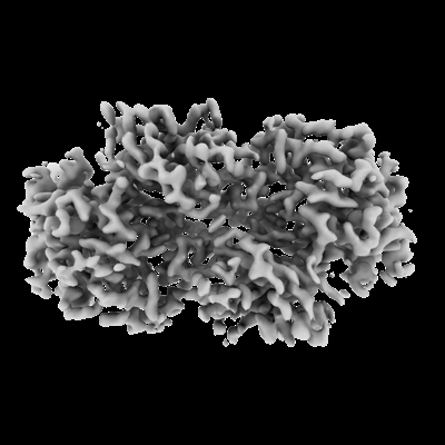
 ダウンロードとリンク
 emd_42804.png
 http://ftp.pdbj.org/pub/emdb/structures/EMD-42804
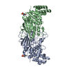


 Z (Sec.)
 Y (Row.)
 X (Col.)


 試料の構成要素
 解析
解析 電子顕微鏡法
 FIELD EMISSION GUN


