response to vitamin K / coagulation factor Xa / platelet alpha granule / Cargo concentration in the ER / Defective factor IX causes thrombophilia / Defective cofactor function of FVIIIa variant / Defective F9 variant does not activate FX / blood circulation / Extrinsic Pathway of Fibrin Clot Formation / COPII-mediated vesicle transport ...response to vitamin K / coagulation factor Xa / platelet alpha granule / Cargo concentration in the ER / Defective factor IX causes thrombophilia / Defective cofactor function of FVIIIa variant / Defective F9 variant does not activate FX / blood circulation / Extrinsic Pathway of Fibrin Clot Formation / COPII-mediated vesicle transport / positive regulation of lipid kinase activity / positive regulation of phospholipase C-activating G protein-coupled receptor signaling pathway / cytolysis by host of symbiont cells / COPII-coated ER to Golgi transport vesicle / thrombospondin receptor activity / Defective factor XII causes hereditary angioedema / thrombin / regulation of blood coagulation / neutrophil-mediated killing of gram-negative bacterium / ligand-gated ion channel signaling pathway / Defective F8 cleavage by thrombin / Platelet Aggregation (Plug Formation) / negative regulation of astrocyte differentiation / negative regulation of platelet activation / positive regulation of collagen biosynthetic process / positive regulation of TOR signaling / negative regulation of cytokine production involved in inflammatory response / positive regulation of blood coagulation / negative regulation of fibrinolysis / Gamma-carboxylation of protein precursors / Transport of gamma-carboxylated protein precursors from the endoplasmic reticulum to the Golgi apparatus / Common Pathway of Fibrin Clot Formation / Removal of aminoterminal propeptides from gamma-carboxylated proteins / fibrinolysis / regulation of cytosolic calcium ion concentration / Intrinsic Pathway of Fibrin Clot Formation / endoplasmic reticulum-Golgi intermediate compartment membrane / Peptide ligand-binding receptors / positive regulation of release of sequestered calcium ion into cytosol / platelet alpha granule lumen / acute-phase response / Regulation of Complement cascade / negative regulation of proteolysis / Cell surface interactions at the vascular wall / lipopolysaccharide binding / Post-translational protein phosphorylation / positive regulation of receptor signaling pathway via JAK-STAT / growth factor activity / positive regulation of insulin secretion / phospholipid binding / platelet activation / response to wounding / positive regulation of protein localization to nucleus / Golgi lumen / Regulation of Insulin-like Growth Factor (IGF) transport and uptake by Insulin-like Growth Factor Binding Proteins (IGFBPs) / antimicrobial humoral immune response mediated by antimicrobial peptide / positive regulation of reactive oxygen species metabolic process / blood coagulation / extracellular vesicle / Thrombin signalling through proteinase activated receptors (PARs) / Platelet degranulation / signaling receptor activity / heparin binding / regulation of cell shape / positive regulation of cell growth / G alpha (q) signalling events / collagen-containing extracellular matrix / positive regulation of phosphatidylinositol 3-kinase/protein kinase B signal transduction / blood microparticle / cell surface receptor signaling pathway / positive regulation of cell migration / positive regulation of protein phosphorylation / G protein-coupled receptor signaling pathway / copper ion binding / endoplasmic reticulum lumen / external side of plasma membrane / serine-type endopeptidase activity / signaling receptor binding / calcium ion binding / positive regulation of cell population proliferation / proteolysis / extracellular space / extracellular exosome / extracellular region / membrane / plasma membrane Similarity search - Function
National Institutes of Health/National Heart, Lung, and Blood Institute (NIH/NHLBI)
HL049413
United States
National Institutes of Health/National Heart, Lung, and Blood Institute (NIH/NHLBI)
HL139554
United States
National Institutes of Health/National Heart, Lung, and Blood Institute (NIH/NHLBI)
HL147821
United States
Childrens Discovery Institute of Washington University and St. Louis Childrens Hospital
CDI-CORE-2015-505
United States
Childrens Discovery Institute of Washington University and St. Louis Childrens Hospital
CDI-CORE-2019-813
United States
Foundation for Barnes-Jewish Hospital
3770
United States
Other private
Washington University Diabetes Research Center DK020579
Other private
The Alvin J. Siteman Cancer Center at Barnes-Jewish Hospital and Washington University School of Medicine CA091842
Citation
Journal: Subcell Biochem / Year: 2024 Title: The Prothrombin-Prothrombinase Interaction. Authors: Bosko M Stojanovski / Bassem M Mohammed / Enrico Di Cera / Abstract: The hemostatic response to vascular injury entails a sequence of proteolytic events where several inactive zymogens of the trypsin family are converted to active proteases. The cascade starts with ...The hemostatic response to vascular injury entails a sequence of proteolytic events where several inactive zymogens of the trypsin family are converted to active proteases. The cascade starts with exposure of tissue factor from the damaged endothelium and culminates with conversion of prothrombin to thrombin in a reaction catalyzed by the prothrombinase complex composed of the enzyme factor Xa, cofactor Va, Ca, and phospholipids. This cofactor-dependent activation is paradigmatic of analogous reactions of the blood coagulation and complement cascades, which makes elucidation of its molecular mechanism of broad significance to the large class of trypsin-like zymogens to which prothrombin belongs. Because of its relevance as the most important reaction in the physiological response to vascular injury, as well as the main trigger of pathological thrombotic complications, the mechanism of prothrombin activation has been studied extensively. However, a molecular interpretation of this mechanism has become available only recently from important developments in structural biology. Here we review current knowledge on the prothrombin-prothrombinase interaction and outline future directions for the study of this key reaction of the coagulation cascade.
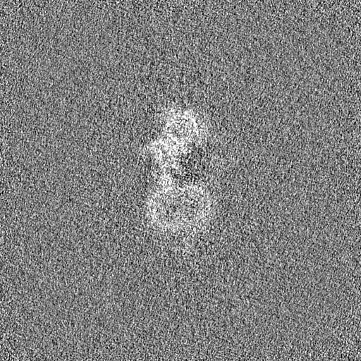
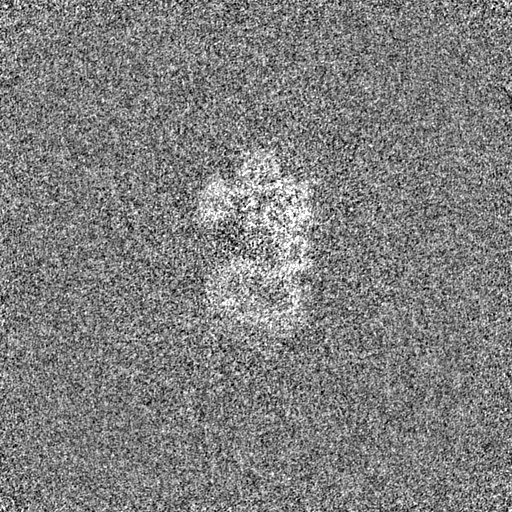
Entire : Prothrombin_Prothrombinase complex on lipid nanodiscs.
Entire
Name: Prothrombin_Prothrombinase complex on lipid nanodiscs.
Components
Complex: Prothrombin_Prothrombinase complex on lipid nanodiscs.
Organelle or cellular component: Coagulation Factor Va
Organelle or cellular component: Coagulation Factor Xa
Organelle or cellular component: Prothrombin
-
Supramolecule #1: Prothrombin_Prothrombinase complex on lipid nanodiscs.
Supramolecule
Name: Prothrombin_Prothrombinase complex on lipid nanodiscs. type: complex / ID: 1 / Parent: 0 / Macromolecule list: #1 Details: The complex is made of coagulation factor Va (derived from human plasma), coagulation factor Xa (Recombinantly expressed in BHK cells) and Prothrombin (Recombinantly expressed in BHK cells). ...Details: The complex is made of coagulation factor Va (derived from human plasma), coagulation factor Xa (Recombinantly expressed in BHK cells) and Prothrombin (Recombinantly expressed in BHK cells). The nanodisc component of the complex is made of the scaffold protein MSP1E3D1 (Recombinantly expressed in bacteria) and the phospholipid component was Porcine Brain phosphatidylserine.
In the structure databanks used in Yorodumi, some data are registered as the other names, "COVID-19 virus" and "2019-nCoV". Here are the details of the virus and the list of structure data.
Jan 31, 2019. EMDB accession codes are about to change! (news from PDBe EMDB page)
EMDB accession codes are about to change! (news from PDBe EMDB page)
The allocation of 4 digits for EMDB accession codes will soon come to an end. Whilst these codes will remain in use, new EMDB accession codes will include an additional digit and will expand incrementally as the available range of codes is exhausted. The current 4-digit format prefixed with “EMD-” (i.e. EMD-XXXX) will advance to a 5-digit format (i.e. EMD-XXXXX), and so on. It is currently estimated that the 4-digit codes will be depleted around Spring 2019, at which point the 5-digit format will come into force.
The EM Navigator/Yorodumi systems omit the EMD- prefix.
Related info.:Q: What is EMD? / ID/Accession-code notation in Yorodumi/EM Navigator
Yorodumi is a browser for structure data from EMDB, PDB, SASBDB, etc.
This page is also the successor to EM Navigator detail page, and also detail information page/front-end page for Omokage search.
The word "yorodu" (or yorozu) is an old Japanese word meaning "ten thousand". "mi" (miru) is to see.
Related info.:EMDB / PDB / SASBDB / Comparison of 3 databanks / Yorodumi Search / Aug 31, 2016. New EM Navigator & Yorodumi / Yorodumi Papers / Jmol/JSmol / Function and homology information / Changes in new EM Navigator and Yorodumi
 Movie
Movie Controller

 Yorodumi
 Open data
 Basic information
 Map data
 Sample
 Keywords
 Function and homology information
 Homo sapiens (human)
 Authors
 United States, 8 items
  Citation
 Structure visualization
 Downloads & links
 emd_42405.png
 http://ftp.pdbj.org/pub/emdb/structures/EMD-42405


 Z
Z Y
Y X
X


 Sample components
 Processing
 Electron microscopy
 FIELD EMISSION GUN