| 登録情報 | データベース: EMDB / ID: EMD-39807
|
|---|
| タイトル | Structure of EG.5.1 RBD in complex with antibody CYFN1006-2. |
|---|
 マップデータ マップデータ | Maybe Masked |
|---|
 試料 試料 | - 複合体: Structure of EG.5.1 RBD in complex with antibody CYFN1006-2.
- タンパク質・ペプチド: Spike glycoprotein,Fibritin,Expression Tag
- タンパク質・ペプチド: CYFN1006-2 light chain
- タンパク質・ペプチド: CYFN1006-2 heavy chain
|
|---|
 キーワード キーワード | antibody / viral protein / VIRAL PROTEIN/IMMUNE SYSTEM / VIRAL PROTEIN-IMMUNE SYSTEM complex |
|---|
| 機能・相同性 |  機能・相同性情報 機能・相同性情報
virion component / symbiont-mediated disruption of host tissue / Maturation of spike protein / Translation of Structural Proteins / Virion Assembly and Release / host cell surface / host extracellular region / symbiont-mediated-mediated suppression of host tetherin activity / Induction of Cell-Cell Fusion / structural constituent of virion ...virion component / symbiont-mediated disruption of host tissue / Maturation of spike protein / Translation of Structural Proteins / Virion Assembly and Release / host cell surface / host extracellular region / symbiont-mediated-mediated suppression of host tetherin activity / Induction of Cell-Cell Fusion / structural constituent of virion / positive regulation of viral entry into host cell / membrane fusion / host cell endoplasmic reticulum-Golgi intermediate compartment membrane / Attachment and Entry / entry receptor-mediated virion attachment to host cell / receptor-mediated virion attachment to host cell / host cell surface receptor binding / symbiont-mediated suppression of host innate immune response / endocytosis involved in viral entry into host cell / receptor ligand activity / fusion of virus membrane with host plasma membrane / fusion of virus membrane with host endosome membrane / viral envelope / symbiont entry into host cell / virion attachment to host cell / host cell plasma membrane / SARS-CoV-2 activates/modulates innate and adaptive immune responses / virion membrane / membrane / identical protein binding / plasma membrane類似検索 - 分子機能 Fibritin C-terminal / Fibritin C-terminal region / Spike (S) protein S1 subunit, receptor-binding domain, SARS-CoV-2 / Spike (S) protein S1 subunit, N-terminal domain, SARS-CoV-like / Coronavirus spike glycoprotein S1, C-terminal / Coronavirus spike glycoprotein S1, C-terminal / Spike glycoprotein, N-terminal domain superfamily / Spike S1 subunit, receptor binding domain superfamily, betacoronavirus / Spike glycoprotein, betacoronavirus / Betacoronavirus spike (S) glycoprotein S1 subunit N-terminal (NTD) domain profile. ...Fibritin C-terminal / Fibritin C-terminal region / Spike (S) protein S1 subunit, receptor-binding domain, SARS-CoV-2 / Spike (S) protein S1 subunit, N-terminal domain, SARS-CoV-like / Coronavirus spike glycoprotein S1, C-terminal / Coronavirus spike glycoprotein S1, C-terminal / Spike glycoprotein, N-terminal domain superfamily / Spike S1 subunit, receptor binding domain superfamily, betacoronavirus / Spike glycoprotein, betacoronavirus / Betacoronavirus spike (S) glycoprotein S1 subunit N-terminal (NTD) domain profile. / Spike glycoprotein S1, N-terminal domain, betacoronavirus-like / Betacoronavirus-like spike glycoprotein S1, N-terminal / Betacoronavirus spike (S) glycoprotein S1 subunit C-terminal (CTD) domain profile. / Spike (S) protein S1 subunit, receptor-binding domain, betacoronavirus / Betacoronavirus spike glycoprotein S1, receptor binding / Spike glycoprotein S2 superfamily, coronavirus / Spike glycoprotein S2, coronavirus, heptad repeat 1 / Spike glycoprotein S2, coronavirus, heptad repeat 2 / Coronavirus spike (S) glycoprotein S2 subunit heptad repeat 1 (HR1) region profile. / Coronavirus spike (S) glycoprotein S2 subunit heptad repeat 2 (HR2) region profile. / Spike glycoprotein S2, coronavirus / Coronavirus spike glycoprotein S2類似検索 - ドメイン・相同性 |
|---|
| 生物種 |   Severe acute respiratory syndrome coronavirus 2 (ウイルス) / synthetic construct (人工物) / Severe acute respiratory syndrome coronavirus 2 (ウイルス) / synthetic construct (人工物) /  Homo sapiens (ヒト) Homo sapiens (ヒト) |
|---|
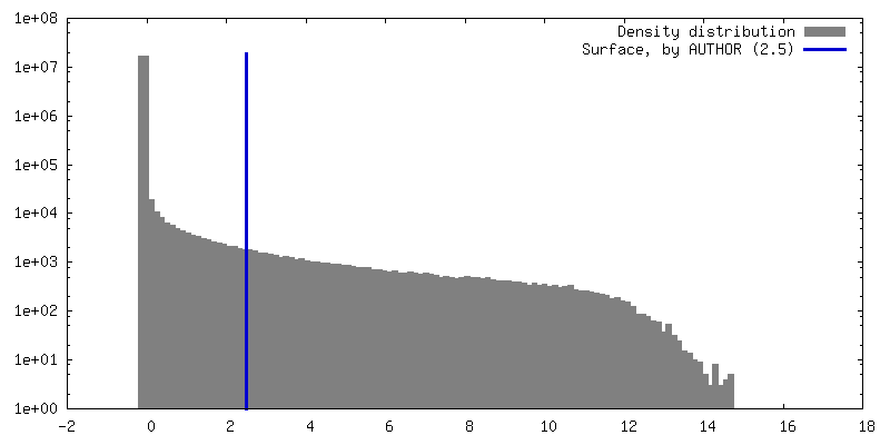
| 手法 | 単粒子再構成法 / クライオ電子顕微鏡法 / 解像度: 2.96 Å |
|---|
 データ登録者 データ登録者 | Wang YJ / Sun L |
|---|
| 資金援助 | 1件 | Organization | Grant number | 国 |
|---|
| Not funded | | |
|
|---|
 引用 引用 | ジャーナル: To Be Published
タイトル: Structure of EG.5.1 RBD in complex with antibody CYFN1006-2.
著者: Wang YJ / Sun L |
|---|
| 履歴 | | 登録 | 2024年4月19日 | - |
|---|
| ヘッダ(付随情報) 公開 | 2025年1月29日 | - |
|---|
| マップ公開 | 2025年1月29日 | - |
|---|
| 更新 | 2025年7月23日 | - |
|---|
| 現状 | 2025年7月23日 | 処理サイト: PDBc / 状態: 公開 |
|---|
|
|---|
 ムービー
ムービー コントローラー


 データを開く
 基本情報
基本情報
 構造の表示
構造の表示
 ダウンロードとリンク
 emd_39807.png
 http://ftp.pdbj.org/pub/emdb/structures/EMD-39807


 Z (Sec.)
 Y (Row.)
 X (Col.)


 試料の構成要素
 解析
解析 電子顕微鏡法
 FIELD EMISSION GUN
