 Movie
Movie Controller


+ Open data

- Basic information
| Entry |  | |||||||||
|---|---|---|---|---|---|---|---|---|---|---|
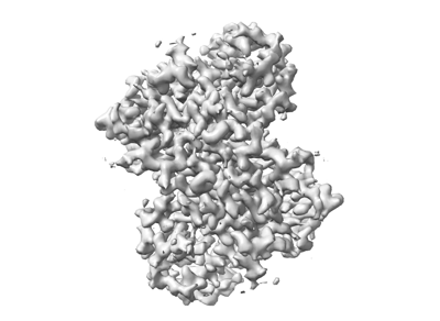
| Title | Structure of prostatic acid phosphatase in human semen | |||||||||
 Map data Map data | ||||||||||
 Sample Sample |
| |||||||||
 Keywords Keywords |  Acid phosphatase / CELL CYCLE Acid phosphatase / CELL CYCLE | |||||||||
| Function / homology |  Function and homology information Function and homology informationthiamine phosphate phosphatase activity / positive regulation of adenosine receptor signaling pathway / thiamine metabolic process / Golgi cisterna / adenosine metabolic process / acid phosphatase / regulation of sensory perception of pain / lysophosphatidic acid phosphatase activity / acid phosphatase activity / XMP 5'-nucleosidase activity ...thiamine phosphate phosphatase activity / positive regulation of adenosine receptor signaling pathway / thiamine metabolic process / Golgi cisterna / adenosine metabolic process / acid phosphatase / regulation of sensory perception of pain / lysophosphatidic acid phosphatase activity / acid phosphatase activity / XMP 5'-nucleosidase activity / 5'-nucleotidase / 5'-nucleotidase activity / vesicle membrane / nucleotide metabolic process / choline binding / azurophil granule membrane / lysosome organization / phosphatase activity / purine nucleobase metabolic process / dephosphorylation / multivesicular body / protein-tyrosine-phosphatase / filopodium / protein tyrosine phosphatase activity / lipid metabolic process / apical part of cell / lysosome / molecular adaptor activity / lysosomal membrane / Neutrophil degranulation / protein homodimerization activity / extracellular space / extracellular exosome / identical protein binding / nucleus / plasma membrane / cytosolSimilarity search - Function | |||||||||
| Biological species |  Homo sapiens (human) Homo sapiens (human) | |||||||||
| Method | single particle reconstruction / cryo EM / Resolution: 3.19 Å | |||||||||
 Authors Authors | Liu XZ / Li JL / Deng D / Wang X | |||||||||
| Funding support |  China, 1 items China, 1 items
| |||||||||
 Citation Citation | Journal: Biochem Biophys Res Commun / Year: 2024 Title: Purification, identification and Cryo-EM structure of prostatic acid phosphatase in human semen. Authors: Xuanzhong Liu / Lin Yu / Zhili Xia / Jialu Li / Wenbo Meng / Ling Min / Fuping Li / Xiang Wang / Abstract: Prostatic acid phosphatase (PAP) is a glycoprotein that plays a crucial role in the hydrolysis of phosphate ester present in prostatic exudates. It is a well-established indicator for prostate cancer ...Prostatic acid phosphatase (PAP) is a glycoprotein that plays a crucial role in the hydrolysis of phosphate ester present in prostatic exudates. It is a well-established indicator for prostate cancer due to its elevated serum levels in disease progression. Despite its abundance in semen, PAP's influence on male fertility has not been extensively studied. In our study, we report a significantly optimized method for purifying human endogenous PAP, achieving remarkably high efficiency and active protein recovery rate. This achievement allowed us to better analyze and understand the PAP protein. We determined the cryo-electron microscopic (Cryo-EM) structure of prostatic acid phosphatase in its physiological state for the first time. Our structural and gel filtration analysis confirmed the formation of a tight homodimer structure of human PAP. This functional homodimer displayed an elongated conformation in the cryo-EM structure compared to the previously reported crystal structure. Additionally, there was a notable 5-degree rotation in the angle between the α domain and α/β domain of each monomer. Through structural analysis, we revealed three potential glycosylation sites: Asn94, Asn220, and Asn333. These sites contained varying numbers and forms of glycosyl units, suggesting sugar moieties influence PAP function. Furthermore, we found that the active sites of PAP, His44 and Asp290, are located between the two protein domains. Overall, our study not only provide an optimized approach for PAP purification, but also offer crucial insights into its structural characteristics. These findings lay the groundwork for further investigations into the physiological function and potential therapeutic applications of this important protein. | |||||||||
| History |
|
- Structure visualization
| Supplemental images |
|---|
- Downloads & links
-EMDB archive
| Map data | emd_38393.map.gz | 28.8 MB | EMDB map data format | |
|---|---|---|---|---|
| Header (meta data) | emd-38393-v30.xmlemd-38393.xml | 14.1 KB 14.1 KB | Display Display | EMDB header |
| FSC (resolution estimation) | emd_38393_fsc.xml | 6.6 KB | Display | FSC data file |
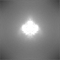
| Images |  emd_38393.png emd_38393.png | 61.1 KB | ||
| Masks | emd_38393_msk_1.map | 30.5 MB | Mask map | |
| Filedesc metadata | emd-38393.cif.gz | 5.4 KB | ||
| Others | emd_38393_half_map_1.map.gzemd_38393_half_map_2.map.gz | 28.2 MB 28.2 MB | ||
| Archive directory |  http://ftp.pdbj.org/pub/emdb/structures/EMD-38393ftp://ftp.pdbj.org/pub/emdb/structures/EMD-38393 http://ftp.pdbj.org/pub/emdb/structures/EMD-38393ftp://ftp.pdbj.org/pub/emdb/structures/EMD-38393 | HTTPS FTP |
-Related structure data
| Related structure data |  8xj4MC M: atomic model generated by this map C: citing same article ( |
|---|---|
| Similar structure data |
-Links
| EMDB pages | EMDB (EBI/PDBe) / EMDataResource |
|---|---|
| Related items in Molecule of the Month |


-Map
| File | Download / File: emd_38393.map.gz / Format: CCP4 / Size: 30.5 MB / Type: IMAGE STORED AS FLOATING POINT NUMBER (4 BYTES) | ||||||||||||||||||||
|---|---|---|---|---|---|---|---|---|---|---|---|---|---|---|---|---|---|---|---|---|---|
| Voxel size | X=Y=Z: 1.1 Å | ||||||||||||||||||||
| Density |
| ||||||||||||||||||||
| Symmetry | Space group: 1 | ||||||||||||||||||||
| Details | EMDB XML:
|
-Supplemental data
-Mask #1
| File | emd_38393_msk_1.map | ||||||||||||
|---|---|---|---|---|---|---|---|---|---|---|---|---|---|
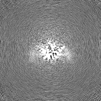
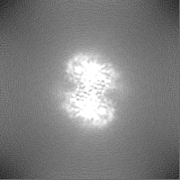
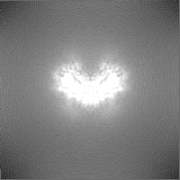
| Projections & Slices |
| ||||||||||||
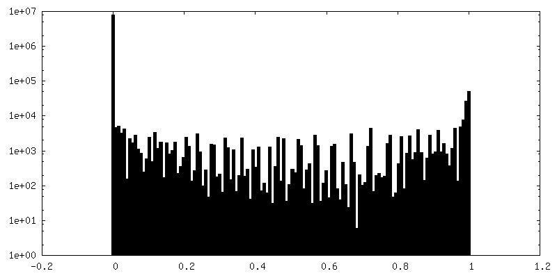
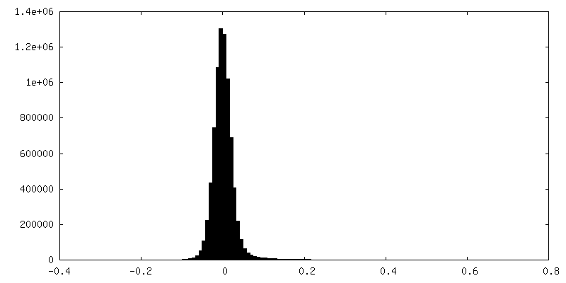
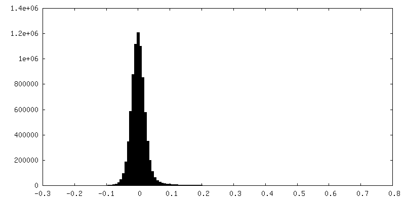
| Density Histograms |
 Z
Z Y
Y X
X


-Half map: #1
| File | emd_38393_half_map_1.map | ||||||||||||
|---|---|---|---|---|---|---|---|---|---|---|---|---|---|
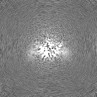
| Projections & Slices |
| ||||||||||||
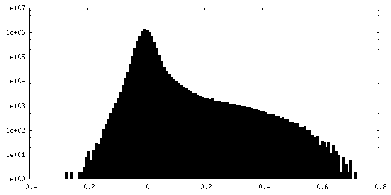
| Density Histograms |


-Half map: #2
| File | emd_38393_half_map_2.map | ||||||||||||
|---|---|---|---|---|---|---|---|---|---|---|---|---|---|
| Projections & Slices |
| ||||||||||||
| Density Histograms |


- Sample components
-Entire : structure of prostatic acid phosphatase in human semen
| Entire | Name: structure of prostatic acid phosphatase in human semen |
|---|---|
| Components |
|
-Supramolecule #1: structure of prostatic acid phosphatase in human semen
| Supramolecule | Name: structure of prostatic acid phosphatase in human semen type: organelle_or_cellular_component / ID: 1 / Parent: 0 / Macromolecule list: #1 |
|---|---|
| Source (natural) | Organism: Homo sapiens (human) |
-Macromolecule #1: Prostatic acid phosphatase
| Macromolecule | Name: Prostatic acid phosphatase / type: protein_or_peptide / ID: 1 / Number of copies: 2 / Enantiomer: LEVO / EC number: acid phosphatase |
|---|---|
| Source (natural) | Organism: Homo sapiens (human) / Tissue: Human seminal plasma |
| Molecular weight | Theoretical: 40.262949 KDa |
| Sequence | String: LAKELKFVTL VFRHGDRSPI DTFPTDPIKE SSWPQGFGQL TQLGMEQHYE LGEYIRKRYR KFLNESYKHE QVYIRSTDVD RTLMSAMTN LAALFPPEGV SIWNPILLWQ PIPVHTVPLS EDQLLYLPFR NCPRFQELES ETLKSEEFQK RLHPYKDFIA T LGKLSGLH ...String: LAKELKFVTL VFRHGDRSPI DTFPTDPIKE SSWPQGFGQL TQLGMEQHYE LGEYIRKRYR KFLNESYKHE QVYIRSTDVD RTLMSAMTN LAALFPPEGV SIWNPILLWQ PIPVHTVPLS EDQLLYLPFR NCPRFQELES ETLKSEEFQK RLHPYKDFIA T LGKLSGLH GQDLFGIWSK VYDPLYCESV HNFTLPSWAT EDTMTKLREL SELSLLSLYG IHKQKEKSRL QGGVLVNEIL NH MKRATQI PSYKKLIMYS AHDTTVSGLQ MALDVYNGLL PPYASCHLTE LYFEKGEYFV EMYYRNETQH EPYPLMLPGC SPS CPLERF AELVGPVIPQ DWSTECMTTN S UniProtKB: Prostatic acid phosphatase |
-Macromolecule #4: 2-acetamido-2-deoxy-beta-D-glucopyranose
| Macromolecule | Name: 2-acetamido-2-deoxy-beta-D-glucopyranose / type: ligand / ID: 4 / Number of copies: 4 / Formula: NAG |
|---|---|
| Molecular weight | Theoretical: 221.208 Da |
| Chemical component information |  ChemComp-NAG: |
-Macromolecule #5: alpha-D-mannopyranose
| Macromolecule | Name: alpha-D-mannopyranose / type: ligand / ID: 5 / Number of copies: 1 / Formula: MAN |
|---|---|
| Molecular weight | Theoretical: 180.156 Da |
| Chemical component information |  ChemComp-MAN: |
-Experimental details
-Structure determination
| Method | cryo EM |
|---|---|
 Processing Processing | single particle reconstruction |
| Aggregation state | particle |
-Sample preparation
| Concentration | 8.5 mg/mL |
|---|---|
| Buffer | pH: 8 |
| Vitrification | Cryogen name: ETHANE |
- Electron microscopy
| Microscope | FEI TITAN KRIOS |
|---|---|
| Electron beam | Acceleration voltage: 300 kV / Electron source: FIELD EMISSION GUN |
| Electron optics | Illumination mode: FLOOD BEAM / Imaging mode: BRIGHT FIELDBright-field microscopy / Nominal defocus max: 1.8 µm / Nominal defocus min: 1.2 µm |
| Image recording | Film or detector model: GATAN K2 SUMMIT (4k x 4k) / Average electron dose: 50.0 e/Å2 |
| Experimental equipment |  Model: Titan Krios / Image courtesy: FEI Company |
-Image processing
| Startup model | Type of model: NONE |
|---|---|
| Initial angle assignment | Type: ANGULAR RECONSTITUTION |
| Final angle assignment | Type: ANGULAR RECONSTITUTION |
| Final reconstruction | Resolution.type: BY AUTHOR / Resolution: 3.19 Å / Resolution method: FSC 0.143 CUT-OFF / Number images used: 75346 |
| FSC plot (resolution estimation) |  |
