[English] 日本語
 Yorodumi
- EMDB-38170: Cryo-EM structure of coxsackievirus A16 empty particle in complex... -
+ Open data
- Basic information
| Entry |  | |||||||||
|---|---|---|---|---|---|---|---|---|---|---|
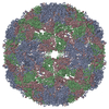
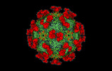
| Title | Cryo-EM structure of coxsackievirus A16 empty particle in complex with Fab h1A6.2 | |||||||||
 Map data Map data | ||||||||||
 Sample Sample |
| |||||||||
 Keywords Keywords | Cryo-EM / coxsackievirus A16 / VIRUS | |||||||||
| Function / homology |  Function and homology information Function and homology informationsymbiont genome entry into host cell via pore formation in plasma membrane / symbiont-mediated suppression of host gene expression / viral capsid / virion attachment to host cell / structural molecule activity Similarity search - Function | |||||||||
| Biological species |    Coxsackievirus A16 Coxsackievirus A16 | |||||||||
| Method | single particle reconstruction / cryo EM / Resolution: 3.36 Å | |||||||||
 Authors Authors | Jiang Y / Huang Y / Zhu R / Zheng Q / Li S / Xia N | |||||||||
| Funding support | 1 items
| |||||||||
 Citation Citation |  Journal: To Be Published Journal: To Be PublishedTitle: Cryo-EM structure of coxsackievirus A16 empty particle in complex with Fab h1A6.2 Authors: Jiang Y / Huang Y / Zhu R / Zheng Q / Li S / Xia N | |||||||||
| History |
|
- Structure visualization
| Supplemental images |
|---|
- Downloads & links
-EMDB archive
| Map data |  emd_38170.map.gz emd_38170.map.gz | 621.8 MB |  EMDB map data format EMDB map data format | |
|---|---|---|---|---|
| Header (meta data) |  emd-38170-v30.xml emd-38170-v30.xml emd-38170.xml emd-38170.xml | 15.6 KB 15.6 KB | Display Display |  EMDB header EMDB header |
| Images |  emd_38170.png emd_38170.png | 74.4 KB | ||
| Filedesc metadata |  emd-38170.cif.gz emd-38170.cif.gz | 5.4 KB | ||
| Others |  emd_38170_half_map_1.map.gz emd_38170_half_map_1.map.gz emd_38170_half_map_2.map.gz emd_38170_half_map_2.map.gz | 156.5 MB 156.5 MB | ||
| Archive directory |  http://ftp.pdbj.org/pub/emdb/structures/EMD-38170 http://ftp.pdbj.org/pub/emdb/structures/EMD-38170 ftp://ftp.pdbj.org/pub/emdb/structures/EMD-38170 ftp://ftp.pdbj.org/pub/emdb/structures/EMD-38170 | HTTPS FTP |
-Validation report
| Summary document |  emd_38170_validation.pdf.gz emd_38170_validation.pdf.gz | 1020.9 KB | Display |  EMDB validaton report EMDB validaton report |
|---|---|---|---|---|
| Full document |  emd_38170_full_validation.pdf.gz emd_38170_full_validation.pdf.gz | 1020.4 KB | Display | |
| Data in XML |  emd_38170_validation.xml.gz emd_38170_validation.xml.gz | 20.5 KB | Display | |
| Data in CIF |  emd_38170_validation.cif.gz emd_38170_validation.cif.gz | 24.5 KB | Display | |
| Arichive directory |  https://ftp.pdbj.org/pub/emdb/validation_reports/EMD-38170 https://ftp.pdbj.org/pub/emdb/validation_reports/EMD-38170 ftp://ftp.pdbj.org/pub/emdb/validation_reports/EMD-38170 ftp://ftp.pdbj.org/pub/emdb/validation_reports/EMD-38170 | HTTPS FTP |
-Related structure data
| Related structure data |  8x9aMC M: atomic model generated by this map C: citing same article ( |
|---|---|
| Similar structure data | Similarity search - Function & homology  F&H Search F&H Search |
- Links
Links
| EMDB pages |  EMDB (EBI/PDBe) / EMDB (EBI/PDBe) /  EMDataResource EMDataResource |
|---|---|
| Related items in Molecule of the Month |
- Map
Map
| File |  Download / File: emd_38170.map.gz / Format: CCP4 / Size: 669.9 MB / Type: IMAGE STORED AS FLOATING POINT NUMBER (4 BYTES) Download / File: emd_38170.map.gz / Format: CCP4 / Size: 669.9 MB / Type: IMAGE STORED AS FLOATING POINT NUMBER (4 BYTES) | ||||||||||||||||||||||||||||||||||||
|---|---|---|---|---|---|---|---|---|---|---|---|---|---|---|---|---|---|---|---|---|---|---|---|---|---|---|---|---|---|---|---|---|---|---|---|---|---|
| Projections & slices | Image control
Images are generated by Spider. | ||||||||||||||||||||||||||||||||||||
| Voxel size | X=Y=Z: 1.12 Å | ||||||||||||||||||||||||||||||||||||
| Density |
| ||||||||||||||||||||||||||||||||||||
| Symmetry | Space group: 1 | ||||||||||||||||||||||||||||||||||||
| Details | EMDB XML:
|
-Supplemental data
-Half map: #2
| File | emd_38170_half_map_1.map | ||||||||||||
|---|---|---|---|---|---|---|---|---|---|---|---|---|---|
| Projections & Slices |
| ||||||||||||
| Density Histograms |
-Half map: #1
| File | emd_38170_half_map_2.map | ||||||||||||
|---|---|---|---|---|---|---|---|---|---|---|---|---|---|
| Projections & Slices |
| ||||||||||||
| Density Histograms |
- Sample components
-Entire : Coxsackievirus A16 empty particle in complex with Fab h1A6.2
| Entire | Name: Coxsackievirus A16 empty particle in complex with Fab h1A6.2 |
|---|---|
| Components |
|
-Supramolecule #1: Coxsackievirus A16 empty particle in complex with Fab h1A6.2
| Supramolecule | Name: Coxsackievirus A16 empty particle in complex with Fab h1A6.2 type: complex / ID: 1 / Parent: 0 / Macromolecule list: all |
|---|
-Supramolecule #3: The Fab of h1A6.2
| Supramolecule | Name: The Fab of h1A6.2 / type: complex / ID: 3 / Parent: 1 |
|---|---|
| Source (natural) | Organism:  |
-Supramolecule #2: Coxsackievirus A16
| Supramolecule | Name: Coxsackievirus A16 / type: virus / ID: 2 / Parent: 1 / Macromolecule list: all / NCBI-ID: 31704 / Sci species name: Coxsackievirus A16 / Virus type: VIRION / Virus isolate: OTHER / Virus enveloped: No / Virus empty: Yes |
|---|
-Macromolecule #1: Capsid protein VP1
| Macromolecule | Name: Capsid protein VP1 / type: protein_or_peptide / ID: 1 / Number of copies: 1 / Enantiomer: LEVO |
|---|---|
| Source (natural) | Organism:   Coxsackievirus A16 Coxsackievirus A16 |
| Molecular weight | Theoretical: 33.106352 KDa |
| Sequence | String: GDPIADMIDQ TVNNQVNRSL TALQVLPTAA NTEASSHRLG TGVVPALQAA ETGASSNASD KNLIETRCVL NHHSTQETAI GNFFSRAGL VSIITMPTTD TQNTDGYVNW DIDLMGYAQL RRKCELFTYM RFDAEFTFVV AKPNGVLVPQ LLQYMYVPPG A PKPTSRDS ...String: GDPIADMIDQ TVNNQVNRSL TALQVLPTAA NTEASSHRLG TGVVPALQAA ETGASSNASD KNLIETRCVL NHHSTQETAI GNFFSRAGL VSIITMPTTD TQNTDGYVNW DIDLMGYAQL RRKCELFTYM RFDAEFTFVV AKPNGVLVPQ LLQYMYVPPG A PKPTSRDS FAWQTATNPS VFVKMTDPPA QVSVPFMSPA SAYQWFYDGY PTFGEHLQAN DLDYGQCPNN MMGTFSIRTV GT EKSPHSI TLRVYMRIKH VRAWIPRPLR NQPYLFKTNP NYKGNDIKCT STSRDKITTL UniProtKB: Genome polyprotein |
-Macromolecule #2: Capsid protein VP2
| Macromolecule | Name: Capsid protein VP2 / type: protein_or_peptide / ID: 2 / Number of copies: 1 / Enantiomer: LEVO |
|---|---|
| Source (natural) | Organism:   Coxsackievirus A16 Coxsackievirus A16 |
| Molecular weight | Theoretical: 27.557104 KDa |
| Sequence | String: SPSAEACGYS DRVAQLTIGN STITTQEAAN IVIAYGEWPE YCPDTDATAV DKPTRPDVSV NRFFTLDTKS WAKDSKGWYW KFPDVLTEV GVFGQNAQFH YLYRSGFCVH VQCNASKFHQ GALLVAVLPE YVLGTIAGGT GNENSHPPYA TTQPGQVGAV L THPYVLDA ...String: SPSAEACGYS DRVAQLTIGN STITTQEAAN IVIAYGEWPE YCPDTDATAV DKPTRPDVSV NRFFTLDTKS WAKDSKGWYW KFPDVLTEV GVFGQNAQFH YLYRSGFCVH VQCNASKFHQ GALLVAVLPE YVLGTIAGGT GNENSHPPYA TTQPGQVGAV L THPYVLDA GIPLSQLTVC PHQWINLRTN NCATIIVPYM NTVPFDSALN HCNFGLLVIP VVPLDFNAGA TSEIPITVTI AP MCAEFAG LRQAVKQ UniProtKB: Genome polyprotein |
-Macromolecule #3: Capsid protein VP3
| Macromolecule | Name: Capsid protein VP3 / type: protein_or_peptide / ID: 3 / Number of copies: 1 / Enantiomer: LEVO |
|---|---|
| Source (natural) | Organism:   Coxsackievirus A16 Coxsackievirus A16 |
| Molecular weight | Theoretical: 26.654295 KDa |
| Sequence | String: GIPTELKPGT NQFLTTDDGV SAPILPGFHP TPPIHIPGEV HNLLEICRVE TILEVNNLKT NETTPMQRLC FPVSVQSKTG ELCAAFRAD PGRDGPWQST ILGQLCRYYT QWSGSLEVTF MFAGSFMATG KMLIAYTPPG GNVPADRITA MLGTHVIWDF G LQSSVTLV ...String: GIPTELKPGT NQFLTTDDGV SAPILPGFHP TPPIHIPGEV HNLLEICRVE TILEVNNLKT NETTPMQRLC FPVSVQSKTG ELCAAFRAD PGRDGPWQST ILGQLCRYYT QWSGSLEVTF MFAGSFMATG KMLIAYTPPG GNVPADRITA MLGTHVIWDF G LQSSVTLV VPWISNTHYR AHARAGYFDY YTTGIITIWY QTNYVVPIGA PTTAYIVALA AAQDNFTMKL CKDTEDIEQT AN IQ UniProtKB: Genome polyprotein |
-Experimental details
-Structure determination
| Method | cryo EM |
|---|---|
 Processing Processing | single particle reconstruction |
| Aggregation state | particle |
- Sample preparation
| Buffer | pH: 7.4 |
|---|---|
| Vitrification | Cryogen name: ETHANE |
- Electron microscopy
| Microscope | FEI TECNAI F30 |
|---|---|
| Image recording | Film or detector model: FEI FALCON III (4k x 4k) / Average electron dose: 40.0 e/Å2 |
| Electron beam | Acceleration voltage: 300 kV / Electron source: OTHER |
| Electron optics | Illumination mode: FLOOD BEAM / Imaging mode: BRIGHT FIELD / Nominal defocus max: 2.8000000000000003 µm / Nominal defocus min: 0.9 µm |
| Experimental equipment |  Model: Tecnai F30 / Image courtesy: FEI Company |
- Image processing
| Startup model | Type of model: OTHER |
|---|---|
| Final reconstruction | Resolution.type: BY AUTHOR / Resolution: 3.36 Å / Resolution method: FSC 0.143 CUT-OFF / Number images used: 32746 |
| Initial angle assignment | Type: MAXIMUM LIKELIHOOD |
| Final angle assignment | Type: MAXIMUM LIKELIHOOD |
 Movie
Movie Controller


 Z (Sec.)
 Y (Row.)
 X (Col.)


