[English] 日本語
 Yorodumi
- EMDB-38151: Cryo-EM structure of a bacteriophage tail- spike protein against ... -
+ Open data
- Basic information
| Entry |  | |||||||||
|---|---|---|---|---|---|---|---|---|---|---|
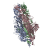
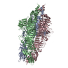
| Title | Cryo-EM structure of a bacteriophage tail- spike protein against Klebsiella pneumoniae K64,ORF41(K64-ORF41) in 5 mM EDTA | |||||||||
 Map data Map data | Cryo-EM structure of a bacteriophage tail- spike protein against Klebsiella pneumoniae K64,ORF41(K64-ORF41) in 5mM EDTA | |||||||||
 Sample Sample |
| |||||||||
 Keywords Keywords | K64-ORF41 / depolymerase / cryo-EM / Klebsiella pneumoniae K64 / capsule polysaccharide / EDTA / LYASE | |||||||||
| Function / homology |  Function and homology information Function and homology informationsymbiont entry into host cell via disruption of host cell glycocalyx / symbiont entry into host cell via disruption of host cell envelope / virus tail / outer membrane / adhesion receptor-mediated virion attachment to host cell Similarity search - Function | |||||||||
| Biological species |  Klebsiella phage SH-Kp 152410 (virus) Klebsiella phage SH-Kp 152410 (virus) | |||||||||
| Method | single particle reconstruction / cryo EM / Resolution: 2.5 Å | |||||||||
 Authors Authors | Xie Y / Huang T / Zhang Z / Tao X | |||||||||
| Funding support |  China, 1 items China, 1 items
| |||||||||
 Citation Citation |  Journal: Int J Biol Macromol / Year: 2024 Journal: Int J Biol Macromol / Year: 2024Title: Structural and functional basis of bacteriophage K64-ORF41 depolymerase for capsular polysaccharide degradation of Klebsiella pneumoniae K64. Authors: Tianyun Huang / Zhuoyuan Zhang / Xin Tao / Xinyu Shi / Peng Lin / Dan Liao / Chenyu Ma / Xinle Cai / Wei Lin / Xiaofan Jiang / Peng Luo / Shan Wu / Yuan Xie /  Abstract: Capsule polysaccharide is an important virulence factor of Klebsiella pneumoniae (K. pneumoniae), which protects bacteria against the host immune response. A promising therapeutic approach is using ...Capsule polysaccharide is an important virulence factor of Klebsiella pneumoniae (K. pneumoniae), which protects bacteria against the host immune response. A promising therapeutic approach is using phage-derived depolymerases to degrade the capsular polysaccharide and expose and sensitize the bacteria to the host immune system. Here we determined the cryo-electron microscopy (cryo-EM) structures of a bacteriophage tail-spike protein against K. pneumoniae K64, ORF41 (K64-ORF41) and ORF41 in EDTA condition (K64-ORF41), at 2.37 Å and 2.50 Å resolution, respectively, for the first time. K64-ORF41 exists as a trimer and each protomer contains a β-helix domain including a right-handed parallel β-sheet helix fold capped at both ends, an insertion domain, and one β-sheet jellyroll domain. Moreover, our structural comparison with other depolymerases of K. pneumoniae suggests that the catalytic residues (Tyr528, His574 and Arg628) are highly conserved although the substrate of capsule polysaccharide is variable. Besides that, we figured out the important residues involved in the substrate binding pocket including Arg405, Tyr526, Trp550 and Phe669. This study establishes the structural and functional basis for the promising phage-derived broad-spectrum activity depolymerase therapeutics and effective CPS-degrading agents for the treatment of carbapenem-resistant K. pneumoniae K64 infections. | |||||||||
| History |
|
- Structure visualization
| Supplemental images |
|---|
- Downloads & links
-EMDB archive
| Map data |  emd_38151.map.gz emd_38151.map.gz | 70.5 MB |  EMDB map data format EMDB map data format | |
|---|---|---|---|---|
| Header (meta data) |  emd-38151-v30.xml emd-38151-v30.xml emd-38151.xml emd-38151.xml | 14.9 KB 14.9 KB | Display Display |  EMDB header EMDB header |
| Images |  emd_38151.png emd_38151.png | 25.3 KB | ||
| Filedesc metadata |  emd-38151.cif.gz emd-38151.cif.gz | 5.8 KB | ||
| Others |  emd_38151_half_map_1.map.gz emd_38151_half_map_1.map.gz emd_38151_half_map_2.map.gz emd_38151_half_map_2.map.gz | 58.5 MB 58.5 MB | ||
| Archive directory |  http://ftp.pdbj.org/pub/emdb/structures/EMD-38151 http://ftp.pdbj.org/pub/emdb/structures/EMD-38151 ftp://ftp.pdbj.org/pub/emdb/structures/EMD-38151 ftp://ftp.pdbj.org/pub/emdb/structures/EMD-38151 | HTTPS FTP |
-Validation report
| Summary document |  emd_38151_validation.pdf.gz emd_38151_validation.pdf.gz | 933.5 KB | Display |  EMDB validaton report EMDB validaton report |
|---|---|---|---|---|
| Full document |  emd_38151_full_validation.pdf.gz emd_38151_full_validation.pdf.gz | 933 KB | Display | |
| Data in XML |  emd_38151_validation.xml.gz emd_38151_validation.xml.gz | 12.5 KB | Display | |
| Data in CIF |  emd_38151_validation.cif.gz emd_38151_validation.cif.gz | 14.9 KB | Display | |
| Arichive directory |  https://ftp.pdbj.org/pub/emdb/validation_reports/EMD-38151 https://ftp.pdbj.org/pub/emdb/validation_reports/EMD-38151 ftp://ftp.pdbj.org/pub/emdb/validation_reports/EMD-38151 ftp://ftp.pdbj.org/pub/emdb/validation_reports/EMD-38151 | HTTPS FTP |
-Related structure data
| Related structure data |  8x8oMC  8x8mC M: atomic model generated by this map C: citing same article ( |
|---|---|
| Similar structure data | Similarity search - Function & homology  F&H Search F&H Search |
- Links
Links
| EMDB pages |  EMDB (EBI/PDBe) / EMDB (EBI/PDBe) /  EMDataResource EMDataResource |
|---|
- Map
Map
| File |  Download / File: emd_38151.map.gz / Format: CCP4 / Size: 75.1 MB / Type: IMAGE STORED AS FLOATING POINT NUMBER (4 BYTES) Download / File: emd_38151.map.gz / Format: CCP4 / Size: 75.1 MB / Type: IMAGE STORED AS FLOATING POINT NUMBER (4 BYTES) | ||||||||||||||||||||||||||||||||||||
|---|---|---|---|---|---|---|---|---|---|---|---|---|---|---|---|---|---|---|---|---|---|---|---|---|---|---|---|---|---|---|---|---|---|---|---|---|---|
| Annotation | Cryo-EM structure of a bacteriophage tail- spike protein against Klebsiella pneumoniae K64,ORF41(K64-ORF41) in 5mM EDTA | ||||||||||||||||||||||||||||||||||||
| Projections & slices | Image control
Images are generated by Spider. | ||||||||||||||||||||||||||||||||||||
| Voxel size | X=Y=Z: 0.851 Å | ||||||||||||||||||||||||||||||||||||
| Density |
| ||||||||||||||||||||||||||||||||||||
| Symmetry | Space group: 1 | ||||||||||||||||||||||||||||||||||||
| Details | EMDB XML:
|
-Supplemental data
-Half map: half1 map of ORF41(K64-ORF41) in 5mM EDTA
| File | emd_38151_half_map_1.map | ||||||||||||
|---|---|---|---|---|---|---|---|---|---|---|---|---|---|
| Annotation | half1 map of ORF41(K64-ORF41) in 5mM EDTA | ||||||||||||
| Projections & Slices |
| ||||||||||||
| Density Histograms |
-Half map: half2 map of ORF41(K64-ORF41) in 5mM EDTA
| File | emd_38151_half_map_2.map | ||||||||||||
|---|---|---|---|---|---|---|---|---|---|---|---|---|---|
| Annotation | half2 map of ORF41(K64-ORF41) in 5mM EDTA | ||||||||||||
| Projections & Slices |
| ||||||||||||
| Density Histograms |
- Sample components
-Entire : K64-ORF41 in 5 mM EDTA
| Entire | Name: K64-ORF41 in 5 mM EDTA |
|---|---|
| Components |
|
-Supramolecule #1: K64-ORF41 in 5 mM EDTA
| Supramolecule | Name: K64-ORF41 in 5 mM EDTA / type: complex / ID: 1 / Parent: 0 / Macromolecule list: all |
|---|---|
| Source (natural) | Organism:  Klebsiella phage SH-Kp 152410 (virus) Klebsiella phage SH-Kp 152410 (virus) |
-Macromolecule #1: Probable tail spike protein
| Macromolecule | Name: Probable tail spike protein / type: protein_or_peptide / ID: 1 / Number of copies: 3 / Enantiomer: LEVO |
|---|---|
| Source (natural) | Organism:  Klebsiella phage SH-Kp 152410 (virus) Klebsiella phage SH-Kp 152410 (virus) |
| Molecular weight | Theoretical: 112.2515 KDa |
| Recombinant expression | Organism:  |
| Sequence | String: MGSSHHHHHH SSGLVPRGSH MDQDTKTIIQ YPTSGDEYDI PFDYLSRKFV RVSLVSNTQR VLLDNITDYR YVSRTRVKLL VSTDGYSRV EIRRFTSASE MVVDFSDGSV LRATDLNVSA LQSAHIAEEA RDLFSTSLSI GQLSYFDAKG LQIKNVAAGV D NTDAVTVQ ...String: MGSSHHHHHH SSGLVPRGSH MDQDTKTIIQ YPTSGDEYDI PFDYLSRKFV RVSLVSNTQR VLLDNITDYR YVSRTRVKLL VSTDGYSRV EIRRFTSASE MVVDFSDGSV LRATDLNVSA LQSAHIAEEA RDLFSTSLSI GQLSYFDAKG LQIKNVAAGV D NTDAVTVQ QLNKIIADVV TTIPDSVADN IRGLWARVLG DIGITLVDGS FETGATITTR TQALWSISGR KCYTWAGALP KV VPENSTP ESTGGISETA WVDSSSKALG VLLAGPSGAE RVGLKQGGTV QDAINWLTFD SFDIVKDGSK DVTADIMAAC VVA NDLGLD IKQNDGTYLV SGNPVWPVYN SLDLNGVTLK LAAGFTGYFA LTQKDSTTVY GPTSPIVQAI NAAGGRTAGS GVLE GLVNS TELNGKFLFM EGADVLYYSR GTAKYWWTNT YLSNRGKLSD NLKYGVSAIT KITAVTPRTK IVYYRLPNLD FGNGP ANNG VIRVLNNTRF IMQGGSISNR PLKDVSKSPV IISLNYCAAF KAYDFFDPYP AFAVDSNNSL VYSYTLNFND IADAVF ENF NSQGYGWGVV GGQRSTNITY RDCNLNRVDM HNPYMGYLKV LDTRLGTWGI NASGMGDMYL ERVTVDLDDS AHGGHRE HE GIINARGDFG GFHDGGLYIK DLTIVGEASA FEATSGHPVA LVSAYSFNAS LAYIPESSPV TPWGFKEVIV EGLHCPFK R TGRRFNSIIS APSIQFTVYH PMRVKLEDCN FNSTAFEKFD LRGWRVTPYN PSKVGIANTL AFRPTNFVDV KDCSMVGLE FTRPTSAYDY SNFDVNLVNV KNVEEHSLSP FTLYTNQCGR YNLVGCGLQQ IVDKSMTSGE RANRRSTFSV TGGTWNSLSG NPTDITYGN GYDIPVVATG VMFVGPYSQT EVTGANLNVA EFVQASGCKF LSSGPTYIQP LLWSGAGGPT GASANFNVAR G NTLGLNIS AVNGETSQVI AATLVIPQGF STGPAAGTTY GFAVEKNINY QLGLNARSLK ANVGLVRCSD TITGVYLNA UniProtKB: Probable tail spike protein |
-Experimental details
-Structure determination
| Method | cryo EM |
|---|---|
 Processing Processing | single particle reconstruction |
| Aggregation state | particle |
- Sample preparation
| Buffer | pH: 8 |
|---|---|
| Vitrification | Cryogen name: ETHANE |
- Electron microscopy
| Microscope | FEI TITAN |
|---|---|
| Image recording | Film or detector model: GATAN K3 BIOQUANTUM (6k x 4k) / Average electron dose: 54.0 e/Å2 |
| Electron beam | Acceleration voltage: 300 kV / Electron source:  FIELD EMISSION GUN FIELD EMISSION GUN |
| Electron optics | Illumination mode: FLOOD BEAM / Imaging mode: BRIGHT FIELD / Nominal defocus max: 1.5 µm / Nominal defocus min: 1.1 µm |
- Image processing
| Startup model | Type of model: OTHER |
|---|---|
| Final reconstruction | Resolution.type: BY AUTHOR / Resolution: 2.5 Å / Resolution method: FSC 0.143 CUT-OFF / Number images used: 371259 |
| Initial angle assignment | Type: PROJECTION MATCHING |
| Final angle assignment | Type: PROJECTION MATCHING |
 Movie
Movie Controller


 Z (Sec.)
 Y (Row.)
 X (Col.)


