National Natural Science Foundation of China (NSFC)
91957201
China
National Natural Science Foundation of China (NSFC)
31821091
China
National Natural Science Foundation of China (NSFC)
31870833
China
Ministry of Science and Technology (MoST, China)
2022YFA1303000
China
Citation
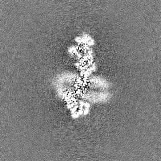
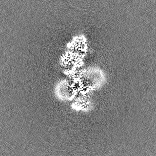
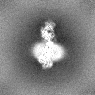
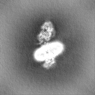
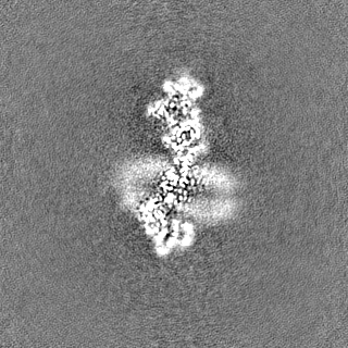
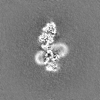
Journal: Nature / Year: 2024 Title: Structure of human phagocyte NADPH oxidase in the activated state. Authors: Xiaoyu Liu / Yiting Shi / Rui Liu / Kangcheng Song / Lei Chen / Abstract: Phagocyte NADPH oxidase, a protein complex with a core made up of NOX2 and p22 subunits, is responsible for transferring electrons from intracellular NADPH to extracellular oxygen. This process ...Phagocyte NADPH oxidase, a protein complex with a core made up of NOX2 and p22 subunits, is responsible for transferring electrons from intracellular NADPH to extracellular oxygen. This process generates superoxide anions that are vital for killing pathogens. The activation of phagocyte NADPH oxidase requires membrane translocation and the binding of several cytosolic factors. However, the exact mechanism by which cytosolic factors bind to and activate NOX2 is not well understood. Here we present the structure of the human NOX2-p22 complex activated by fragments of three cytosolic factors: p47, p67 and Rac1. The structure reveals that the p67-Rac1 complex clamps onto the dehydrogenase domain of NOX2 and induces its contraction, which stabilizes the binding of NADPH and results in a reduction of the distance between the NADPH-binding domain and the flavin adenine dinucleotide (FAD)-binding domain. Furthermore, the dehydrogenase domain docks onto the bottom of the transmembrane domain of NOX2, which reduces the distance between FAD and the inner haem. These structural rearrangements might facilitate the efficient transfer of electrons between the redox centres in NOX2 and lead to the activation of phagocyte NADPH oxidase.
In the structure databanks used in Yorodumi, some data are registered as the other names, "COVID-19 virus" and "2019-nCoV". Here are the details of the virus and the list of structure data.
Jan 31, 2019. EMDB accession codes are about to change! (news from PDBe EMDB page)
EMDB accession codes are about to change! (news from PDBe EMDB page)
The allocation of 4 digits for EMDB accession codes will soon come to an end. Whilst these codes will remain in use, new EMDB accession codes will include an additional digit and will expand incrementally as the available range of codes is exhausted. The current 4-digit format prefixed with “EMD-” (i.e. EMD-XXXX) will advance to a 5-digit format (i.e. EMD-XXXXX), and so on. It is currently estimated that the 4-digit codes will be depleted around Spring 2019, at which point the 5-digit format will come into force.
The EM Navigator/Yorodumi systems omit the EMD- prefix.
Related info.:Q: What is EMD? / ID/Accession-code notation in Yorodumi/EM Navigator
Yorodumi is a browser for structure data from EMDB, PDB, SASBDB, etc.
This page is also the successor to EM Navigator detail page, and also detail information page/front-end page for Omokage search.
The word "yorodu" (or yorozu) is an old Japanese word meaning "ten thousand". "mi" (miru) is to see.
Related info.:EMDB / PDB / SASBDB / Comparison of 3 databanks / Yorodumi Search / Aug 31, 2016. New EM Navigator & Yorodumi / Yorodumi Papers / Jmol/JSmol / Function and homology information / Changes in new EM Navigator and Yorodumi
 Movie
Movie Controller

 Yorodumi
 Open data
 Basic information
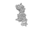
 Map data
 Sample
 Keywords
 Function and homology information
 Homo sapiens (human) /
 Authors
 China, 4 items
  Citation
 Structure visualization
 Downloads & links
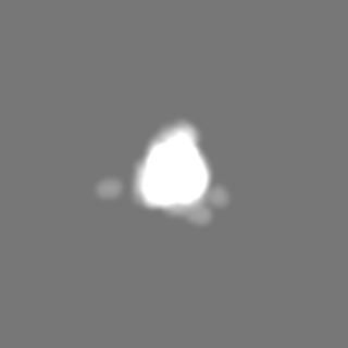
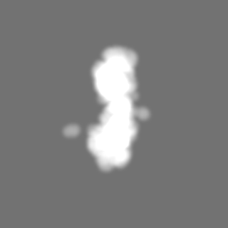
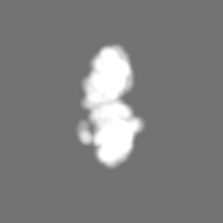
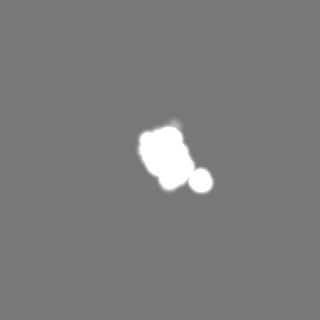
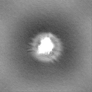
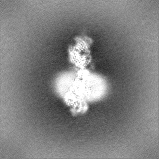
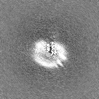
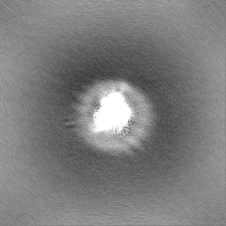
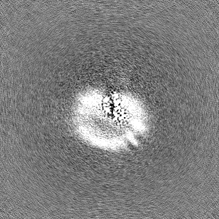
 emd_38016.png
 http://ftp.pdbj.org/pub/emdb/structures/EMD-38016
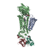


 Z
Z Y
Y X
X


 Sample components


 Processing
 Electron microscopy
 FIELD EMISSION GUN