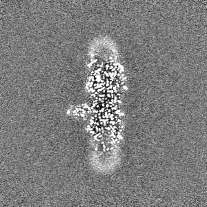
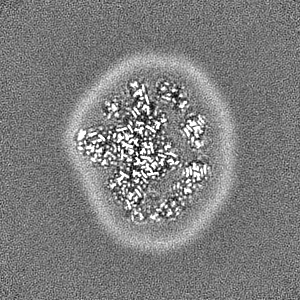
- EMDB-37513: Cryo-EM structure of the red-shifted Fittonia albivenis PSI-LHCI -
+
データを開く
IDまたはキーワード:
読み込み中...
-
基本情報
登録情報
データベース: EMDB / ID: EMD-37513
タイトル
Cryo-EM structure of the red-shifted Fittonia albivenis PSI-LHCI
マップデータ
試料
複合体: Photosystem I complex
タンパク質・ペプチド: x 18種
リガンド: x 12種
キーワード
Complex / PHOTOSYNTHESIS
機能・相同性
機能・相同性情報
plastoglobule / chloroplast thylakoid / thylakoid / chloroplast envelope / photosystem I reaction center / photosystem I / photosynthetic electron transport in photosystem I / photosystem I / chlorophyll binding / chloroplast thylakoid membrane ...plastoglobule / chloroplast thylakoid / thylakoid / chloroplast envelope / photosystem I reaction center / photosystem I / photosynthetic electron transport in photosystem I / photosystem I / chlorophyll binding / chloroplast thylakoid membrane / photosynthesis / chloroplast / 4 iron, 4 sulfur cluster binding / electron transfer activity / oxidoreductase activity / protein domain specific binding / mRNA binding / magnesium ion binding / metal ion binding / nucleus 類似検索 - 分子機能
4Fe-4S dicluster domain / Photosystem I reaction centre subunit VIII / Photosystem I reaction centre subunit VIII / Photosystem I reaction centre subunit VIII superfamily / Photosystem I PsaF, reaction centre subunit III / Photosystem I PsaF, reaction centre subunit III superfamily / Photosystem I reaction centre subunit III / Photosystem I PsaJ, reaction centre subunit IX superfamily / Photosystem I PsaJ, reaction centre subunit IX / Photosystem I reaction centre subunit IX / PsaJ ...4Fe-4S dicluster domain / Photosystem I reaction centre subunit VIII / Photosystem I reaction centre subunit VIII / Photosystem I reaction centre subunit VIII superfamily / Photosystem I PsaF, reaction centre subunit III / Photosystem I PsaF, reaction centre subunit III superfamily / Photosystem I reaction centre subunit III / Photosystem I PsaJ, reaction centre subunit IX superfamily / Photosystem I PsaJ, reaction centre subunit IX / Photosystem I reaction centre subunit IX / PsaJ / Photosystem I PsaA / Photosystem I protein PsaC / Photosystem I PsaB / Photosystem I PsaA/PsaB, conserved site / Photosystem I psaA and psaB proteins signature. / : / Photosystem I PsaA/PsaB / Photosystem I PsaA/PsaB superfamily / Photosystem I psaA/psaB protein / 4Fe-4S ferredoxin, iron-sulphur binding, conserved site / 4Fe-4S ferredoxin-type iron-sulfur binding region signature. / 4Fe-4S ferredoxin-type iron-sulfur binding domain profile. / 4Fe-4S ferredoxin-type, iron-sulphur binding domain 類似検索 - ドメイン・相同性
Photosystem I P700 chlorophyll a apoprotein A1 / Photosystem I reaction center subunit VIII / Photosystem I iron-sulfur center / Photosystem I P700 chlorophyll a apoprotein A2 / Photosystem I reaction center subunit IX / Photosystem I reaction center subunit III, chloroplastic 類似検索 - 構成要素
ジャーナル: Nat Commun / 年: 2024 タイトル: Structure of the red-shifted Fittonia albivenis photosystem I. 著者: Xiuxiu Li / Guoqiang Huang / Lixia Zhu / Chenyang Hao / Sen-Fang Sui / Xiaochun Qin / 要旨: Photosystem I (PSI) from Fittonia albivenis, an Acanthaceae ornamental plant, is notable among green plants for its red-shifted emission spectrum. Here, we solved the structure of a PSI-light ...Photosystem I (PSI) from Fittonia albivenis, an Acanthaceae ornamental plant, is notable among green plants for its red-shifted emission spectrum. Here, we solved the structure of a PSI-light harvesting complex I (LHCI) supercomplex from F. albivenis at 2.46-Å resolution using cryo-electron microscopy. The supercomplex contains a core complex of 14 subunits and an LHCI belt with four antenna subunits (Lhca1-4) similar to previously reported angiosperm PSI-LHCI structures; however, Lhca3 differs in three regions surrounding a dimer of low-energy chlorophylls (Chls) termed red Chls, which absorb far-red beyond visible light. The unique amino acid sequences within these regions are exclusively shared by plants with strongly red-shifted fluorescence emission, suggesting candidate structural elements for regulating the energy state of red Chls. These results provide a structural basis for unraveling the mechanisms of light harvest and transfer in PSI-LHCI of under canopy plants and for designing Lhc to harness longer-wavelength light in the far-red spectral range.
MAASVAAQSP VAVFRPRFLT GAPGKLNRPS VTVKQAVSSR GSFKVEAEKG EWLPGLPSPA YLDGSLPGDN GFDPLGLAED PENLKWYIQ AELVNSRWAM LGVAGMLLPE VFTYLGIINV PKWYDAGKSE YFASSSTLFV IEFILFHYVE IRRWQDIKNP G CVNQDPIF KNYSLPPHEC GYPGSVFNPL NFEPTLEAKE KELANGRLAM LAFLGFIVQH NVTGKGPFDN LVQHVADPWH NT IINTIRG Y
+
分子 #5: Photosystem I P700 chlorophyll a apoprotein A1
分子
名称: Photosystem I P700 chlorophyll a apoprotein A1 / タイプ: protein_or_peptide / ID: 5 詳細: Sequence reference for Fittonia albivenis is not available in UniProt at the time of biocuration. Current sequence reference is from UniProt id A0A8A0WPY6. コピー数: 1 / 光学異性体: LEVO / EC番号: photosystem I
UniProtKB: Photosystem I P700 chlorophyll a apoprotein A1
+
分子 #6: Photosystem I P700 chlorophyll a apoprotein A2
分子
名称: Photosystem I P700 chlorophyll a apoprotein A2 / タイプ: protein_or_peptide / ID: 6 詳細: Sequence reference for Fittonia albivenis is not available in UniProt at the time of biocuration. Current sequence reference is from UniProt id G9IB61. コピー数: 1 / 光学異性体: LEVO / EC番号: photosystem I
UniProtKB: Photosystem I P700 chlorophyll a apoprotein A2
+
分子 #7: Photosystem I iron-sulfur center
分子
名称: Photosystem I iron-sulfur center / タイプ: protein_or_peptide / ID: 7 詳細: Sequence reference for Fittonia albivenis is not available in UniProt at the time of biocuration. Current sequence reference is from UniProt id A4QJG7. コピー数: 1 / 光学異性体: LEVO / EC番号: photosystem I
由来(天然)
生物種: Fittonia albivenis (植物)
分子量
理論値: 9.049509 KDa
配列
文字列:
MSHSVKIYDT CIGCTQCVRA CPTDVLEMIP WDGCKAKQIA SAPRTEDCVG CKRCESACPT DFLSVRVYLW HETTRSMGLA Y
UniProtKB: Photosystem I iron-sulfur center
+
分子 #8: Photosystem I reaction center subunit II
分子
名称: Photosystem I reaction center subunit II / タイプ: protein_or_peptide / ID: 8 / コピー数: 1 / 光学異性体: LEVO
分子 #10: Photosystem I reaction center subunit III, chloroplastic
分子
名称: Photosystem I reaction center subunit III, chloroplastic タイプ: protein_or_peptide / ID: 10 詳細: Sequence reference for Fittonia albivenis is not available in UniProt at the time of biocuration. Current sequence reference is from UniProt id Q9SHE8. コピー数: 1 / 光学異性体: LEVO
分子 #13: Photosystem I reaction center subunit VIII
分子
名称: Photosystem I reaction center subunit VIII / タイプ: protein_or_peptide / ID: 13 詳細: Sequence reference for Fittonia albivenis is not available in UniProt at the time of biocuration. Current sequence reference is from UniProt id A0A8A9WIB9. コピー数: 1 / 光学異性体: LEVO
由来(天然)
生物種: Fittonia albivenis (植物)
分子量
理論値: 3.930826 KDa
配列
文字列:
MTAFNLPSIF VPLVGLVFPA IAMASLFLHV QKNKIV
UniProtKB: Photosystem I reaction center subunit VIII
+
分子 #14: Photosystem I reaction center subunit IX
分子
名称: Photosystem I reaction center subunit IX / タイプ: protein_or_peptide / ID: 14 詳細: Sequence reference for Fittonia albivenis is not available in UniProt at the time of biocuration. Current sequence reference is from UniProt id Q3C1K9. コピー数: 1 / 光学異性体: LEVO
由来(天然)
生物種: Fittonia albivenis (植物)
分子量
理論値: 5.029914 KDa
配列
文字列:
MRDLKTYLSV APVLSTLWFG ALAGLLIEIN RFFPDALTFP FFSF
UniProtKB: Photosystem I reaction center subunit IX
+
分子 #15: Photosystem I reaction center subunit psaK
分子
名称: Photosystem I reaction center subunit psaK / タイプ: protein_or_peptide / ID: 15 / コピー数: 1 / 光学異性体: LEVO
 ムービー
ムービー コントローラー


 データを開く
 基本情報
基本情報
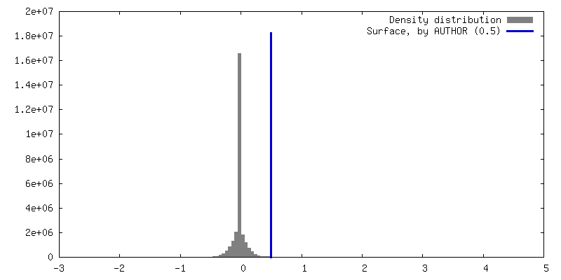
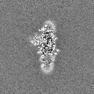
 マップデータ
 試料
試料 キーワード
キーワード 機能・相同性情報
 Fittonia albivenis (植物)
 データ登録者
 引用
引用
 構造の表示
構造の表示
 ダウンロードとリンク
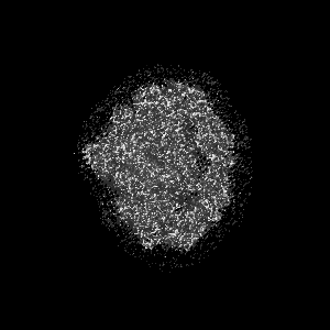
 emd_37513.png
 http://ftp.pdbj.org/pub/emdb/structures/EMD-37513
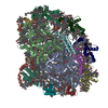


 Z (Sec.)
 Y (Row.)
 X (Col.)


 試料の構成要素


 解析
解析 電子顕微鏡法
 FIELD EMISSION GUN
