Movie
Movie Controller

[English] 日本語
 Yorodumi
- EMDB-37466: Photosynthetic LH1-RC complex from the purple sulfur bacterium Al... -
+ Open data

- Basic information
| Entry |  | |||||||||||||||||||||
|---|---|---|---|---|---|---|---|---|---|---|---|---|---|---|---|---|---|---|---|---|---|---|
| Title | Photosynthetic LH1-RC complex from the purple sulfur bacterium Allochromatium vinosum purified by Ca2+-DEAE | |||||||||||||||||||||
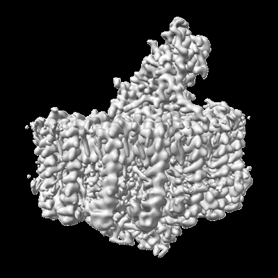
 Map data Map data | ||||||||||||||||||||||
 Sample Sample |
| |||||||||||||||||||||
 Keywords Keywords | LH1-RC COMPLEX / PHOTOSYNTHESIS / PURPLE SULFUR BACTERIA | |||||||||||||||||||||
| Function / homology |  Function and homology information Function and homology informationorganelle inner membrane / plasma membrane-derived chromatophore membrane / plasma membrane light-harvesting complex / bacteriochlorophyll binding / photosynthesis, light reaction / electron transporter, transferring electrons within the cyclic electron transport pathway of photosynthesis activity / photosynthetic electron transport in photosystem II / electron transfer activity / iron ion binding / heme binding ...organelle inner membrane / plasma membrane-derived chromatophore membrane / plasma membrane light-harvesting complex / bacteriochlorophyll binding / photosynthesis, light reaction / electron transporter, transferring electrons within the cyclic electron transport pathway of photosynthesis activity / photosynthetic electron transport in photosystem II / electron transfer activity / iron ion binding / heme binding / membrane / metal ion binding / plasma membrane Similarity search - Function | |||||||||||||||||||||
| Biological species |  Allochromatium vinosum DSM 180 (bacteria) Allochromatium vinosum DSM 180 (bacteria) | |||||||||||||||||||||
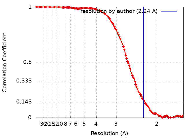
| Method | single particle reconstruction / cryo EM / Resolution: 2.24 Å | |||||||||||||||||||||
 Authors Authors | Tani K / Kanno R / Harada A / Kobayashi A / Minamino A / Nakamura N / Ji X-C / Purba ER / Hall M / Yu L-J ...Tani K / Kanno R / Harada A / Kobayashi A / Minamino A / Nakamura N / Ji X-C / Purba ER / Hall M / Yu L-J / Madigan MT / Mizoguchi A / Iwasaki K / Humbel BM / Kimura Y / Wang-Otomo Z-Y | |||||||||||||||||||||
| Funding support |  Japan, 6 items Japan, 6 items
| |||||||||||||||||||||
 Citation Citation | Journal: Commun Biol / Year: 2024 Title: High-resolution structure and biochemical properties of the LH1-RC photocomplex from the model purple sulfur bacterium, Allochromatium vinosum. Authors: Kazutoshi Tani / Ryo Kanno / Ayaka Harada / Yuki Kobayashi / Akane Minamino / Shinji Takenaka / Natsuki Nakamura / Xuan-Cheng Ji / Endang R Purba / Malgorzata Hall / Long-Jiang Yu / Michael ...Authors: Kazutoshi Tani / Ryo Kanno / Ayaka Harada / Yuki Kobayashi / Akane Minamino / Shinji Takenaka / Natsuki Nakamura / Xuan-Cheng Ji / Endang R Purba / Malgorzata Hall / Long-Jiang Yu / Michael T Madigan / Akira Mizoguchi / Kenji Iwasaki / Bruno M Humbel / Yukihiro Kimura / Zheng-Yu Wang-Otomo /   Abstract: The mesophilic purple sulfur phototrophic bacterium Allochromatium (Alc.) vinosum (bacterial family Chromatiaceae) has been a favored model for studies of bacterial photosynthesis and sulfur ...The mesophilic purple sulfur phototrophic bacterium Allochromatium (Alc.) vinosum (bacterial family Chromatiaceae) has been a favored model for studies of bacterial photosynthesis and sulfur metabolism, and its core light-harvesting (LH1) complex has been a focus of numerous studies of photosynthetic light reactions. However, despite intense efforts, no high-resolution structure and thorough biochemical analysis of the Alc. vinosum LH1 complex have been reported. Here we present cryo-EM structures of the Alc. vinosum LH1 complex associated with reaction center (RC) at 2.24 Å resolution. The overall structure of the Alc. vinosum LH1 resembles that of its moderately thermophilic relative Alc. tepidum in that it contains multiple pigment-binding α- and β-polypeptides. Unexpectedly, however, six Ca ions were identified in the Alc. vinosum LH1 bound to certain α1/β1- or α1/β3-polypeptides through a different Ca-binding motif from that seen in Alc. tepidum and other Chromatiaceae that contain Ca-bound LH1 complexes. Two water molecules were identified as additional Ca-coordinating ligands. Based on these results, we reexamined biochemical and spectroscopic properties of the Alc. vinosum LH1-RC. While modest but distinct effects of Ca were detected in the absorption spectrum of the Alc. vinosum LH1 complex, a marked decrease in thermostability of its LH1-RC complex was observed upon removal of Ca. The presence of Ca in the photocomplex of Alc. vinosum suggests that Ca-binding to LH1 complexes may be a common adaptation in species of Chromatiaceae for conferring spectral and thermal flexibility on this key component of their photosynthetic machinery. | |||||||||||||||||||||
| History |
|
- Structure visualization
| Supplemental images |
|---|
- Downloads & links
-EMDB archive
| Map data | emd_37466.map.gz | 164.8 MB | EMDB map data format | |
|---|---|---|---|---|
| Header (meta data) | emd-37466-v30.xmlemd-37466.xml | 32.3 KB 32.3 KB | Display Display | EMDB header |
| FSC (resolution estimation) | emd_37466_fsc.xml | 12.3 KB | Display | FSC data file |
| Images |  emd_37466.png emd_37466.png | 95.4 KB | ||
| Filedesc metadata | emd-37466.cif.gz | 8.1 KB | ||
| Others | emd_37466_half_map_1.map.gzemd_37466_half_map_2.map.gz | 140.3 MB 140.2 MB | ||
| Archive directory |  http://ftp.pdbj.org/pub/emdb/structures/EMD-37466ftp://ftp.pdbj.org/pub/emdb/structures/EMD-37466 http://ftp.pdbj.org/pub/emdb/structures/EMD-37466ftp://ftp.pdbj.org/pub/emdb/structures/EMD-37466 | HTTPS FTP |
-Validation report
| Summary document | emd_37466_validation.pdf.gz | 878.3 KB | Display | EMDB validaton report |
|---|---|---|---|---|
| Full document | emd_37466_full_validation.pdf.gz | 877.8 KB | Display | |
| Data in XML | emd_37466_validation.xml.gz | 20.2 KB | Display | |
| Data in CIF | emd_37466_validation.cif.gz | 26.6 KB | Display | |
| Arichive directory | https://ftp.pdbj.org/pub/emdb/validation_reports/EMD-37466ftp://ftp.pdbj.org/pub/emdb/validation_reports/EMD-37466 | HTTPS FTP |
-Related structure data
| Related structure data |  8wdvMC  8wduC M: atomic model generated by this map C: citing same article ( |
|---|---|
| Similar structure data |

-Links
| EMDB pages | EMDB (EBI/PDBe) / EMDataResource |
|---|---|
| Related items in Molecule of the Month |


-Map
| File | Download / File: emd_37466.map.gz / Format: CCP4 / Size: 178 MB / Type: IMAGE STORED AS FLOATING POINT NUMBER (4 BYTES) | ||||||||||||||||||||
|---|---|---|---|---|---|---|---|---|---|---|---|---|---|---|---|---|---|---|---|---|---|
| Voxel size | X=Y=Z: 0.82 Å | ||||||||||||||||||||
| Density |
| ||||||||||||||||||||
| Symmetry | Space group: 1 | ||||||||||||||||||||
| Details | EMDB XML:
|
-Supplemental data
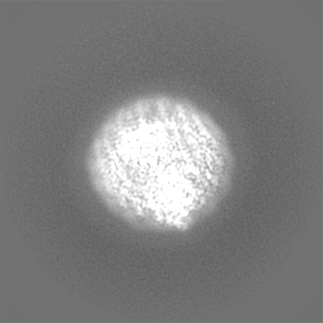
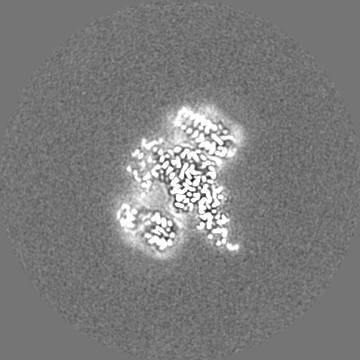
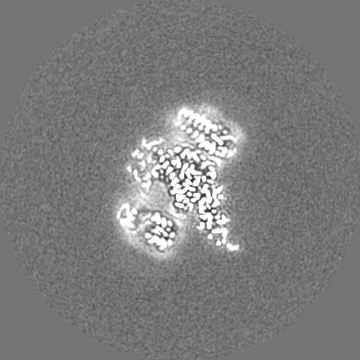
-Half map: Odd half map
| File | emd_37466_half_map_1.map | ||||||||||||
|---|---|---|---|---|---|---|---|---|---|---|---|---|---|
| Annotation | Odd half map | ||||||||||||
| Projections & Slices |
| ||||||||||||
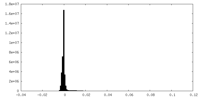
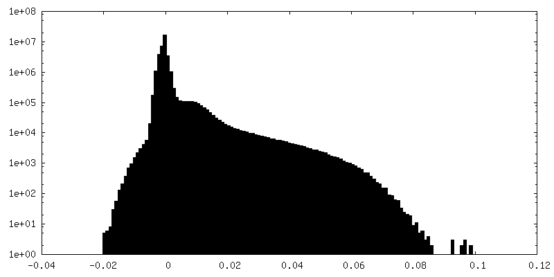
| Density Histograms |
 Z
Z Y
Y X
X

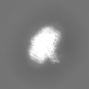


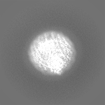
-Half map: Even half map
| File | emd_37466_half_map_2.map | ||||||||||||
|---|---|---|---|---|---|---|---|---|---|---|---|---|---|
| Annotation | Even half map | ||||||||||||
| Projections & Slices |
| ||||||||||||
| Density Histograms |


- Sample components
+Entire : Photosynthetic LH1-RC complex from the purple sulfur phototrophic...
+Supramolecule #1: Photosynthetic LH1-RC complex from the purple sulfur phototrophic...
+Macromolecule #1: Photosynthetic reaction center cytochrome c subunit
+Macromolecule #2: Reaction center protein L chain
+Macromolecule #3: Reaction center protein M chain
+Macromolecule #4: Photosynthetic reaction center H subunit
+Macromolecule #5: Antenna complex alpha/beta subunit
+Macromolecule #6: Antenna complex alpha/beta subunit
+Macromolecule #7: Antenna complex alpha/beta subunit
+Macromolecule #8: Antenna complex alpha/beta subunit
+Macromolecule #9: Antenna complex alpha/beta subunit
+Macromolecule #10: PROTOPORPHYRIN IX CONTAINING FE

+Macromolecule #11: MAGNESIUM ION
+Macromolecule #12: CALCIUM ION
+Macromolecule #13: (2S)-3-hydroxypropane-1,2-diyl dihexadecanoate
+Macromolecule #14: PALMITIC ACID

+Macromolecule #15: (1R)-2-{[{[(2S)-2,3-DIHYDROXYPROPYL]OXY}(HYDROXY)PHOSPHORYL]OXY}-...

+Macromolecule #16: BACTERIOCHLOROPHYLL A

+Macromolecule #17: BACTERIOPHEOPHYTIN A

+Macromolecule #18: Ubiquinone-8

+Macromolecule #19: FE (III) ION
+Macromolecule #20: MENAQUINONE 8

+Macromolecule #21: SPIRILLOXANTHIN

+Macromolecule #22: CARDIOLIPIN

+Macromolecule #23: LAURYL DIMETHYLAMINE-N-OXIDE

+Macromolecule #24: DODECYL-BETA-D-MALTOSIDE

+Macromolecule #25: water

-Experimental details
-Structure determination
| Method | cryo EM |
|---|---|
 Processing Processing | single particle reconstruction |
| Aggregation state | particle |
-Sample preparation
| Concentration | 5.2 mg/mL |
|---|---|
| Buffer | pH: 7.5 |
| Vitrification | Cryogen name: ETHANE / Chamber humidity: 90 % / Chamber temperature: 277 K / Instrument: LEICA EM GP |
| Details | This sample was monodisperse. |
- Electron microscopy
| Microscope | FEI TITAN KRIOS |
|---|---|
| Image recording | Film or detector model: FEI FALCON III (4k x 4k) / Detector mode: COUNTING / Average exposure time: 30.15 sec. / Average electron dose: 40.0 e/Å2 |
| Electron beam | Acceleration voltage: 300 kV / Electron source:  FIELD EMISSION GUN FIELD EMISSION GUN |
| Electron optics | Illumination mode: FLOOD BEAM / Imaging mode: BRIGHT FIELD / Cs: 2.7 mm / Nominal defocus max: 2.6 µm / Nominal defocus min: 0.5 µm |
| Sample stage | Specimen holder model: FEI TITAN KRIOS AUTOGRID HOLDER / Cooling holder cryogen: NITROGEN |
| Experimental equipment |  Model: Titan Krios / Image courtesy: FEI Company |
+Image processing
-Atomic model buiding 1
| Initial model | PDB ID: Chain - Source name: PDB / Chain - Initial model type: experimental model |
|---|---|
| Refinement | Space: REAL / Protocol: RIGID BODY FIT / Overall B value: 63 / Target criteria: Correlation coefficient |
| Output model | PDB-8wdv: |