Movie
Movie Controller


+ Open data

- Basic information
| Entry |  | |||||||||
|---|---|---|---|---|---|---|---|---|---|---|
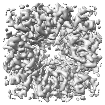
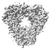
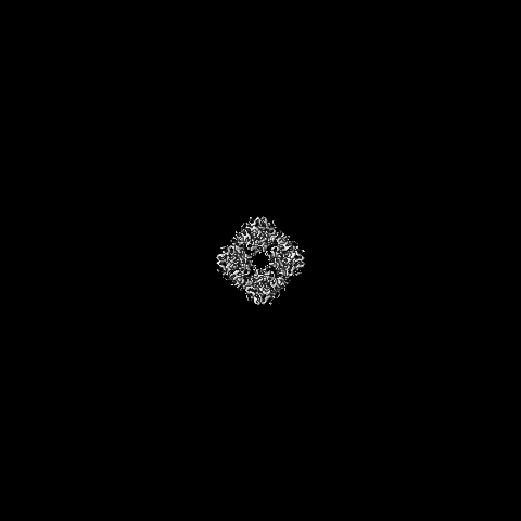
| Title | FCP tetramer in Chaetoceros gracilis | |||||||||
 Map data Map data | ||||||||||
 Sample Sample |
| |||||||||
 Keywords Keywords | FCP tetramers / PHOTOSYNTHESIS | |||||||||
| Function / homology |  Function and homology information Function and homology informationlight-harvesting complex / photosynthesis, light harvesting in photosystem I / plastid / chlorophyll binding / response to light stimulus / membrane Similarity search - Function | |||||||||
| Biological species |  Chaetoceros neogracilis (Diatom) Chaetoceros neogracilis (Diatom) | |||||||||
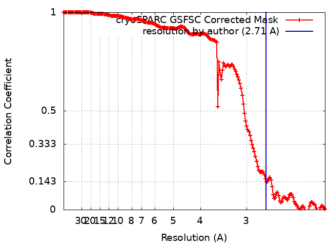
| Method | single particle reconstruction / cryo EM / Resolution: 2.71 Å | |||||||||
 Authors Authors | Feng Y / Li Z / Zhou C / Shen J-R / Liu C / Wang W | |||||||||
| Funding support |  China, 1 items China, 1 items
| |||||||||
 Citation Citation | Journal: Plant Commun / Year: 2024 Title: Structural and spectroscopic insights into fucoxanthin chlorophyll a/c-binding proteins of diatoms in diverse oligomeric states. Authors: Cuicui Zhou / Yue Feng / Zhenhua Li / Lili Shen / Xiaoyi Li / Yumei Wang / Guangye Han / Tingyun Kuang / Cheng Liu / Jian-Ren Shen / Wenda Wang /  Abstract: Diatoms, a group of prevalent marine algae, significantly contribute to global primary productivity. Their substantial biomass is linked to enhanced absorption of blue-green light underwater, ...Diatoms, a group of prevalent marine algae, significantly contribute to global primary productivity. Their substantial biomass is linked to enhanced absorption of blue-green light underwater, facilitated by fucoxanthin chlorophyll a/c-binding proteins (FCPs), exhibiting oligomeric diversity across diatom species. Utilizing mild CN-PAGE analysis on solubilized thylakoid membranes, we displayed monomeric, dimeric, trimeric, tetrameric and pentameric FCPs in diatoms. Mass spectrometry analysis revealed each oligomeric FCP has specific protein compositions, constituting a large Lhcf family of FCP antennas. In addition, we resolved the structures of Thalassiosira pseudonana FCP (Tp-FCP) homotrimer and Chaetoceros gracilis FCP (Cg-FCP) pentamer by cryo-electron microscopy at 2.73 Å and 2.65 Å resolutions, respectively. The distinct pigment composition and organization in various oligomeric FCPs change their blue-green light-harvesting, excitation energy transfer pathways. In comparison to dimeric and trimeric FCPs, Cg-FCP tetramer and Cg-FCP pentamer exhibit stronger absorption by Chls c, red-shifted and broader Chl a fluorescence emission, as well as more robust circular dichroism signals originating from Chl a-carotenoid dimers. These spectroscopic characteristics indicate that Chl a molecules in Cg-FCP tetramer and Cg-FCP pentamer are more heterogeneous than in both dimers and Tp-FCP trimer. The structural and spectroscopic insights provided by this study contribute to a better understanding of the mechanisms that empower diatoms to adapt to fluctuating light environments. | |||||||||
| History |
|
- Structure visualization
| Supplemental images |
|---|
- Downloads & links
-EMDB archive
| Map data | emd_37441.map.gz | 211.5 MB | EMDB map data format | |
|---|---|---|---|---|
| Header (meta data) | emd-37441-v30.xmlemd-37441.xml | 14.7 KB 14.7 KB | Display Display | EMDB header |
| FSC (resolution estimation) | emd_37441_fsc.xml | 15.7 KB | Display | FSC data file |
| Images |  emd_37441.png emd_37441.png | 149 KB | ||
| Filedesc metadata | emd-37441.cif.gz | 5.2 KB | ||
| Others | emd_37441_half_map_1.map.gzemd_37441_half_map_2.map.gz | 391.5 MB 391.5 MB | ||
| Archive directory |  http://ftp.pdbj.org/pub/emdb/structures/EMD-37441ftp://ftp.pdbj.org/pub/emdb/structures/EMD-37441 http://ftp.pdbj.org/pub/emdb/structures/EMD-37441ftp://ftp.pdbj.org/pub/emdb/structures/EMD-37441 | HTTPS FTP |
-Validation report
| Summary document | emd_37441_validation.pdf.gz | 645.6 KB | Display | EMDB validaton report |
|---|---|---|---|---|
| Full document | emd_37441_full_validation.pdf.gz | 645.2 KB | Display | |
| Data in XML | emd_37441_validation.xml.gz | 23.5 KB | Display | |
| Data in CIF | emd_37441_validation.cif.gz | 30.3 KB | Display | |
| Arichive directory | https://ftp.pdbj.org/pub/emdb/validation_reports/EMD-37441ftp://ftp.pdbj.org/pub/emdb/validation_reports/EMD-37441 | HTTPS FTP |
-Related structure data
| Related structure data | 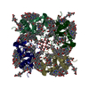 8wckMC  8jp3C 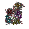 8wclC M: atomic model generated by this map C: citing same article ( |
|---|---|
| Similar structure data |
-Links
| EMDB pages | EMDB (EBI/PDBe) / EMDataResource |
|---|---|
| Related items in Molecule of the Month |


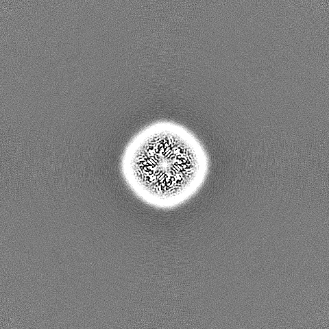
-Map
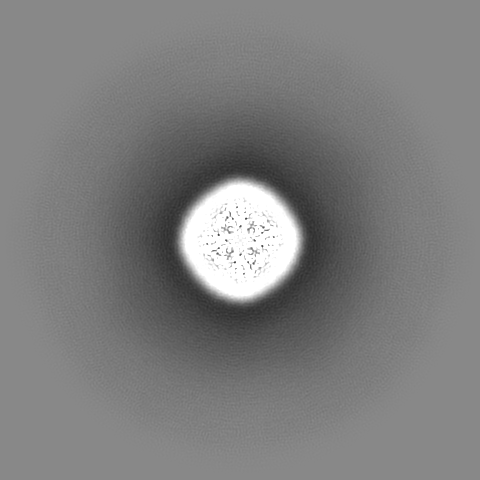
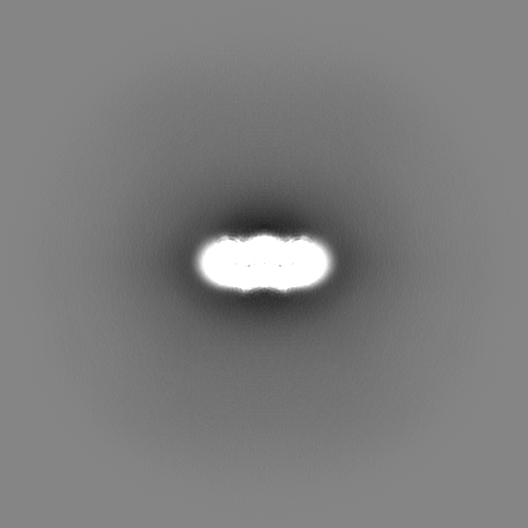
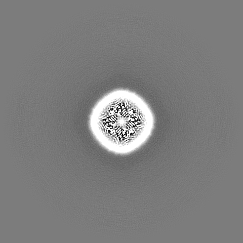
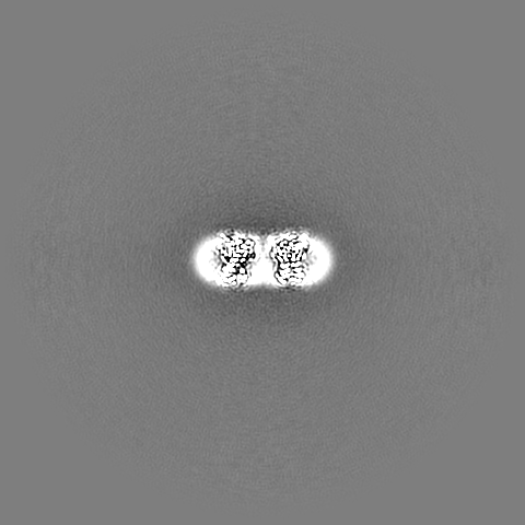
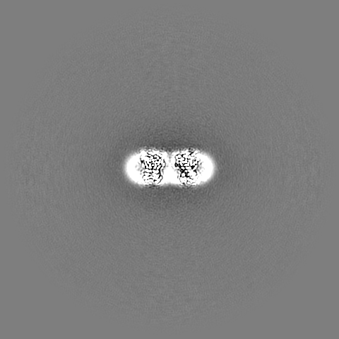
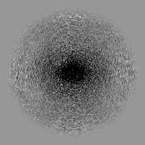
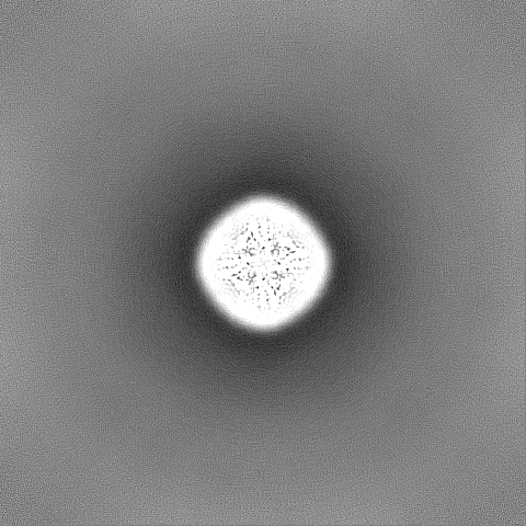
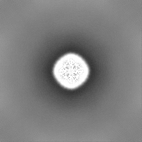
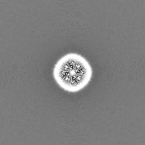
| File | Download / File: emd_37441.map.gz / Format: CCP4 / Size: 421.9 MB / Type: IMAGE STORED AS FLOATING POINT NUMBER (4 BYTES) | ||||||||||||||||||||||||||||||||||||
|---|---|---|---|---|---|---|---|---|---|---|---|---|---|---|---|---|---|---|---|---|---|---|---|---|---|---|---|---|---|---|---|---|---|---|---|---|---|
| Projections & slices | Image control
Images are generated by Spider. | ||||||||||||||||||||||||||||||||||||
| Voxel size | X=Y=Z: 1.04 Å | ||||||||||||||||||||||||||||||||||||
| Density |
| ||||||||||||||||||||||||||||||||||||
| Symmetry | Space group: 1 | ||||||||||||||||||||||||||||||||||||
| Details | EMDB XML:
|
 Z (Sec.)
 Y (Row.)
 X (Col.)


-Supplemental data
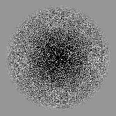
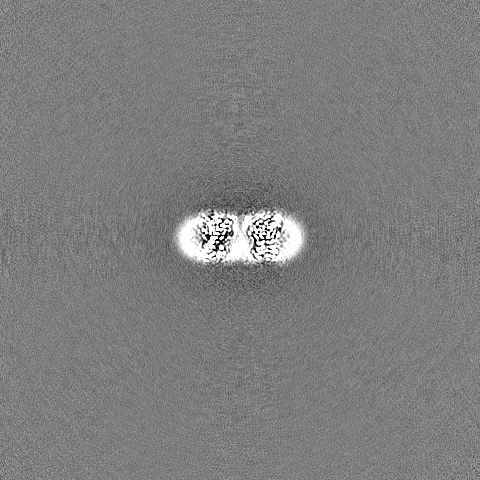
-Half map: #2
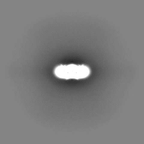
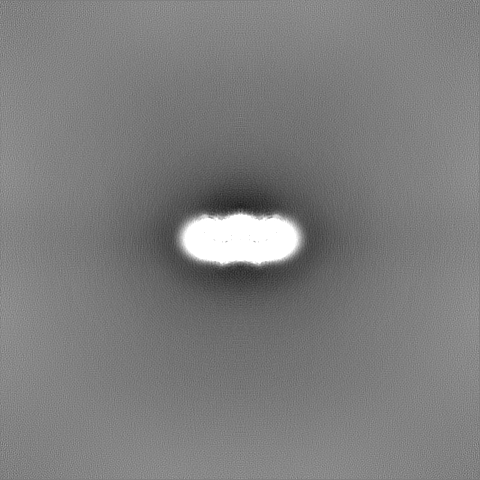
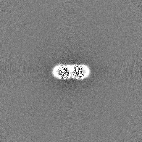
| File | emd_37441_half_map_1.map | ||||||||||||
|---|---|---|---|---|---|---|---|---|---|---|---|---|---|
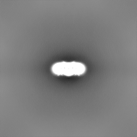
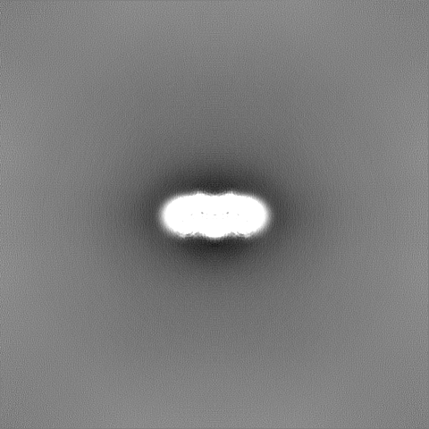
| Projections & Slices |
| ||||||||||||
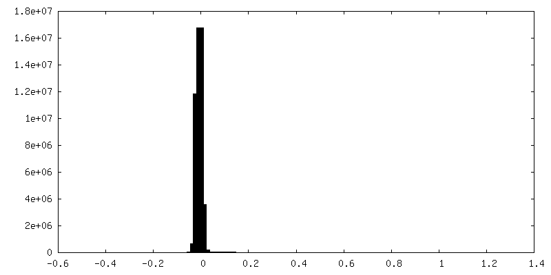
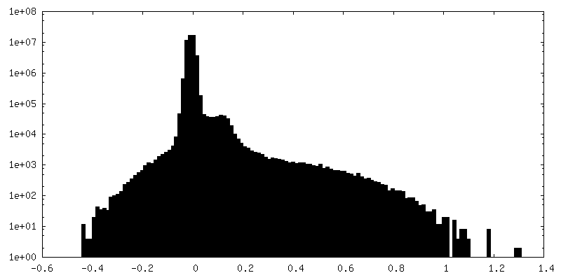
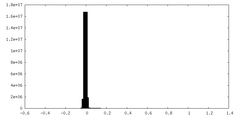
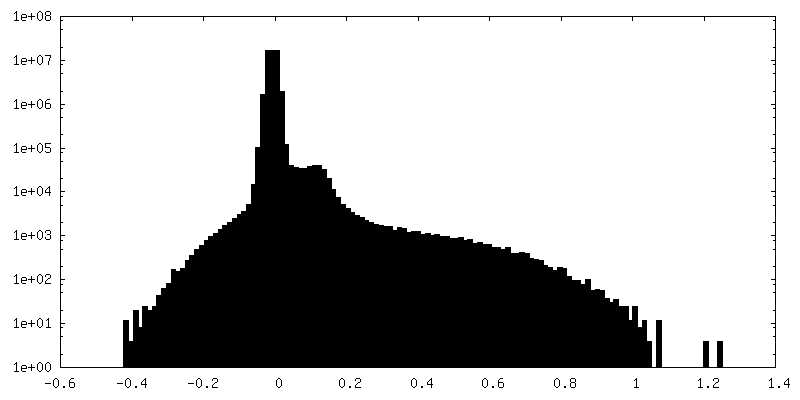
| Density Histograms |
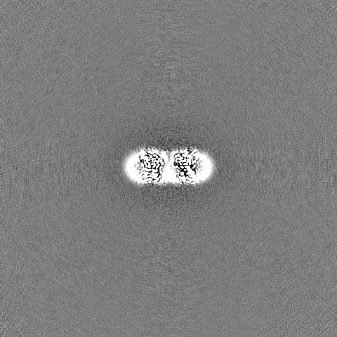
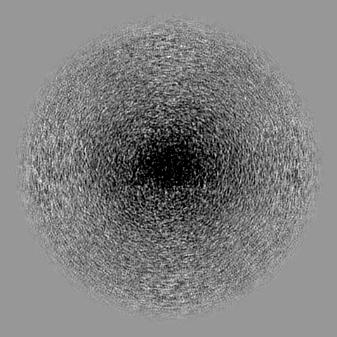
-Half map: #1
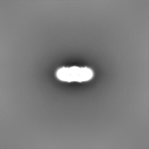
| File | emd_37441_half_map_2.map | ||||||||||||
|---|---|---|---|---|---|---|---|---|---|---|---|---|---|
| Projections & Slices |
| ||||||||||||
| Density Histograms |

- Sample components
-Entire : FCP tetramer
| Entire | Name: FCP tetramer |
|---|---|
| Components |
|
-Supramolecule #1: FCP tetramer
| Supramolecule | Name: FCP tetramer / type: complex / ID: 1 / Parent: 0 / Macromolecule list: #1 |
|---|---|
| Source (natural) | Organism: Chaetoceros neogracilis (Diatom) |
-Macromolecule #1: Chlorophyll a/b-binding protein
| Macromolecule | Name: Chlorophyll a/b-binding protein / type: protein_or_peptide / ID: 1 / Number of copies: 4 / Enantiomer: LEVO |
|---|---|
| Source (natural) | Organism: Chaetoceros neogracilis (Diatom) |
| Molecular weight | Theoretical: 22.098182 KDa |
| Sequence | String: MKLAVAALLV ASAAAFAPAP ASKASTSLKV SEIELGVTEP LGVYDPLGWL ESEPEAFERR RAVERKHGRV AMAAVVGTIV HNNHIVFDG YLSPSNNLKF SDIPTGVDGI RAIPTAGLAQ ILAFFALVEL AWMPASKYDG DYGVGYFGTD IKDPEEKARK L NVELNNGR ...String: MKLAVAALLV ASAAAFAPAP ASKASTSLKV SEIELGVTEP LGVYDPLGWL ESEPEAFERR RAVERKHGRV AMAAVVGTIV HNNHIVFDG YLSPSNNLKF SDIPTGVDGI RAIPTAGLAQ ILAFFALVEL AWMPASKYDG DYGVGYFGTD IKDPEEKARK L NVELNNGR AAMMGIMGNM VAEVLTGQTM YEQYASGHIS PFGDGQGVF UniProtKB: Chlorophyll a/b-binding protein |
-Macromolecule #2: (3S,3'S,5R,5'R,6S,6'R,8'R)-3,5'-dihydroxy-8-oxo-6',7'-didehydro-5...
| Macromolecule | Name: (3S,3'S,5R,5'R,6S,6'R,8'R)-3,5'-dihydroxy-8-oxo-6',7'-didehydro-5,5',6,6',7,8-hexahydro-5,6-epoxy-beta,beta-caroten-3'- yl acetate type: ligand / ID: 2 / Number of copies: 24 / Formula: A86 |
|---|---|
| Molecular weight | Theoretical: 658.906 Da |
-Macromolecule #3: CHLOROPHYLL A
| Macromolecule | Name: CHLOROPHYLL A / type: ligand / ID: 3 / Number of copies: 24 / Formula: CLA |
|---|---|
| Molecular weight | Theoretical: 893.489 Da |
| Chemical component information |  ChemComp-CLA: |
-Macromolecule #4: Chlorophyll c2
| Macromolecule | Name: Chlorophyll c2 / type: ligand / ID: 4 / Number of copies: 8 / Formula: KC2 |
|---|---|
| Molecular weight | Theoretical: 608.926 Da |
| Chemical component information |  ChemComp-KC2: |
-Macromolecule #5: Chlorophyll c1
| Macromolecule | Name: Chlorophyll c1 / type: ligand / ID: 5 / Number of copies: 8 / Formula: KC1 |
|---|---|
| Molecular weight | Theoretical: 610.941 Da |
| Chemical component information |  ChemComp-KC1: |
-Macromolecule #6: DODECYL-BETA-D-MALTOSIDE
| Macromolecule | Name: DODECYL-BETA-D-MALTOSIDE / type: ligand / ID: 6 / Number of copies: 8 / Formula: LMT |
|---|---|
| Molecular weight | Theoretical: 510.615 Da |
| Chemical component information |  ChemComp-LMT: |
-Experimental details
-Structure determination
| Method | cryo EM |
|---|---|
 Processing Processing | single particle reconstruction |
| Aggregation state | particle |
-Sample preparation
| Buffer | pH: 6.5 |
|---|---|
| Vitrification | Cryogen name: ETHANE |
- Electron microscopy
| Microscope | FEI TITAN KRIOS |
|---|---|
| Image recording | Film or detector model: GATAN K3 (6k x 4k) / Average electron dose: 60.0 e/Å2 |
| Electron beam | Acceleration voltage: 300 kV / Electron source:  FIELD EMISSION GUN FIELD EMISSION GUN |
| Electron optics | Illumination mode: FLOOD BEAM / Imaging mode: BRIGHT FIELD / Nominal defocus max: 2.0 µm / Nominal defocus min: 1.0 µm |
| Experimental equipment |  Model: Titan Krios / Image courtesy: FEI Company |