 Movie
Movie Controller


+ Open data

- Basic information
| Entry |  | |||||||||
|---|---|---|---|---|---|---|---|---|---|---|
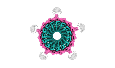
| Title | Cyanophage A-1(L) neck/gp5-neck fiber | |||||||||
 Map data Map data | ||||||||||
 Sample Sample |
| |||||||||
 Keywords Keywords | baseplate / virus / viral protein | |||||||||
| Biological species |  unclassified Caudoviricetes (virus) unclassified Caudoviricetes (virus) | |||||||||
| Method | single particle reconstruction / cryo EM / Resolution: 3.66 Å | |||||||||
 Authors Authors | Yu RC / Li Q / Zhou CZ | |||||||||
| Funding support |  China, 1 items China, 1 items
| |||||||||
 Citation Citation | Journal: Nat Commun / Year: 2024 Title: Structure of the intact tail machine of Anabaena myophage A-1(L). Authors: Rong-Cheng Yu / Feng Yang / Hong-Yan Zhang / Pu Hou / Kang Du / Jie Zhu / Ning Cui / Xudong Xu / Yuxing Chen / Qiong Li / Cong-Zhao Zhou / Abstract: The Myoviridae cyanophage A-1(L) specifically infects the model cyanobacteria Anabaena sp. PCC 7120. Following our recent report on the capsid structure of A-1(L), here we present the high-resolution ...The Myoviridae cyanophage A-1(L) specifically infects the model cyanobacteria Anabaena sp. PCC 7120. Following our recent report on the capsid structure of A-1(L), here we present the high-resolution cryo-EM structure of its intact tail machine including the neck, tail and attached fibers. Besides the dodecameric portal, the neck contains a canonical hexamer connected to a unique pentadecamer that anchors five extended bead-chain-like neck fibers. The 1045-Å-long contractile tail is composed of a helical bundle of tape measure proteins surrounded by a layer of tube proteins and a layer of sheath proteins, ended with a five-component baseplate. The six long and six short tail fibers are folded back pairwise, each with one end anchoring to the baseplate and the distal end pointing to the capsid. Structural analysis combined with biochemical assays further enable us to identify the dual hydrolytic activities of the baseplate hub, in addition to two host receptor binding domains in the tail fibers. Moreover, the structure of the intact A-1(L) also helps us to reannotate its genome. These findings will facilitate the application of A-1(L) as a chassis cyanophage in synthetic biology. | |||||||||
| History |
|
- Structure visualization
| Supplemental images |
|---|
- Downloads & links
-EMDB archive
| Map data | emd_37155.map.gz | 111.5 MB |  EMDB map data format EMDB map data format | |
|---|---|---|---|---|
| Header (meta data) | emd-37155-v30.xmlemd-37155.xml | 14.5 KB 14.5 KB | Display Display | EMDB header |
| Images |  emd_37155.png emd_37155.png | 43.3 KB | ||
| Filedesc metadata | emd-37155.cif.gz | 5.2 KB | ||
| Others | emd_37155_half_map_1.map.gzemd_37155_half_map_2.map.gz | 97.5 MB 97.4 MB | ||
| Archive directory |  http://ftp.pdbj.org/pub/emdb/structures/EMD-37155ftp://ftp.pdbj.org/pub/emdb/structures/EMD-37155 http://ftp.pdbj.org/pub/emdb/structures/EMD-37155ftp://ftp.pdbj.org/pub/emdb/structures/EMD-37155 | HTTPS FTP |
-Validation report
| Summary document | emd_37155_validation.pdf.gz | 975 KB | Display | EMDB validaton report |
|---|---|---|---|---|
| Full document | emd_37155_full_validation.pdf.gz | 974.5 KB | Display | |
| Data in XML | emd_37155_validation.xml.gz | 13.7 KB | Display | |
| Data in CIF | emd_37155_validation.cif.gz | 16.2 KB | Display | |
| Arichive directory | https://ftp.pdbj.org/pub/emdb/validation_reports/EMD-37155ftp://ftp.pdbj.org/pub/emdb/validation_reports/EMD-37155 | HTTPS FTP |
-Related structure data
| Related structure data |  8kegMC  8ke9C  8keaC 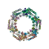 8kecC 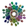 8keeC 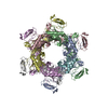 8kefC  8ts6C M: atomic model generated by this map C: citing same article ( |
|---|


-Links
| EMDB pages | EMDB (EBI/PDBe) / EMDataResource |
|---|
-Map
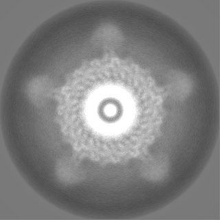
| File | Download / File: emd_37155.map.gz / Format: CCP4 / Size: 125 MB / Type: IMAGE STORED AS FLOATING POINT NUMBER (4 BYTES) | ||||||||||||||||||||
|---|---|---|---|---|---|---|---|---|---|---|---|---|---|---|---|---|---|---|---|---|---|
| Voxel size | X=Y=Z: 1.07 Å | ||||||||||||||||||||
| Density |
| ||||||||||||||||||||
| Symmetry | Space group: 1 | ||||||||||||||||||||
| Details | EMDB XML:
|
-Supplemental data
-Half map: #1
| File | emd_37155_half_map_1.map | ||||||||||||
|---|---|---|---|---|---|---|---|---|---|---|---|---|---|
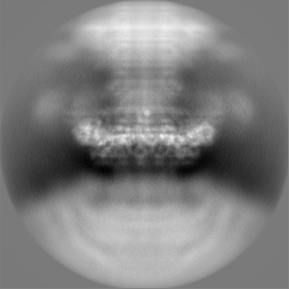
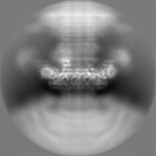
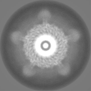
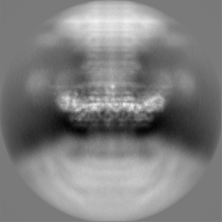
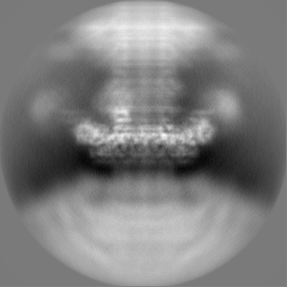
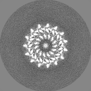
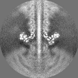
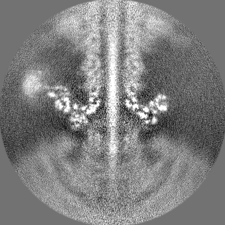
| Projections & Slices |
| ||||||||||||
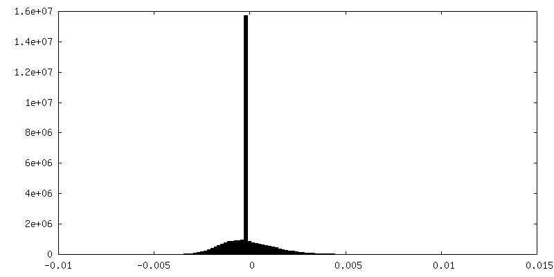
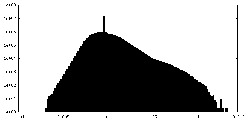
| Density Histograms |
 Z
Z Y
Y X
X


-Half map: #2
| File | emd_37155_half_map_2.map | ||||||||||||
|---|---|---|---|---|---|---|---|---|---|---|---|---|---|
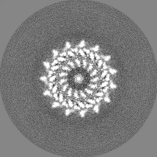
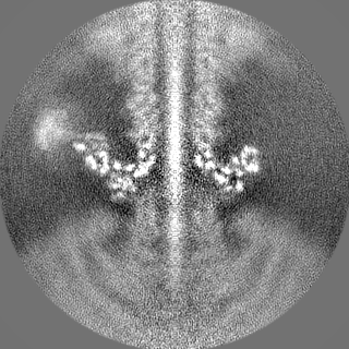
| Projections & Slices |
| ||||||||||||
| Density Histograms |


- Sample components
-Entire : unclassified Caudoviricetes
| Entire | Name: unclassified Caudoviricetes (virus) |
|---|---|
| Components |
|
-Supramolecule #1: unclassified Caudoviricetes
| Supramolecule | Name: unclassified Caudoviricetes / type: virus / ID: 1 / Parent: 0 / Macromolecule list: all / NCBI-ID: 2788787 / Sci species name: unclassified Caudoviricetes / Virus type: VIRION / Virus isolate: OTHER / Virus enveloped: No / Virus empty: No |
|---|---|
| Host (natural) | Organism:  Nostoc sp. PCC 7120 = FACHB-418 (bacteria) Nostoc sp. PCC 7120 = FACHB-418 (bacteria) |
-Macromolecule #1: Neck gp5
| Macromolecule | Name: Neck gp5 / type: protein_or_peptide / ID: 1 / Number of copies: 15 / Enantiomer: LEVO |
|---|---|
| Source (natural) | Organism: unclassified Caudoviricetes (virus) |
| Molecular weight | Theoretical: 18.293848 KDa |
| Sequence | String: VTPGAILVTH PLESYYNQFL DGLPTSELEE LEIEFVAMCI GFPRSQNNFV NALLDLSTIE VYTIRTSVKE ILRLDKLNKE SIENRQVTK VEDITMNMNY IGDIKDELVT EVKKLARILK LPVLDNLYVS YDDVEGRIGA NGLLEPLNRG SGFGSYLTGY T VR |
-Macromolecule #2: neck fiber gp82N
| Macromolecule | Name: neck fiber gp82N / type: protein_or_peptide / ID: 2 / Number of copies: 15 / Enantiomer: LEVO |
|---|---|
| Source (natural) | Organism: unclassified Caudoviricetes (virus) |
| Molecular weight | Theoretical: 26.44942 KDa |
| Sequence | String: MRRLKGTIRH LDDQPWINVS LFLTLINGTF NSANQYPIDT KHAKTDQNGE FVFNVVPNVG IDQSYYILTT PDNKKHSFTV PDGTSDIEF SVVREAGIIA TDPEYTNVLN YLEDYIDDAI ANIQASSVIA EIFTCGQTIS ALKALRFDSS TGKVFYASSS D ATHLNKCV ...String: MRRLKGTIRH LDDQPWINVS LFLTLINGTF NSANQYPIDT KHAKTDQNGE FVFNVVPNVG IDQSYYILTT PDNKKHSFTV PDGTSDIEF SVVREAGIIA TDPEYTNVLN YLEDYIDDAI ANIQASSVIA EIFTCGQTIS ALKALRFDSS TGKVFYASSS D ATHLNKCV GVSSQSGVLN DNIQVVTSGY LSDQSWNWTI GSPIFFDSGG TLTHTPGSSY YQVIGIPVTT NKVLISVEQP IK L |
-Experimental details
-Structure determination
| Method | cryo EM |
|---|---|
 Processing Processing | single particle reconstruction |
| Aggregation state | particle |
-Sample preparation
| Buffer | pH: 7.5 |
|---|---|
| Vitrification | Cryogen name: ETHANE / Chamber humidity: 100 % / Chamber temperature: 300 K |
- Electron microscopy
| Microscope | FEI TITAN KRIOS |
|---|---|
| Image recording | Film or detector model: GATAN K2 SUMMIT (4k x 4k) / Average electron dose: 50.0 e/Å2 |
| Electron beam | Acceleration voltage: 300 kV / Electron source:  FIELD EMISSION GUN FIELD EMISSION GUN |
| Electron optics | Illumination mode: SPOT SCAN / Imaging mode: BRIGHT FIELD / Nominal defocus max: 2.5 µm / Nominal defocus min: 1.5 µm |
| Experimental equipment |  Model: Titan Krios / Image courtesy: FEI Company |
-Image processing
| Startup model | Type of model: NONE |
|---|---|
| Final reconstruction | Resolution.type: BY AUTHOR / Resolution: 3.66 Å / Resolution method: FSC 0.143 CUT-OFF / Software - Name: RELION (ver. 3.1) / Number images used: 36158 |
| Initial angle assignment | Type: RANDOM ASSIGNMENT |
| Final angle assignment | Type: RANDOM ASSIGNMENT |
