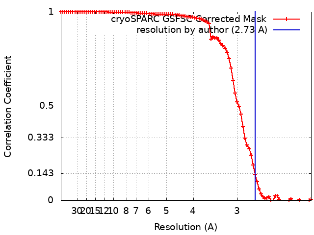
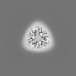
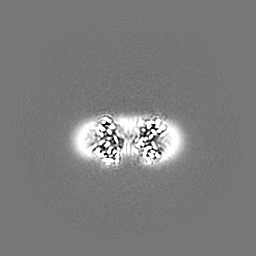
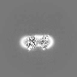
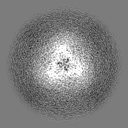
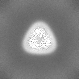
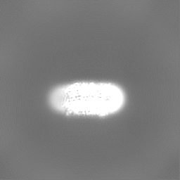
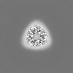
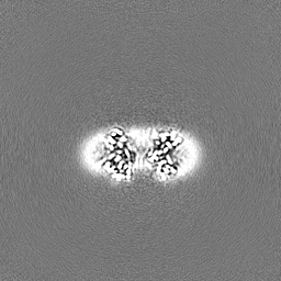
ジャーナル: Plant Commun / 年: 2024 タイトル: Structural and spectroscopic insights into fucoxanthin chlorophyll a/c-binding proteins of diatoms in diverse oligomeric states. 著者: Cuicui Zhou / Yue Feng / Zhenhua Li / Lili Shen / Xiaoyi Li / Yumei Wang / Guangye Han / Tingyun Kuang / Cheng Liu / Jian-Ren Shen / Wenda Wang / 要旨: Diatoms, a group of prevalent marine algae, contribute significantly to global primary productivity. Their substantial biomass is linked to enhanced absorption of blue-green light underwater, ...Diatoms, a group of prevalent marine algae, contribute significantly to global primary productivity. Their substantial biomass is linked to enhanced absorption of blue-green light underwater, facilitated by fucoxanthin chlorophyll (Chl) a/c-binding proteins (FCPs), which exhibit oligomeric diversity across diatom species. Using mild clear native PAGE analysis of solubilized thylakoid membranes, we displayed monomeric, dimeric, trimeric, tetrameric, and pentameric FCPs in diatoms. Mass spectrometry analysis revealed that each oligomeric FCP has a specific protein composition, and together they constitute a large Lhcf family of FCP antennas. In addition, we resolved the structures of the Thalassiosira pseudonana FCP (Tp-FCP) homotrimer and the Chaetoceros gracilis FCP (Cg-FCP) pentamer by cryoelectron microscopy at 2.73-Å and 2.65-Å resolution, respectively. The distinct pigment compositions and organizations of various oligomeric FCPs affect their blue-green light-harvesting, excitation energy transfer pathways. Compared with dimeric and trimeric FCPs, the Cg-FCP tetramer and Cg-FCP pentamer exhibit stronger absorption by Chl c, redshifted and broader Chl a fluorescence emission, and more robust circular dichroism signals originating from Chl a-carotenoid dimers. These spectroscopic characteristics indicate that Chl a molecules in the Cg-FCP tetramer and Cg-FCP pentamer are more heterogeneous than in both dimers and the Tp-FCP trimer. The structural and spectroscopic insights provided by this study contribute to a better understanding of the mechanisms that empower diatoms to adapt to fluctuating light environments.
 ムービー
ムービー コントローラー


 データを開く
 基本情報
基本情報
 マップデータ
 試料
試料 キーワード
キーワード 機能・相同性情報
 Thalassiosira pseudonana CCMP1335 (珪藻)
 データ登録者
 中国, 1件
  引用
引用
 構造の表示
構造の表示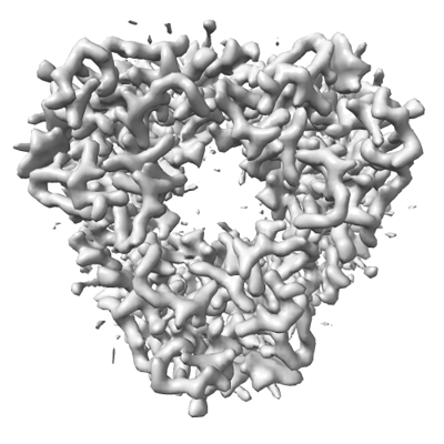
 ダウンロードとリンク
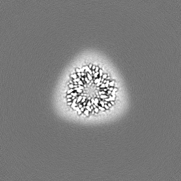
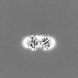
 emd_36466.png
 http://ftp.pdbj.org/pub/emdb/structures/EMD-36466


 Z (Sec.)
 Y (Row.)
 X (Col.)


 試料の構成要素


 解析
解析 電子顕微鏡法
 FIELD EMISSION GUN