 Movie
Movie Controller


+ Open data

- Basic information
| Entry |  | |||||||||
|---|---|---|---|---|---|---|---|---|---|---|
| Title | The cryoEM structure of H7N9-HA protein | |||||||||
 Map data Map data | The cryoEM structure of HA protein | |||||||||
 Sample Sample |
| |||||||||
 Keywords Keywords | H7N9 / HA / Trimer / complex / MEMBRANE PROTEIN | |||||||||
| Function / homology |  Function and homology information Function and homology informationviral budding from plasma membrane / clathrin-dependent endocytosis of virus by host cell / host cell surface receptor binding / apical plasma membrane / fusion of virus membrane with host plasma membrane / fusion of virus membrane with host endosome membrane / viral envelope / virion attachment to host cell / host cell plasma membrane / virion membrane Similarity search - Function | |||||||||
| Biological species |  H7N9 subtype (virus) H7N9 subtype (virus) | |||||||||
| Method | single particle reconstruction / cryo EM / Resolution: 3.1 Å | |||||||||
 Authors Authors | Zhao BB / Sun ZZ | |||||||||
| Funding support |  China, 1 items China, 1 items
| |||||||||
 Citation Citation | Journal: To Be Published Title: The cryoEM structure of H7N9-HA protein Authors: Zhao BB / Sun ZZ | |||||||||
| History |
|
- Structure visualization
| Supplemental images |
|---|

- Downloads & links
-EMDB archive
| Map data | emd_35729.map.gz | 230.2 MB | EMDB map data format | |
|---|---|---|---|---|
| Header (meta data) | emd-35729-v30.xmlemd-35729.xml | 13 KB 13 KB | Display Display | EMDB header |
| FSC (resolution estimation) | emd_35729_fsc.xml | 13.1 KB | Display | FSC data file |
| Images |  emd_35729.png emd_35729.png | 74.3 KB | ||
| Filedesc metadata | emd-35729.cif.gz | 5.4 KB | ||
| Others | emd_35729_half_map_1.map.gzemd_35729_half_map_2.map.gz | 226.5 MB 226.5 MB | ||
| Archive directory |  http://ftp.pdbj.org/pub/emdb/structures/EMD-35729ftp://ftp.pdbj.org/pub/emdb/structures/EMD-35729 http://ftp.pdbj.org/pub/emdb/structures/EMD-35729ftp://ftp.pdbj.org/pub/emdb/structures/EMD-35729 | HTTPS FTP |
-Validation report
| Summary document | emd_35729_validation.pdf.gz | 997.2 KB | Display | EMDB validaton report |
|---|---|---|---|---|
| Full document | emd_35729_full_validation.pdf.gz | 996.8 KB | Display | |
| Data in XML | emd_35729_validation.xml.gz | 22.2 KB | Display | |
| Data in CIF | emd_35729_validation.cif.gz | 28.8 KB | Display | |
| Arichive directory | https://ftp.pdbj.org/pub/emdb/validation_reports/EMD-35729ftp://ftp.pdbj.org/pub/emdb/validation_reports/EMD-35729 | HTTPS FTP |
-Related structure data
| Related structure data |  8iuxMC M: atomic model generated by this map C: citing same article ( |
|---|---|
| Similar structure data |
-Links
| EMDB pages | EMDB (EBI/PDBe) / EMDataResource |
|---|---|
| Related items in Molecule of the Month |


-Map
| File | Download / File: emd_35729.map.gz / Format: CCP4 / Size: 244.1 MB / Type: IMAGE STORED AS FLOATING POINT NUMBER (4 BYTES) | ||||||||||||||||||||||||||||||||||||
|---|---|---|---|---|---|---|---|---|---|---|---|---|---|---|---|---|---|---|---|---|---|---|---|---|---|---|---|---|---|---|---|---|---|---|---|---|---|
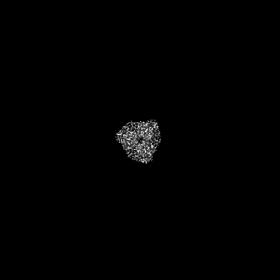
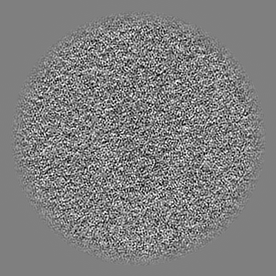
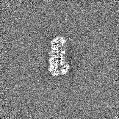
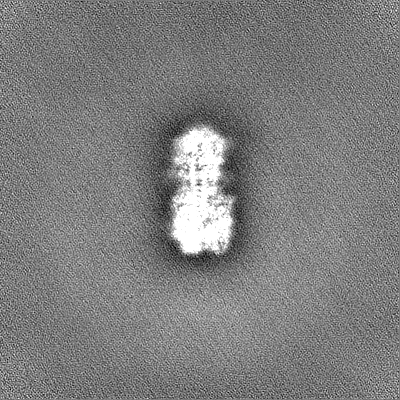
| Annotation | The cryoEM structure of HA protein | ||||||||||||||||||||||||||||||||||||
| Projections & slices | Image control
Images are generated by Spider. | ||||||||||||||||||||||||||||||||||||
| Voxel size | X=Y=Z: 1.076 Å | ||||||||||||||||||||||||||||||||||||
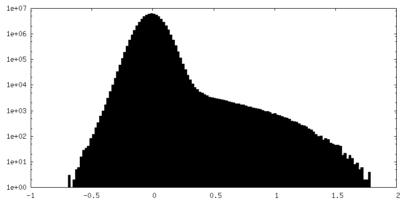
| Density |
| ||||||||||||||||||||||||||||||||||||
| Symmetry | Space group: 1 | ||||||||||||||||||||||||||||||||||||
| Details | EMDB XML:
|
 Z (Sec.)
 Y (Row.)
 X (Col.)


-Supplemental data
-Half map: The half A cryoEM structure of HA protein
| File | emd_35729_half_map_1.map | ||||||||||||
|---|---|---|---|---|---|---|---|---|---|---|---|---|---|
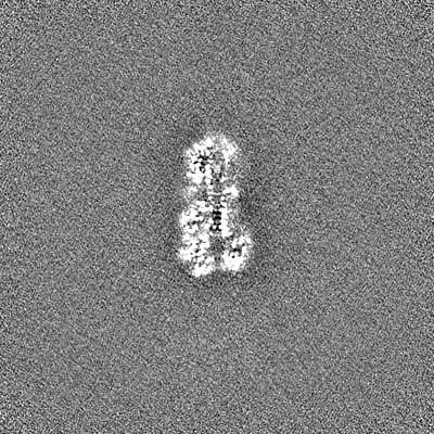
| Annotation | The half A cryoEM structure of HA protein | ||||||||||||
| Projections & Slices |
| ||||||||||||
| Density Histograms |


-Half map: The half B cryoEM structure of HA protein
| File | emd_35729_half_map_2.map | ||||||||||||
|---|---|---|---|---|---|---|---|---|---|---|---|---|---|
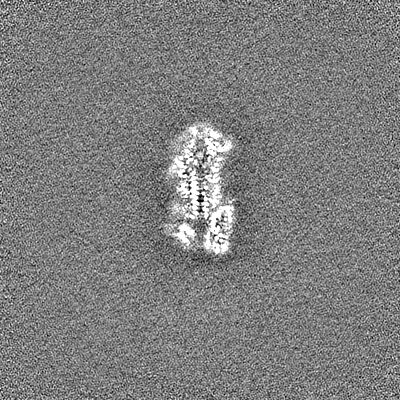
| Annotation | The half B cryoEM structure of HA protein | ||||||||||||
| Projections & Slices |
| ||||||||||||
| Density Histograms |


- Sample components
-Entire : High Five Cell
| Entire | Name: High Five Cell |
|---|---|
| Components |
|
-Supramolecule #1: High Five Cell
| Supramolecule | Name: High Five Cell / type: cell / ID: 1 / Parent: 0 / Macromolecule list: #1 |
|---|---|
| Source (natural) | Organism: H7N9 subtype (virus) |
-Macromolecule #1: Hemagglutinin
| Macromolecule | Name: Hemagglutinin / type: protein_or_peptide / ID: 1 / Number of copies: 1 / Enantiomer: LEVO |
|---|---|
| Source (natural) | Organism: H7N9 subtype (virus) |
| Molecular weight | Theoretical: 54.963367 KDa |
| Recombinant expression | Organism: Baculovirus expression vector pFastBac1-HM |
| Sequence | String: DKICLGHHAV SNGTKVNTLT ERGVEVVNAT ETVERTNTPR ICSKGKRTVD LGQCGLLGTI TGPPQCDQFL EFSADLIIER REGSDVCYP GKFVNEEALR QILRESGGID KESMGFTYNG IRTNGVTSAC RRSGSSFYAE MKWLLSNTDN AAFPQMTKSY K NTRESPAI ...String: DKICLGHHAV SNGTKVNTLT ERGVEVVNAT ETVERTNTPR ICSKGKRTVD LGQCGLLGTI TGPPQCDQFL EFSADLIIER REGSDVCYP GKFVNEEALR QILRESGGID KESMGFTYNG IRTNGVTSAC RRSGSSFYAE MKWLLSNTDN AAFPQMTKSY K NTRESPAI IVWGIHHSVS TAEQTKLYGS GNKLVTVGSS NYQQSFVPSP GARPQVNGLS GRIDFHWLIL NPNDTVTFSF NG AFIAPDR ASFLRGKSMG IQSGVQVDAN CEGDCYHSGG TIISNLPFQN IDSRAVGKCP RYVKQRSLLL ATGMKNVPEV PKG KRTARG LFGAIAGFIE NGWEGLIDGW YGFRHQNAQG EGTAADYKST QSAIDQITGK LNRLIAKTNQ QFKLIDNEFN EVEK QIGNV INWTRDSITE VWSYNAELLV AMENQHTIDL ADSEMDKLYE RVKRQLRENA EEDGTGCFEI FHKCDDDCMA SIRNN TYDH RKYREEAMQN UniProtKB: Hemagglutinin |
-Macromolecule #2: 2-acetamido-2-deoxy-beta-D-glucopyranose
| Macromolecule | Name: 2-acetamido-2-deoxy-beta-D-glucopyranose / type: ligand / ID: 2 / Number of copies: 3 / Formula: NAG |
|---|---|
| Molecular weight | Theoretical: 221.208 Da |
| Chemical component information |  ChemComp-NAG: |
-Experimental details
-Structure determination
| Method | cryo EM |
|---|---|
 Processing Processing | single particle reconstruction |
| Aggregation state | cell |
-Sample preparation
| Buffer | pH: 8 |
|---|---|
| Vitrification | Cryogen name: NITROGEN |
- Electron microscopy
| Microscope | FEI TITAN KRIOS |
|---|---|
| Image recording | Film or detector model: GATAN K2 SUMMIT (4k x 4k) / Detector mode: SUPER-RESOLUTION / Average electron dose: 50.0 e/Å2 |
| Electron beam | Acceleration voltage: 300 kV / Electron source:  FIELD EMISSION GUN FIELD EMISSION GUN |
| Electron optics | Illumination mode: FLOOD BEAM / Imaging mode: BRIGHT FIELD / Nominal defocus max: 3.0 µm / Nominal defocus min: 1.5 µm |
| Experimental equipment |  Model: Titan Krios / Image courtesy: FEI Company |

