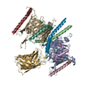
+ Open data
- Basic information
| Entry |  | |||||||||
|---|---|---|---|---|---|---|---|---|---|---|
| Title | TOM-TIM23 supercomplex with a GFP containing substrate | |||||||||
 Map data Map data | ||||||||||
 Sample Sample |
| |||||||||
 Keywords Keywords | Mitochondrial protein import / TOM40 / TIM23 / PROTEIN TRANSPORT | |||||||||
| Biological species |  | |||||||||
| Method | single particle reconstruction / cryo EM / Resolution: 9.36 Å | |||||||||
 Authors Authors | Zhou XY / Yang YQ / Wang GP / Wang SS | |||||||||
| Funding support |  China, 1 items China, 1 items
| |||||||||
 Citation Citation |  Journal: Nat Struct Mol Biol / Year: 2023 Journal: Nat Struct Mol Biol / Year: 2023Title: Molecular pathway of mitochondrial preprotein import through the TOM-TIM23 supercomplex. Authors: Xueyin Zhou / Yuqi Yang / Guopeng Wang / Shanshan Wang / Dongjie Sun / Xiaomin Ou / Yuke Lian / Long Li /  Abstract: Over half of mitochondrial proteins are imported from the cytosol via the pre-sequence pathway, controlled by the TOM complex in the outer membrane and the TIM23 complex in the inner membrane. The ...Over half of mitochondrial proteins are imported from the cytosol via the pre-sequence pathway, controlled by the TOM complex in the outer membrane and the TIM23 complex in the inner membrane. The mechanisms through which proteins are translocated via the TOM and TIM23 complexes remain unclear. Here we report the assembly of the active TOM-TIM23 supercomplex of Saccharomyces cerevisiae with translocating polypeptide substrates. Electron cryo-microscopy analyses reveal that the polypeptide substrates pass the TOM complex through the center of a Tom40 subunit, interacting with a glutamine-rich region. Structural and biochemical analyses show that the TIM23 complex contains a heterotrimer of the subunits Tim23, Tim17 and Mgr2. The polypeptide substrates are shielded from lipids by Mgr2 and Tim17, which creates a translocation pathway characterized by a negatively charged entrance and a central hydrophobic region. These findings reveal an unexpected pre-sequence pathway through the TOM-TIM23 supercomplex spanning the double membranes of mitochondria. | |||||||||
| History |
|
- Structure visualization
| Supplemental images |
|---|
- Downloads & links
-EMDB archive
| Map data |  emd_34661.map.gz emd_34661.map.gz | 8.9 MB |  EMDB map data format EMDB map data format | |
|---|---|---|---|---|
| Header (meta data) |  emd-34661-v30.xml emd-34661-v30.xml emd-34661.xml emd-34661.xml | 12.9 KB 12.9 KB | Display Display |  EMDB header EMDB header |
| FSC (resolution estimation) |  emd_34661_fsc.xml emd_34661_fsc.xml | 10.1 KB | Display |  FSC data file FSC data file |
| Images |  emd_34661.png emd_34661.png | 22.9 KB | ||
| Filedesc metadata |  emd-34661.cif.gz emd-34661.cif.gz | 4 KB | ||
| Others |  emd_34661_half_map_1.map.gz emd_34661_half_map_1.map.gz emd_34661_half_map_2.map.gz emd_34661_half_map_2.map.gz | 65.3 MB 65.3 MB | ||
| Archive directory |  http://ftp.pdbj.org/pub/emdb/structures/EMD-34661 http://ftp.pdbj.org/pub/emdb/structures/EMD-34661 ftp://ftp.pdbj.org/pub/emdb/structures/EMD-34661 ftp://ftp.pdbj.org/pub/emdb/structures/EMD-34661 | HTTPS FTP |
-Validation report
| Summary document |  emd_34661_validation.pdf.gz emd_34661_validation.pdf.gz | 644.4 KB | Display |  EMDB validaton report EMDB validaton report |
|---|---|---|---|---|
| Full document |  emd_34661_full_validation.pdf.gz emd_34661_full_validation.pdf.gz | 643.9 KB | Display | |
| Data in XML |  emd_34661_validation.xml.gz emd_34661_validation.xml.gz | 16.5 KB | Display | |
| Data in CIF |  emd_34661_validation.cif.gz emd_34661_validation.cif.gz | 21.2 KB | Display | |
| Arichive directory |  https://ftp.pdbj.org/pub/emdb/validation_reports/EMD-34661 https://ftp.pdbj.org/pub/emdb/validation_reports/EMD-34661 ftp://ftp.pdbj.org/pub/emdb/validation_reports/EMD-34661 ftp://ftp.pdbj.org/pub/emdb/validation_reports/EMD-34661 | HTTPS FTP |
-Related structure data
- Links
Links
| EMDB pages |  EMDB (EBI/PDBe) / EMDB (EBI/PDBe) /  EMDataResource EMDataResource |
|---|
- Map
Map
| File |  Download / File: emd_34661.map.gz / Format: CCP4 / Size: 83.7 MB / Type: IMAGE STORED AS FLOATING POINT NUMBER (4 BYTES) Download / File: emd_34661.map.gz / Format: CCP4 / Size: 83.7 MB / Type: IMAGE STORED AS FLOATING POINT NUMBER (4 BYTES) | ||||||||||||||||||||||||||||||||||||
|---|---|---|---|---|---|---|---|---|---|---|---|---|---|---|---|---|---|---|---|---|---|---|---|---|---|---|---|---|---|---|---|---|---|---|---|---|---|
| Projections & slices | Image control
Images are generated by Spider. | ||||||||||||||||||||||||||||||||||||
| Voxel size | X=Y=Z: 1.07 Å | ||||||||||||||||||||||||||||||||||||
| Density |
| ||||||||||||||||||||||||||||||||||||
| Symmetry | Space group: 1 | ||||||||||||||||||||||||||||||||||||
| Details | EMDB XML:
|
-Supplemental data
-Half map: #1
| File | emd_34661_half_map_1.map | ||||||||||||
|---|---|---|---|---|---|---|---|---|---|---|---|---|---|
| Projections & Slices |
| ||||||||||||
| Density Histograms |
-Half map: #2
| File | emd_34661_half_map_2.map | ||||||||||||
|---|---|---|---|---|---|---|---|---|---|---|---|---|---|
| Projections & Slices |
| ||||||||||||
| Density Histograms |
- Sample components
-Entire : TOM-TIM23 supercomplex with a GFP containing substrate
| Entire | Name: TOM-TIM23 supercomplex with a GFP containing substrate |
|---|---|
| Components |
|
-Supramolecule #1: TOM-TIM23 supercomplex with a GFP containing substrate
| Supramolecule | Name: TOM-TIM23 supercomplex with a GFP containing substrate type: complex / ID: 1 / Parent: 0 / Macromolecule list: #1 |
|---|---|
| Source (natural) | Organism:  |
-Experimental details
-Structure determination
| Method | cryo EM |
|---|---|
 Processing Processing | single particle reconstruction |
| Aggregation state | particle |
- Sample preparation
| Buffer | pH: 8 |
|---|---|
| Grid | Model: Quantifoil R1.2/1.3 / Material: GOLD / Mesh: 300 |
| Vitrification | Cryogen name: ETHANE |
- Electron microscopy
| Microscope | FEI TITAN KRIOS |
|---|---|
| Specialist optics | Energy filter - Name: GIF Bioquantum / Energy filter - Slit width: 20 eV |
| Image recording | Film or detector model: GATAN K3 BIOQUANTUM (6k x 4k) / Average electron dose: 60.0 e/Å2 |
| Electron beam | Acceleration voltage: 300 kV / Electron source:  FIELD EMISSION GUN FIELD EMISSION GUN |
| Electron optics | Illumination mode: FLOOD BEAM / Imaging mode: BRIGHT FIELD / Cs: 2.7 mm / Nominal defocus max: 1.2 µm / Nominal defocus min: 1.0 µm / Nominal magnification: 81000 |
| Experimental equipment |  Model: Titan Krios / Image courtesy: FEI Company |
 Movie
Movie Controller


 Z (Sec.)
 Y (Row.)
 X (Col.)


