National Natural Science Foundation of China (NSFC)
No. 31771490
China
Citation
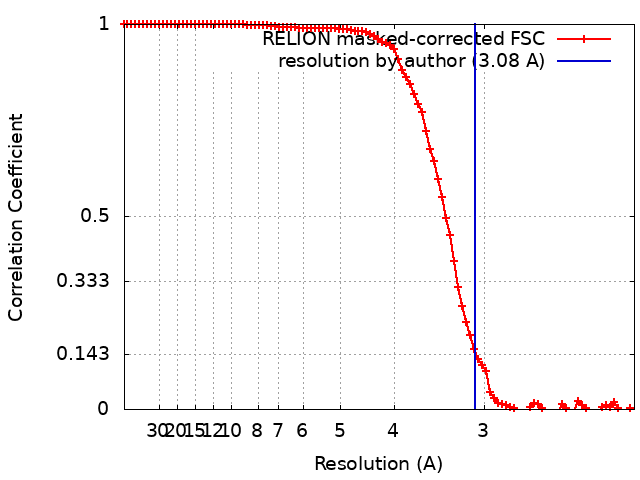
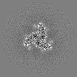
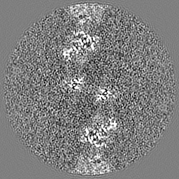
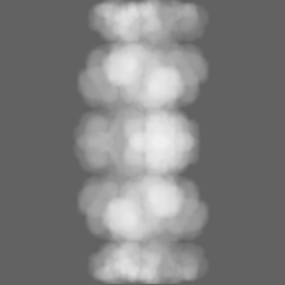
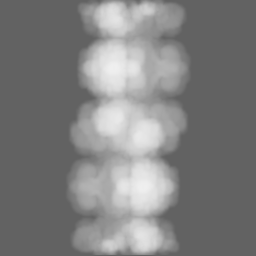
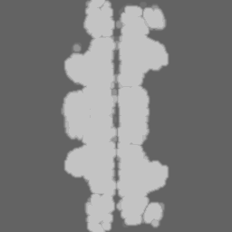
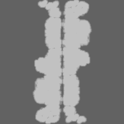
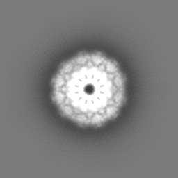
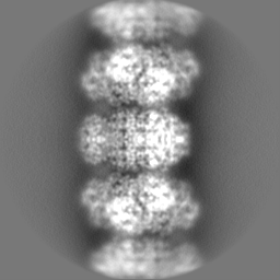
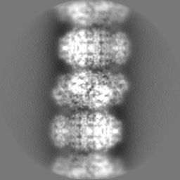
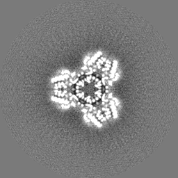
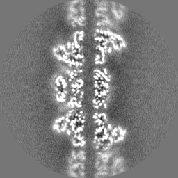
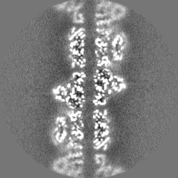
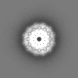
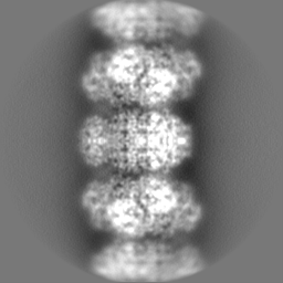
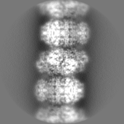
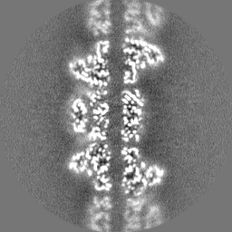
Journal: Cell Biosci / Year: 2023 Title: Structural basis of human PRPS2 filaments. Authors: Guang-Ming Lu / Huan-Huan Hu / Chia-Chun Chang / Jiale Zhong / Xian Zhou / Chen-Jun Guo / Tianyi Zhang / Yi-Lan Li / Boqi Yin / Ji-Long Liu / Abstract: BACKGROUND: PRPP synthase (PRPS) transfers the pyrophosphate groups from ATP to ribose-5-phosphate to produce 5-phosphate ribose-1-pyrophosphate (PRPP), a key intermediate in the biosynthesis of ...BACKGROUND: PRPP synthase (PRPS) transfers the pyrophosphate groups from ATP to ribose-5-phosphate to produce 5-phosphate ribose-1-pyrophosphate (PRPP), a key intermediate in the biosynthesis of several metabolites including nucleotides, dinucleotides and some amino acids. There are three PRPS isoforms encoded in human genome. While human PRPS1 (hPRPS1) and human PRPS2 (hPRPS2) are expressed in most tissues, human PRPS3 (hPRPS3) is exclusively expressed in testis. Although hPRPS1 and hPRPS2 share 95% sequence identity, hPRPS2 has been shown to be less sensitive to allosteric inhibition and specifically upregulated in certain cancers in the translational level. Recent studies demonstrate that PRPS can form a subcellular compartment termed the cytoophidium in multiple organisms across prokaryotes and eukaryotes. Forming filaments and cytoophidia is considered as a distinctive mechanism involving the polymerization of the protein. Previously we solved the filament structures of Escherichia coli PRPS (ecPRPS) using cryo-electron microscopy (cryo-EM) . RESULTS: Order to investigate the function and molecular mechanism of hPRPS2 polymerization, here we solve the polymer structure of hPRPS2 at 3.08 Å resolution. hPRPS2 hexamers stack into polymers ...RESULTS: Order to investigate the function and molecular mechanism of hPRPS2 polymerization, here we solve the polymer structure of hPRPS2 at 3.08 Å resolution. hPRPS2 hexamers stack into polymers in the conditions with the allosteric/competitive inhibitor ADP. The binding modes of ADP at the canonical allosteric site and at the catalytic active site are clearly determined. A point mutation disrupting the inter-hexamer interaction prevents hPRPS2 polymerization and results in significantly reduced catalytic activity. CONCLUSION: Findings suggest that the regulation of hPRPS2 polymer is distinct from ecPRPS polymer and provide new insights to the regulation of hPRPS2 with structural basis.
Film or detector model: GATAN K3 BIOQUANTUM (6k x 4k) / Number grids imaged: 1 / Number real images: 2403 / Average exposure time: 4.0 sec. / Average electron dose: 60.0 e/Å2
Electron beam
Acceleration voltage: 300 kV / Electron source: FIELD EMISSION GUN
In the structure databanks used in Yorodumi, some data are registered as the other names, "COVID-19 virus" and "2019-nCoV". Here are the details of the virus and the list of structure data.
Jan 31, 2019. EMDB accession codes are about to change! (news from PDBe EMDB page)
EMDB accession codes are about to change! (news from PDBe EMDB page)
The allocation of 4 digits for EMDB accession codes will soon come to an end. Whilst these codes will remain in use, new EMDB accession codes will include an additional digit and will expand incrementally as the available range of codes is exhausted. The current 4-digit format prefixed with “EMD-” (i.e. EMD-XXXX) will advance to a 5-digit format (i.e. EMD-XXXXX), and so on. It is currently estimated that the 4-digit codes will be depleted around Spring 2019, at which point the 5-digit format will come into force.
The EM Navigator/Yorodumi systems omit the EMD- prefix.
Related info.:Q: What is EMD? / ID/Accession-code notation in Yorodumi/EM Navigator
Yorodumi is a browser for structure data from EMDB, PDB, SASBDB, etc.
This page is also the successor to EM Navigator detail page, and also detail information page/front-end page for Omokage search.
The word "yorodu" (or yorozu) is an old Japanese word meaning "ten thousand". "mi" (miru) is to see.
Related info.:EMDB / PDB / SASBDB / Comparison of 3 databanks / Yorodumi Search / Aug 31, 2016. New EM Navigator & Yorodumi / Yorodumi Papers / Jmol/JSmol / Function and homology information / Changes in new EM Navigator and Yorodumi
 Movie
Movie Controller


 Open data
 Basic information
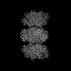
 Map data
 Sample
 Keywords
 Function and homology information
 Homo sapiens (human)
 Authors
 China, 2 items
  Citation
 Structure visualization
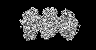
 Downloads & links
 emd_33883.png
 http://ftp.pdbj.org/pub/emdb/structures/EMD-33883
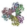

 Z (Sec.)
 Y (Row.)
 X (Col.)


 Sample components


 Processing
 Electron microscopy
 FIELD EMISSION GUN