 Movie
Movie Controller

[English] 日本語
 Yorodumi
- EMDB-33241: Cryo-EM Structure of Human Niacin Receptor HCA2-Gi protein complex -
+ Open data

- Basic information
| Entry |  | ||||||||||||||||||
|---|---|---|---|---|---|---|---|---|---|---|---|---|---|---|---|---|---|---|---|
| Title | Cryo-EM Structure of Human Niacin Receptor HCA2-Gi protein complex | ||||||||||||||||||
 Map data Map data | |||||||||||||||||||
 Sample Sample |
| ||||||||||||||||||
 Keywords Keywords | Niacin receptor / hydroxycarboxylic acid receptor 2 / HCA2 / Gi complex / MEMBRANE PROTEIN | ||||||||||||||||||
| Function / homology |  Function and homology information Function and homology informationneutrophil apoptotic process / nicotinic acid receptor activity / Hydroxycarboxylic acid-binding receptors / positive regulation of neutrophil apoptotic process / Class A/1 (Rhodopsin-like receptors) / positive regulation of adiponectin secretion / negative regulation of lipid catabolic process / adenylate cyclase inhibitor activity / positive regulation of protein localization to cell cortex / T cell migration ...neutrophil apoptotic process / nicotinic acid receptor activity / Hydroxycarboxylic acid-binding receptors / positive regulation of neutrophil apoptotic process / Class A/1 (Rhodopsin-like receptors) / positive regulation of adiponectin secretion / negative regulation of lipid catabolic process / adenylate cyclase inhibitor activity / positive regulation of protein localization to cell cortex / T cell migration / positive regulation of relaxation of smooth muscle / Adenylate cyclase inhibitory pathway / D2 dopamine receptor binding / adenylate cyclase-inhibiting serotonin receptor signaling pathway / G protein-coupled serotonin receptor binding / cellular response to forskolin / regulation of mitotic spindle organization / chemokine-mediated signaling pathway / Regulation of insulin secretion / neuropeptide signaling pathway / response to prostaglandin E / positive regulation of cholesterol biosynthetic process / negative regulation of insulin secretion / G protein-coupled receptor binding / response to peptide hormone / adenylate cyclase-modulating G protein-coupled receptor signaling pathway / G-protein beta/gamma-subunit complex binding / centriolar satellite / adenylate cyclase-inhibiting G protein-coupled receptor signaling pathway / Olfactory Signaling Pathway / Activation of the phototransduction cascade / G protein-coupled acetylcholine receptor signaling pathway / G beta:gamma signalling through PLC beta / Presynaptic function of Kainate receptors / Thromboxane signalling through TP receptor / Activation of G protein gated Potassium channels / Inhibition of voltage gated Ca2+ channels via Gbeta/gamma subunits / G-protein activation / Glucagon signaling in metabolic regulation / G beta:gamma signalling through CDC42 / Prostacyclin signalling through prostacyclin receptor / G beta:gamma signalling through BTK / Synthesis, secretion, and inactivation of Glucagon-like Peptide-1 (GLP-1) / photoreceptor disc membrane / ADP signalling through P2Y purinoceptor 12 / Sensory perception of sweet, bitter, and umami (glutamate) taste / GDP binding / Glucagon-type ligand receptors / cell junction / Adrenaline,noradrenaline inhibits insulin secretion / Vasopressin regulates renal water homeostasis via Aquaporins / Glucagon-like Peptide-1 (GLP1) regulates insulin secretion / G alpha (z) signalling events / cellular response to catecholamine stimulus / ADP signalling through P2Y purinoceptor 1 / ADORA2B mediated anti-inflammatory cytokines production / G beta:gamma signalling through PI3Kgamma / adenylate cyclase-activating dopamine receptor signaling pathway / Cooperation of PDCL (PhLP1) and TRiC/CCT in G-protein beta folding / GPER1 signaling / cellular response to prostaglandin E stimulus / heterotrimeric G-protein complex / G alpha (12/13) signalling events / Inactivation, recovery and regulation of the phototransduction cascade / G-protein beta-subunit binding / extracellular vesicle / sensory perception of taste / adenylate cyclase-activating G protein-coupled receptor signaling pathway / sperm principal piece / Thrombin signalling through proteinase activated receptors (PARs) / signaling receptor complex adaptor activity / retina development in camera-type eye / GTPase binding / G protein activity / fibroblast proliferation / Ca2+ pathway / midbody / cell cortex / High laminar flow shear stress activates signaling by PIEZO1 and PECAM1:CDH5:KDR in endothelial cells / G alpha (i) signalling events / G alpha (s) signalling events / phospholipase C-activating G protein-coupled receptor signaling pathway / G alpha (q) signalling events / Hydrolases; Acting on acid anhydrides; Acting on GTP to facilitate cellular and subcellular movement / Ras protein signal transduction / cell population proliferation / Extra-nuclear estrogen signaling / ciliary basal body / G protein-coupled receptor signaling pathway / cell division / lysosomal membrane / GTPase activity / centrosome / synapse / GTP binding / protein-containing complex binding / nucleolus / magnesium ion binding / Golgi apparatus / signal transduction Similarity search - Function | ||||||||||||||||||
| Biological species |  Homo sapiens (human) Homo sapiens (human) | ||||||||||||||||||
| Method | single particle reconstruction / cryo EM / Resolution: 3.1 Å | ||||||||||||||||||
 Authors Authors | Yang Y / Kang HJ / Gao RG / Wang JJ / Han GW / DiBerto JF / Wu LJ / Tong JH / Qu L / Wu YR ...Yang Y / Kang HJ / Gao RG / Wang JJ / Han GW / DiBerto JF / Wu LJ / Tong JH / Qu L / Wu YR / Pileski R / Li XM / Zhang XC / Zhao SW / Kenakin T / Wang Q / Stevens RC / Peng W / Roth BL / Rao ZH / Liu ZJ | ||||||||||||||||||
| Funding support |  China, 5 items China, 5 items
| ||||||||||||||||||
 Citation Citation | Journal: Nat Commun / Year: 2023 Title: Structural insights into the human niacin receptor HCA2-G signalling complex. Authors: Yang Yang / Hye Jin Kang / Ruogu Gao / Jingjing Wang / Gye Won Han / Jeffrey F DiBerto / Lijie Wu / Jiahui Tong / Lu Qu / Yiran Wu / Ryan Pileski / Xuemei Li / Xuejun Cai Zhang / Suwen Zhao ...Authors: Yang Yang / Hye Jin Kang / Ruogu Gao / Jingjing Wang / Gye Won Han / Jeffrey F DiBerto / Lijie Wu / Jiahui Tong / Lu Qu / Yiran Wu / Ryan Pileski / Xuemei Li / Xuejun Cai Zhang / Suwen Zhao / Terry Kenakin / Quan Wang / Raymond C Stevens / Wei Peng / Bryan L Roth / Zihe Rao / Zhi-Jie Liu /   Abstract: The hydroxycarboxylic acid receptor 2 (HCA2) agonist niacin has been used as treatment for dyslipidemia for several decades albeit with skin flushing as a common side-effect in treated individuals. ...The hydroxycarboxylic acid receptor 2 (HCA2) agonist niacin has been used as treatment for dyslipidemia for several decades albeit with skin flushing as a common side-effect in treated individuals. Extensive efforts have been made to identify HCA2 targeting lipid lowering agents with fewer adverse effects, despite little being known about the molecular basis of HCA2 mediated signalling. Here, we report the cryo-electron microscopy structure of the HCA2-G signalling complex with the potent agonist MK-6892, along with crystal structures of HCA2 in inactive state. These structures, together with comprehensive pharmacological analysis, reveal the ligand binding mode and activation and signalling mechanisms of HCA2. This study elucidates the structural determinants essential for HCA2 mediated signalling and provides insights into ligand discovery for HCA2 and related receptors. | ||||||||||||||||||
| History |
|
- Structure visualization
| Supplemental images |
|---|

- Downloads & links
-EMDB archive
| Map data | emd_33241.map.gz | 59.7 MB | EMDB map data format | |
|---|---|---|---|---|
| Header (meta data) | emd-33241-v30.xmlemd-33241.xml | 25.8 KB 25.8 KB | Display Display | EMDB header |
| FSC (resolution estimation) | emd_33241_fsc.xml | 9.3 KB | Display | FSC data file |
| Images | 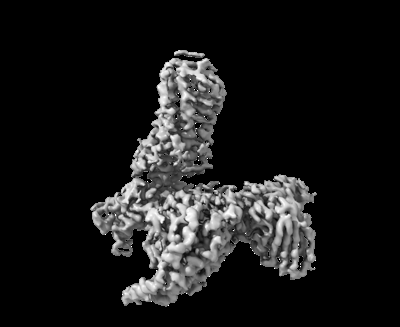 emd_33241.png emd_33241.png | 49 KB | ||
| Masks | emd_33241_msk_1.map | 64 MB | Mask map | |
| Filedesc metadata | emd-33241.cif.gz | 7.6 KB | ||
| Others | emd_33241_half_map_1.map.gzemd_33241_half_map_2.map.gz | 59.4 MB 59.4 MB | ||
| Archive directory |  http://ftp.pdbj.org/pub/emdb/structures/EMD-33241ftp://ftp.pdbj.org/pub/emdb/structures/EMD-33241 http://ftp.pdbj.org/pub/emdb/structures/EMD-33241ftp://ftp.pdbj.org/pub/emdb/structures/EMD-33241 | HTTPS FTP |
-Related structure data
| Related structure data |  7xk2MC  7zl9C  7zlyC M: atomic model generated by this map C: citing same article ( |
|---|---|
| Similar structure data |
-Links
| EMDB pages | EMDB (EBI/PDBe) / EMDataResource |
|---|---|
| Related items in Molecule of the Month |


-Map
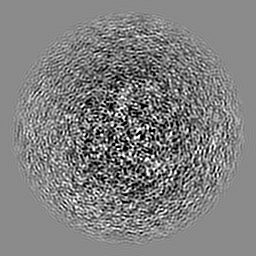
| File | Download / File: emd_33241.map.gz / Format: CCP4 / Size: 64 MB / Type: IMAGE STORED AS FLOATING POINT NUMBER (4 BYTES) | ||||||||||||||||||||||||||||||||||||
|---|---|---|---|---|---|---|---|---|---|---|---|---|---|---|---|---|---|---|---|---|---|---|---|---|---|---|---|---|---|---|---|---|---|---|---|---|---|
| Projections & slices | Image control
Images are generated by Spider. | ||||||||||||||||||||||||||||||||||||
| Voxel size | X=Y=Z: 1.04 Å | ||||||||||||||||||||||||||||||||||||
| Density |
| ||||||||||||||||||||||||||||||||||||
| Symmetry | Space group: 1 | ||||||||||||||||||||||||||||||||||||
| Details | EMDB XML:
|
 Z (Sec.)
 Y (Row.)
 X (Col.)


-Supplemental data
-Mask #1
| File | emd_33241_msk_1.map | ||||||||||||
|---|---|---|---|---|---|---|---|---|---|---|---|---|---|
| Projections & Slices |
| ||||||||||||
| Density Histograms |


-Half map: #2
| File | emd_33241_half_map_1.map | ||||||||||||
|---|---|---|---|---|---|---|---|---|---|---|---|---|---|
| Projections & Slices |
| ||||||||||||
| Density Histograms |


-Half map: #1
| File | emd_33241_half_map_2.map | ||||||||||||
|---|---|---|---|---|---|---|---|---|---|---|---|---|---|
| Projections & Slices |
| ||||||||||||
| Density Histograms |


- Sample components
-Entire : Niacin Receptor HCA2-Gi complex
| Entire | Name: Niacin Receptor HCA2-Gi complex |
|---|---|
| Components |
|
-Supramolecule #1: Niacin Receptor HCA2-Gi complex
| Supramolecule | Name: Niacin Receptor HCA2-Gi complex / type: complex / ID: 1 / Parent: 0 / Macromolecule list: #1-#2, #4-#5 |
|---|---|
| Source (natural) | Organism: Homo sapiens (human) |
-Macromolecule #1: Guanine nucleotide-binding protein G(i) subunit alpha-1
| Macromolecule | Name: Guanine nucleotide-binding protein G(i) subunit alpha-1 type: protein_or_peptide / ID: 1 / Number of copies: 1 / Enantiomer: LEVO |
|---|---|
| Source (natural) | Organism: Homo sapiens (human) |
| Molecular weight | Theoretical: 40.55916 KDa |
| Recombinant expression | Organism: Homo sapiens (human) |
| Sequence | String: GSMGCTLSAE DKAAVERSKM IDRNLREDGE KAAREVKLLL LGAGESGKST IVKQMKIIHE AGYSEEECKQ YKAVVYSNTI QSIIAIIRA MGRLKIDFGD SARADDARQL FVLAGAAEEG FMTAELAGVI KRLWKDSGVQ ACFNRSREYQ LNDSAAYYLN D LDRIAQPN ...String: GSMGCTLSAE DKAAVERSKM IDRNLREDGE KAAREVKLLL LGAGESGKST IVKQMKIIHE AGYSEEECKQ YKAVVYSNTI QSIIAIIRA MGRLKIDFGD SARADDARQL FVLAGAAEEG FMTAELAGVI KRLWKDSGVQ ACFNRSREYQ LNDSAAYYLN D LDRIAQPN YIPTQQDVLR TRVKTTGIVE THFTFKDLHF KMFDVGGQRS ERKKWIHCFE GVTAIIFCVA LSDYDLVLAE DE EMNRMHE SMKLFDSICN NKWFTDTSII LFLNKKDLFE EKIKKSPLTI CYPEYAGSNT YEEAAAYIQC QFEDLNKRKD TKE IYTHFT CATDTKNVQF VFDAVTDVII KNNLKDCGLF UniProtKB: Guanine nucleotide-binding protein G(i) subunit alpha-1 |
-Macromolecule #2: Guanine nucleotide-binding protein G(I)/G(S)/G(T) subunit beta-1
| Macromolecule | Name: Guanine nucleotide-binding protein G(I)/G(S)/G(T) subunit beta-1 type: protein_or_peptide / ID: 2 / Number of copies: 1 / Enantiomer: LEVO |
|---|---|
| Source (natural) | Organism: Homo sapiens (human) |
| Molecular weight | Theoretical: 37.41693 KDa |
| Recombinant expression | Organism: Homo sapiens (human) |
| Sequence | String: MSELDQLRQE AEQLKNQIRD ARKACADATL SQITNNIDPV GRIQMRTRRT LRGHLAKIYA MHWGTDSRLL VSASQDGKLI IWDSYTTNK VHAIPLRSSW VMTCAYAPSG NYVACGGLDN ICSIYNLKTR EGNVRVSREL AGHTGYLSCC RFLDDNQIVT S SGDTTCAL ...String: MSELDQLRQE AEQLKNQIRD ARKACADATL SQITNNIDPV GRIQMRTRRT LRGHLAKIYA MHWGTDSRLL VSASQDGKLI IWDSYTTNK VHAIPLRSSW VMTCAYAPSG NYVACGGLDN ICSIYNLKTR EGNVRVSREL AGHTGYLSCC RFLDDNQIVT S SGDTTCAL WDIETGQQTT TFTGHTGDVM SLSLAPDTRL FVSGACDASA KLWDVREGMC RQTFTGHESD INAICFFPNG NA FATGSDD ATCRLFDLRA DQELMTYSHD NIICGITSVS FSKSGRLLLA GYDDFNCNVW DALKADRAGV LAGHDNRVSC LGV TDDGMA VATGSWDSFL KIWN UniProtKB: Guanine nucleotide-binding protein G(I)/G(S)/G(T) subunit beta-1 |
-Macromolecule #3: Hydroxycarboxylic acid receptor 2
| Macromolecule | Name: Hydroxycarboxylic acid receptor 2 / type: protein_or_peptide / ID: 3 / Number of copies: 1 / Enantiomer: LEVO |
|---|---|
| Source (natural) | Organism: Homo sapiens (human) |
| Molecular weight | Theoretical: 38.010254 KDa |
| Recombinant expression | Organism: Homo sapiens (human) |
| Sequence | String: MNRHHLQDHF LEIDKKNCCV FRDDFIVKVL PPVLGLEFIF GLLGNGLALW IFCFHLKSWK SSRIFLFNLA VADFLLIICL PFLMDNYVR RWDWKFGDIP CRLMLFMLAM NRQGSIIFLT VVAVDRYFRV VHPHHALNKI SNRTAAIISC LLWGITIGLT V HLLKKKMP ...String: MNRHHLQDHF LEIDKKNCCV FRDDFIVKVL PPVLGLEFIF GLLGNGLALW IFCFHLKSWK SSRIFLFNLA VADFLLIICL PFLMDNYVR RWDWKFGDIP CRLMLFMLAM NRQGSIIFLT VVAVDRYFRV VHPHHALNKI SNRTAAIISC LLWGITIGLT V HLLKKKMP IQNGGANLCS SFSICHTFQW HEAMFLLEFF LPLGIILFCS ARIIWSLRQR QMDRHAKIKR AITFIMVVAI VF VICFLPS VVVRIRIFWL LHTSGTQNCE VYRSVDLAFF ITLSFTYMNS MLDPVVYYFS SPSFPNFFST LINRCLQRKM TGE PDNNR UniProtKB: Hydroxycarboxylic acid receptor 2 |
-Macromolecule #4: scFv16
| Macromolecule | Name: scFv16 / type: protein_or_peptide / ID: 4 / Number of copies: 1 / Enantiomer: LEVO |
|---|---|
| Source (natural) | Organism: Homo sapiens (human) |
| Molecular weight | Theoretical: 26.466486 KDa |
| Recombinant expression | Organism: Homo sapiens (human) |
| Sequence | String: DVQLVESGGG LVQPGGSRKL SCSASGFAFS SFGMHWVRQA PEKGLEWVAY ISSGSGTIYY ADTVKGRFTI SRDDPKNTLF LQMTSLRSE DTAMYYCVRS IYYYGSSPFD FWGQGTTLTV SSGGGGSGGG GSGGGGSDIV MTQATSSVPV TPGESVSISC R SSKSLLHS ...String: DVQLVESGGG LVQPGGSRKL SCSASGFAFS SFGMHWVRQA PEKGLEWVAY ISSGSGTIYY ADTVKGRFTI SRDDPKNTLF LQMTSLRSE DTAMYYCVRS IYYYGSSPFD FWGQGTTLTV SSGGGGSGGG GSGGGGSDIV MTQATSSVPV TPGESVSISC R SSKSLLHS NGNTYLYWFL QRPGQSPQLL IYRMSNLASG VPDRFSGSGS GTAFTLTISR LEAEDVGVYY CMQHLEYPLT FG AGTKLEL K |
-Macromolecule #5: Guanine nucleotide-binding protein G(I)/G(S)/G(O) subunit gamma-2
| Macromolecule | Name: Guanine nucleotide-binding protein G(I)/G(S)/G(O) subunit gamma-2 type: protein_or_peptide / ID: 5 / Number of copies: 1 / Enantiomer: LEVO |
|---|---|
| Source (natural) | Organism: Homo sapiens (human) |
| Molecular weight | Theoretical: 7.861143 KDa |
| Recombinant expression | Organism: Homo sapiens (human) |
| Sequence | String: MASNNTASIA QARKLVEQLK MEANIDRIKV SKAAADLMAY CEAHAKEDPL LTPVPASENP FREKKFFCAI L UniProtKB: Guanine nucleotide-binding protein G(I)/G(S)/G(O) subunit gamma-2 |
-Macromolecule #6: 2-[[2,2-dimethyl-3-[3-(5-oxidanylpyridin-2-yl)-1,2,4-oxadiazol-5-...
| Macromolecule | Name: 2-[[2,2-dimethyl-3-[3-(5-oxidanylpyridin-2-yl)-1,2,4-oxadiazol-5-yl]propanoyl]amino]cyclohexene-1-carboxylic acid type: ligand / ID: 6 / Number of copies: 1 / Formula: FI7 |
|---|---|
| Molecular weight | Theoretical: 386.402 Da |
| Chemical component information |  ChemComp-FI7: |
-Experimental details
-Structure determination
| Method | cryo EM |
|---|---|
 Processing Processing | single particle reconstruction |
| Aggregation state | particle |
-Sample preparation
| Buffer | pH: 7.5 |
|---|---|
| Vitrification | Cryogen name: ETHANE |
- Electron microscopy
| Microscope | FEI TITAN KRIOS |
|---|---|
| Image recording | Film or detector model: GATAN K2 QUANTUM (4k x 4k) / Detector mode: SUPER-RESOLUTION / Average electron dose: 60.0 e/Å2 |
| Electron beam | Acceleration voltage: 300 kV / Electron source:  FIELD EMISSION GUN FIELD EMISSION GUN |
| Electron optics | Illumination mode: FLOOD BEAM / Imaging mode: BRIGHT FIELD / Nominal defocus max: 2.0 µm / Nominal defocus min: 1.0 µm |
| Experimental equipment |  Model: Titan Krios / Image courtesy: FEI Company |

