 ムービー
ムービー コントローラー


+ データを開く

- 基本情報
基本情報
| 登録情報 | 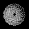 | |||||||||
|---|---|---|---|---|---|---|---|---|---|---|
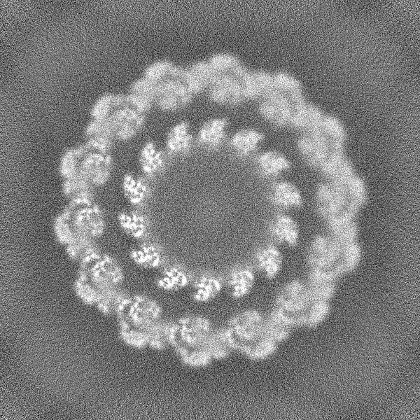
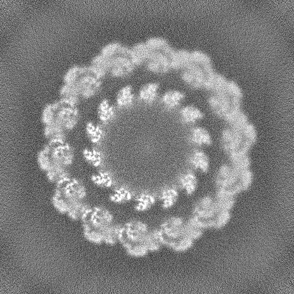
| タイトル | structure of vp51 in white spot syndrome virus capsid | |||||||||
 マップデータ マップデータ | ||||||||||
 試料 試料 |
| |||||||||
 キーワード キーワード | Complex / C14 / capsid / disc / VIRAL PROTEIN | |||||||||
| 機能・相同性 | viral envelope / DNA binding / Capsid protein 機能・相同性情報 機能・相同性情報 | |||||||||
| 生物種 |  White spot syndrome virus (ウイルス) White spot syndrome virus (ウイルス) | |||||||||
| 手法 | 単粒子再構成法 / クライオ電子顕微鏡法 / ネガティブ染色法 / 解像度: 3.73 Å | |||||||||
 データ登録者 データ登録者 | Shan H / Liu MD / Shen QT | |||||||||
| 資金援助 |  中国, 1件 中国, 1件
| |||||||||
 引用 引用 | ジャーナル: To Be Published タイトル: structure of vp51 in white spot syndrome virus capsid 著者: Hong S / Shen QT | |||||||||
| 履歴 |
|
- 構造の表示
構造の表示
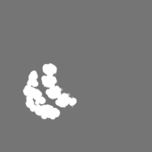
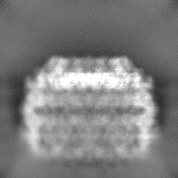
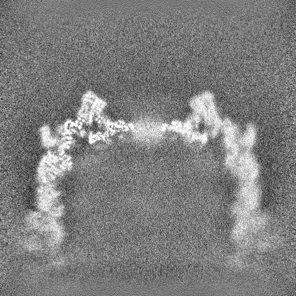
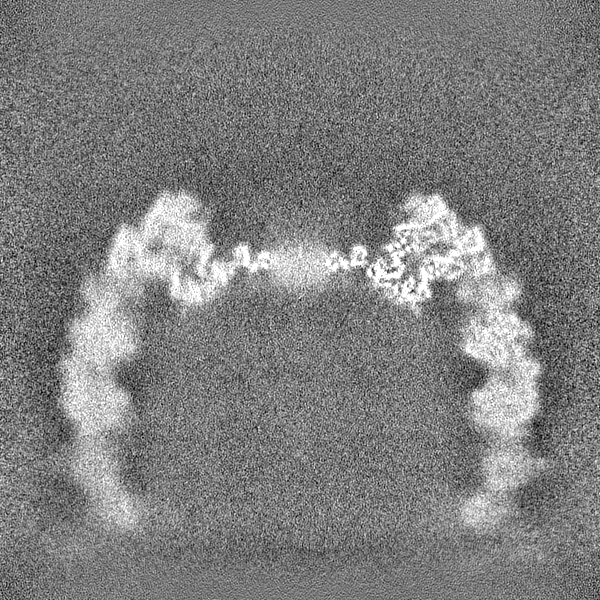
| 添付画像 |
|---|
- ダウンロードとリンク
-EMDBアーカイブ
| マップデータ | emd_33168.map.gz | 408 MB | EMDBマップデータ形式 | |
|---|---|---|---|---|
| ヘッダ (付随情報) | emd-33168-v30.xmlemd-33168.xml | 15.5 KB 15.5 KB | 表示 表示 | EMDBヘッダ |
| 画像 |  emd_33168.png emd_33168.png | 143.9 KB | ||
| マスクデータ | emd_33168_msk_1.map | 824 MB | マスクマップ | |
| Filedesc metadata | emd-33168.cif.gz | 5.6 KB | ||
| その他 | emd_33168_half_map_1.map.gzemd_33168_half_map_2.map.gz | 764.6 MB 764.6 MB | ||
| アーカイブディレクトリ |  http://ftp.pdbj.org/pub/emdb/structures/EMD-33168ftp://ftp.pdbj.org/pub/emdb/structures/EMD-33168 http://ftp.pdbj.org/pub/emdb/structures/EMD-33168ftp://ftp.pdbj.org/pub/emdb/structures/EMD-33168 | HTTPS FTP |
-関連構造データ

-リンク
| EMDBのページ | EMDB (EBI/PDBe) / EMDataResource |
|---|
-マップ
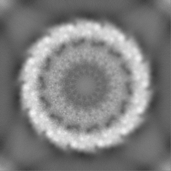
| ファイル | ダウンロード / ファイル: emd_33168.map.gz / 形式: CCP4 / 大きさ: 824 MB / タイプ: IMAGE STORED AS FLOATING POINT NUMBER (4 BYTES) | ||||||||||||||||||||||||||||||||||||
|---|---|---|---|---|---|---|---|---|---|---|---|---|---|---|---|---|---|---|---|---|---|---|---|---|---|---|---|---|---|---|---|---|---|---|---|---|---|
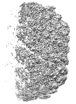
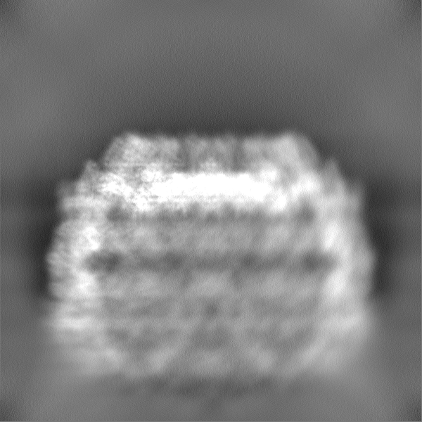
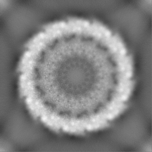
| 投影像・断面図 | 画像のコントロール
画像は Spider により作成 | ||||||||||||||||||||||||||||||||||||
| ボクセルのサイズ | X=Y=Z: 1.34 Å | ||||||||||||||||||||||||||||||||||||
| 密度 |
| ||||||||||||||||||||||||||||||||||||
| 対称性 | 空間群: 1 | ||||||||||||||||||||||||||||||||||||
| 詳細 | EMDB XML:
|
 Z (Sec.)
 Y (Row.)
 X (Col.)


-添付データ
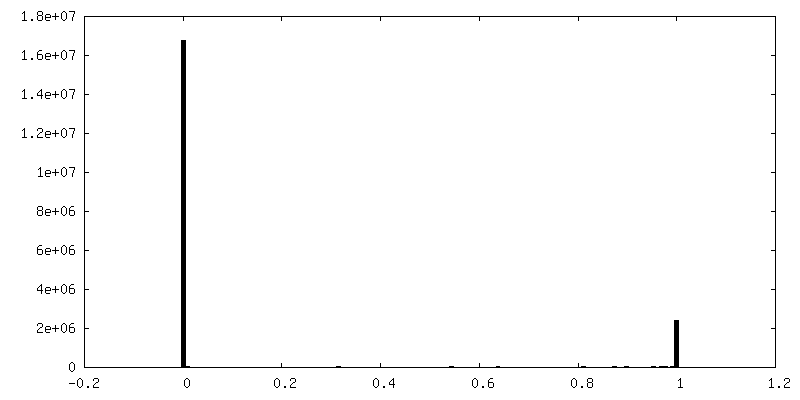
-マスク #1
| ファイル | emd_33168_msk_1.map | ||||||||||||
|---|---|---|---|---|---|---|---|---|---|---|---|---|---|
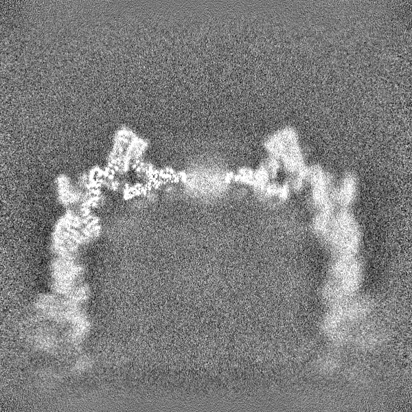
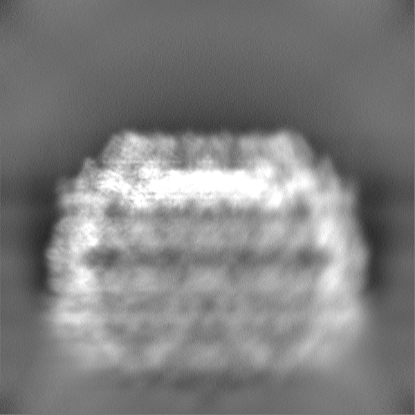
| 投影像・断面図 |
| ||||||||||||
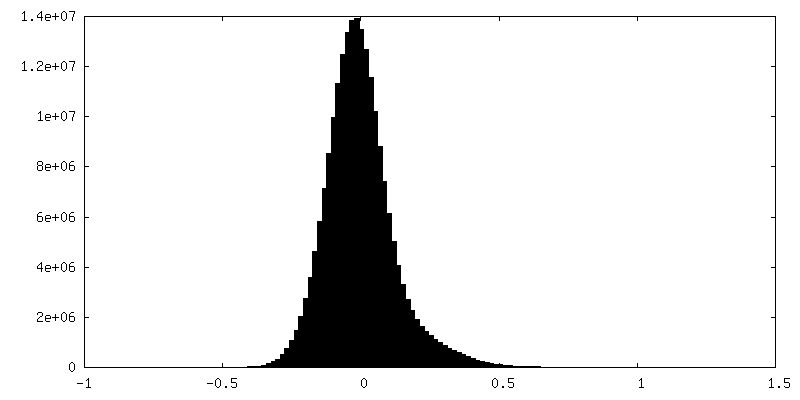
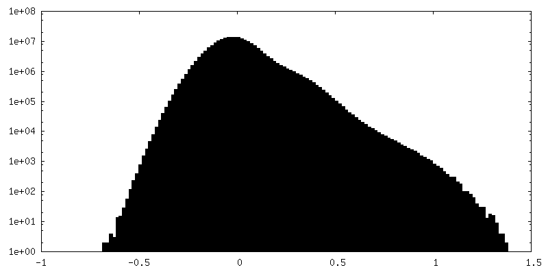
| 密度ヒストグラム |


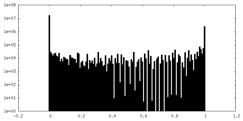
-ハーフマップ: #2
| ファイル | emd_33168_half_map_1.map | ||||||||||||
|---|---|---|---|---|---|---|---|---|---|---|---|---|---|
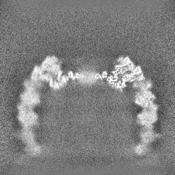
| 投影像・断面図 |
| ||||||||||||
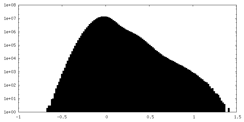
| 密度ヒストグラム |

-ハーフマップ: #1
| ファイル | emd_33168_half_map_2.map | ||||||||||||
|---|---|---|---|---|---|---|---|---|---|---|---|---|---|
| 投影像・断面図 |
| ||||||||||||
| 密度ヒストグラム |

- 試料の構成要素
-全体 : white spot syndrome virus
| 全体 | 名称: white spot syndrome virus (ウイルス) |
|---|---|
| 要素 |
|
-超分子 #1: white spot syndrome virus
| 超分子 | 名称: white spot syndrome virus / タイプ: complex / ID: 1 / 親要素: 0 / 含まれる分子: all |
|---|---|
| 由来(天然) | 生物種: White spot syndrome virus (ウイルス) |
-分子 #1: Capsid protein
| 分子 | 名称: Capsid protein / タイプ: protein_or_peptide / ID: 1 / コピー数: 2 / 光学異性体: LEVO |
|---|---|
| 由来(天然) | 生物種: White spot syndrome virus (ウイルス) |
| 分子量 | 理論値: 51.965145 KDa |
| 配列 | 文字列: MSASLILDEY LKKTASAVLD VADSFEKIKG EIQSPEEAAA LSVALYGAPP KPSASAVASI ITGERTSLND KYLSDNVLLK MSVARVGQE NNRKRADQAA DEIRTIMEDI TGSLSGAYRQ YSPLEEENKV HIGIMNNKTP SIVCGYYTMD TSISSEPLSL T DFQNPTVI ...文字列: MSASLILDEY LKKTASAVLD VADSFEKIKG EIQSPEEAAA LSVALYGAPP KPSASAVASI ITGERTSLND KYLSDNVLLK MSVARVGQE NNRKRADQAA DEIRTIMEDI TGSLSGAYRQ YSPLEEENKV HIGIMNNKTP SIVCGYYTMD TSISSEPLSL T DFQNPTVI ANVTKRMESI FSKVDSARST RFDAFVNGVA NNMDIKSSID WANMVENVIK LPDSTPNPCS VDTIVSRDAS VV KTAVNDI YASVGKSYCR PATQLTFMSE IEKLRKAAVV CFEALMSDTR ERAFVEFLFY VSFKEDASNT NSKLFVQNKL SSM SGNPRQ PIKLVRRSAE ETLFGLCFMF KVMPPEFMNC IFNFPTIPHS TQYHGLYGTC LTPLLRKYGS SFEKSWAHFE EILS ERANA VKKFGVNDTR IDCLDAVANL TGPVYVLILD LVRTLSAQRS CSTKFLREIK ENYLLWNRFV S UniProtKB: Capsid protein |
-実験情報
-構造解析
| 手法 | ネガティブ染色法, クライオ電子顕微鏡法 |
|---|---|
 解析 解析 | 単粒子再構成法 |
| 試料の集合状態 | particle |
-試料調製
| 緩衝液 | pH: 8 |
|---|---|
| 染色 | タイプ: NEGATIVE / 材質: uranyl acetate |
| 凍結 | 凍結剤: ETHANE |
- 電子顕微鏡法
| 顕微鏡 | FEI TITAN KRIOS |
|---|---|
| 撮影 | フィルム・検出器のモデル: GATAN K3 BIOQUANTUM (6k x 4k) 平均電子線量: 1.25 e/Å2 |
| 電子線 | 加速電圧: 300 kV / 電子線源:  FIELD EMISSION GUN FIELD EMISSION GUN |
| 電子光学系 | C2レンズ絞り径: 50.0 µm / 照射モード: FLOOD BEAM / 撮影モード: BRIGHT FIELD / Cs: 2.7 mm / 最大 デフォーカス(公称値): 1.8 µm / 最小 デフォーカス(公称値): 1.0 µm |
| 実験機器 |  モデル: Titan Krios / 画像提供: FEI Company |
