 Movie
Movie Controller

[English] 日本語
 Yorodumi
- EMDB-3280: Biochemical and structural characterization of the yeast Sdo1p su... -
+ Open data

- Basic information
| Entry | Database: EMDB / ID: EMD-3280 | |||||||||
|---|---|---|---|---|---|---|---|---|---|---|
| Title | Biochemical and structural characterization of the yeast Sdo1p suggests a surveillance role in the 60S ribosomal subunit maturation | |||||||||
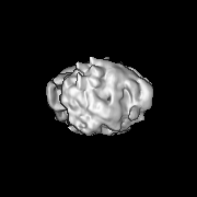
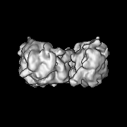
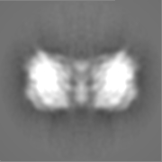
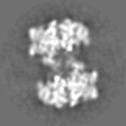
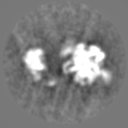
 Map data Map data | Reconstruction of 60S-Sdo1p complex. | |||||||||
 Sample Sample |
| |||||||||
 Keywords Keywords | Ribosome biogenesis / SBDS / SDS / Sdo1 / cryo-electron microscopy (cryo-EM) | |||||||||
| Biological species |  | |||||||||
| Method | single particle reconstruction / cryo EM / Resolution: 14.0 Å | |||||||||
 Authors Authors | Ma CY / Yan KG / Tan D / Li NN / Zhang YX / Yuan Y / Li ZF / Dong MQ / Lei JL / Gao N | |||||||||
 Citation Citation | Journal: Protein Cell / Year: 2016 Title: Structural dynamics of the yeast Shwachman-Diamond syndrome protein (Sdo1) on the ribosome and its implication in the 60S subunit maturation. Authors: Chengying Ma / Kaige Yan / Dan Tan / Ningning Li / Yixiao Zhang / Yi Yuan / Zhifei Li / Meng-Qiu Dong / Jianlin Lei / Ning Gao /  Abstract: The human Shwachman-Diamond syndrome (SDS) is an autosomal recessive disease caused by mutations in a highly conserved ribosome assembly factor SBDS. The functional role of SBDS is to cooperate with ...The human Shwachman-Diamond syndrome (SDS) is an autosomal recessive disease caused by mutations in a highly conserved ribosome assembly factor SBDS. The functional role of SBDS is to cooperate with another assembly factor, elongation factor 1-like (Efl1), to promote the release of eukaryotic initiation factor 6 (eIF6) from the late-stage cytoplasmic 60S precursors. In the present work, we characterized, both biochemically and structurally, the interaction between the 60S subunit and SBDS protein (Sdo1p) from yeast. Our data show that Sdo1p interacts tightly with the mature 60S subunit in vitro through its domain I and II, and is capable of bridging two 60S subunits to form a stable 2:2 dimer. Structural analysis indicates that Sdo1p bind to the ribosomal P-site, in the proximity of uL16 and uL5, and with direct contact to H69 and H38. The dynamic nature of Sdo1p on the 60S subunit, together with its strategic binding position, suggests a surveillance role of Sdo1p in monitoring the conformational maturation of the ribosomal P-site. Altogether, our data support a conformational signal-relay cascade during late-stage 60S maturation, involving uL16, Sdo1p, and Efl1p, which interrogates the functional P-site to control the departure of the anti-association factor eIF6. | |||||||||
| History |
|
- Structure visualization
| Movie |
 Movie viewer Movie viewer |
|---|---|
| Structure viewer | EM map: SurfViewMolmilJmol/JSmol |
| Supplemental images |


- Downloads & links
-EMDB archive
| Map data | emd_3280.map.gz | 16.1 MB | EMDB map data format | |
|---|---|---|---|---|
| Header (meta data) | emd-3280-v30.xmlemd-3280.xml | 8.7 KB 8.7 KB | Display Display | EMDB header |
| FSC (resolution estimation) | emd_3280_fsc.xml | 6.3 KB | Display | FSC data file |
| Images | 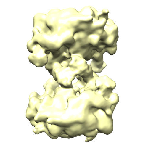 EMD-3280_60S-Sdo1p.jpg EMD-3280_60S-Sdo1p.jpg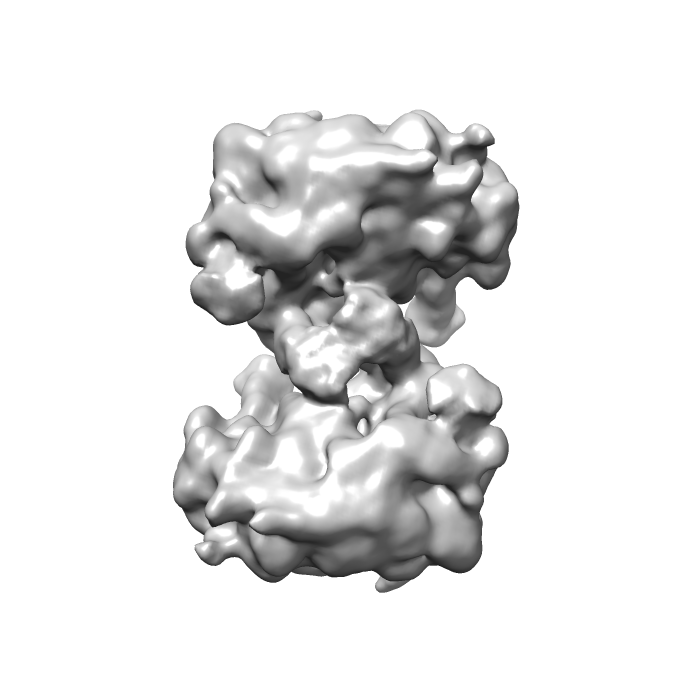 emd_3280.png emd_3280.png | 115.8 KB 104.6 KB | ||
| Archive directory |  http://ftp.pdbj.org/pub/emdb/structures/EMD-3280ftp://ftp.pdbj.org/pub/emdb/structures/EMD-3280 http://ftp.pdbj.org/pub/emdb/structures/EMD-3280ftp://ftp.pdbj.org/pub/emdb/structures/EMD-3280 | HTTPS FTP |
-Validation report
| Summary document | emd_3280_validation.pdf.gz | 257.5 KB | Display | EMDB validaton report |
|---|---|---|---|---|
| Full document | emd_3280_full_validation.pdf.gz | 256.6 KB | Display | |
| Data in XML | emd_3280_validation.xml.gz | 9.3 KB | Display | |
| Arichive directory | https://ftp.pdbj.org/pub/emdb/validation_reports/EMD-3280ftp://ftp.pdbj.org/pub/emdb/validation_reports/EMD-3280 | HTTPS FTP |
-Related structure data
| Similar structure data |
|---|
-Links
| EMDB pages | EMDB (EBI/PDBe) / EMDataResource |
|---|---|
| Related items in Molecule of the Month |

-Map
| File | Download / File: emd_3280.map.gz / Format: CCP4 / Size: 21.7 MB / Type: IMAGE STORED AS FLOATING POINT NUMBER (4 BYTES) | ||||||||||||||||||||||||||||||||||||||||||||||||||||||||||||
|---|---|---|---|---|---|---|---|---|---|---|---|---|---|---|---|---|---|---|---|---|---|---|---|---|---|---|---|---|---|---|---|---|---|---|---|---|---|---|---|---|---|---|---|---|---|---|---|---|---|---|---|---|---|---|---|---|---|---|---|---|---|
| Annotation | Reconstruction of 60S-Sdo1p complex. | ||||||||||||||||||||||||||||||||||||||||||||||||||||||||||||
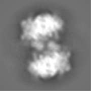
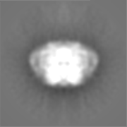
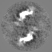
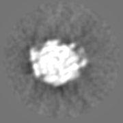
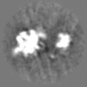
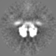
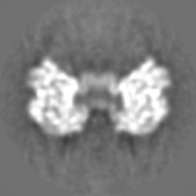
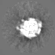
| Projections & slices | Image control
Images are generated by Spider. | ||||||||||||||||||||||||||||||||||||||||||||||||||||||||||||
| Voxel size | X=Y=Z: 2.76 Å | ||||||||||||||||||||||||||||||||||||||||||||||||||||||||||||
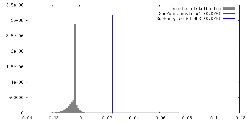
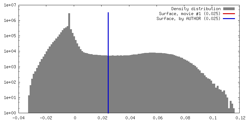
| Density |
| ||||||||||||||||||||||||||||||||||||||||||||||||||||||||||||
| Symmetry | Space group: 1 | ||||||||||||||||||||||||||||||||||||||||||||||||||||||||||||
| Details | EMDB XML:
CCP4 map header:
| ||||||||||||||||||||||||||||||||||||||||||||||||||||||||||||
 Z (Sec.)
 Y (Row.)
 X (Col.)


-Supplemental data
- Sample components
-Entire : The dimeric structure of the yeast 60S ribosomal subunits bound w...
| Entire | Name: The dimeric structure of the yeast 60S ribosomal subunits bound with Sdo1p |
|---|---|
| Components |
|
-Supramolecule #1000: The dimeric structure of the yeast 60S ribosomal subunits bound w...
| Supramolecule | Name: The dimeric structure of the yeast 60S ribosomal subunits bound with Sdo1p type: sample / ID: 1000 / Oligomeric state: two Sdo1p binding to two 60S subunit / Number unique components: 2 |
|---|
-Supramolecule #1: Sdo1p bound 60S subunit
| Supramolecule | Name: Sdo1p bound 60S subunit / type: complex / ID: 1 / Recombinant expression: No / Ribosome-details: ribosome-eukaryote: LSU 60S |
|---|---|
| Source (natural) | Organism: |
-Macromolecule #1: Sdo1p
| Macromolecule | Name: Sdo1p / type: protein_or_peptide / ID: 1 / Recombinant expression: Yes |
|---|---|
| Source (natural) | Organism: |
-Experimental details
-Structure determination
| Method | cryo EM |
|---|---|
 Processing Processing | single particle reconstruction |
| Aggregation state | particle |
-Sample preparation
| Vitrification | Cryogen name: ETHANE / Instrument: FEI VITROBOT MARK IV |
|---|
- Electron microscopy
| Microscope | FEI TECNAI F20 |
|---|---|
| Date | Dec 8, 2011 |
| Image recording | Category: CCD / Film or detector model: FEI EAGLE (4k x 4k) |
| Electron beam | Acceleration voltage: 200 kV / Electron source:  FIELD EMISSION GUN FIELD EMISSION GUN |
| Electron optics | Illumination mode: FLOOD BEAM / Imaging mode: BRIGHT FIELD / Cs: 2.0 mm |
| Sample stage | Specimen holder: Gatan CT3500 / Specimen holder model: OTHER |
| Experimental equipment |  Model: Tecnai F20 / Image courtesy: FEI Company |
-Image processing
| CTF correction | Details: ctfindIII |
|---|---|
| Final reconstruction | Applied symmetry - Point group: C2 (2 fold cyclic) / Resolution.type: BY AUTHOR / Resolution: 14.0 Å / Resolution method: OTHER / Software - Name: Spider, relion / Number images used: 13219 |
| FSC plot (resolution estimation) |  |
-Atomic model buiding 1
| Initial model | PDB ID:  3o58 |
|---|---|
| Software | Name: Chimera |
| Refinement | Space: REAL / Protocol: RIGID BODY FIT |
