[English] 日本語
Yorodumi
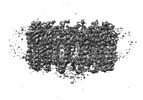
- EMDB-32062: Rba sphaeroides PufX-KO RC-LH1 -

+
Open data


ID or keywords:

Loading...

-
Basic information

Entry
Database: EMDB / ID: EMD-32062
TitleRba sphaeroides PufX-KO RC-LH1
Map data
Sample
  • Complex: Rhodobacter sphaeroides PufX-KO RC-LH1
    • Protein or peptide: Light-harvesting protein B-875 alpha chain
    • Protein or peptide: Light-harvesting protein B-875 beta chain
    • Protein or peptide: Reaction center protein L chain
    • Protein or peptide: Reaction center protein M chain
    • Protein or peptide: Reaction center protein H chain
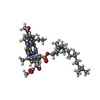
  • Ligand: BACTERIOCHLOROPHYLL A
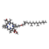
  • Ligand: BACTERIOPHEOPHYTIN A
  • Ligand: UBIQUINONE-10
  • Ligand: FE (II) ION
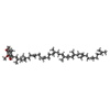
  • Ligand: SPEROIDENONE
KeywordsPhotosystem / Complex / PHOTOSYNTHESIS / Mutant
Function / homology
Function and homology information


organelle inner membrane / plasma membrane-derived chromatophore membrane / plasma membrane light-harvesting complex / bacteriochlorophyll binding / photosynthesis, light reaction / photosynthetic electron transport in photosystem II / electron transporter, transferring electrons within the cyclic electron transport pathway of photosynthesis activity / metal ion binding / plasma membrane
Similarity search - Function
Antenna complex, beta subunit, conserved site / Antenna complexes beta subunits signature. / Antenna complex, alpha subunit / Antenna complex, alpha subunit conserved site / Antenna complexes alpha subunits signature. / Antenna complex, alpha/beta subunit / Light-harvesting protein B beta chain / Antenna complex, beta domain superfamily / Antenna complex alpha/beta subunit / Light-harvesting complex ...Antenna complex, beta subunit, conserved site / Antenna complexes beta subunits signature. / Antenna complex, alpha subunit / Antenna complex, alpha subunit conserved site / Antenna complexes alpha subunits signature. / Antenna complex, alpha/beta subunit / Light-harvesting protein B beta chain / Antenna complex, beta domain superfamily / Antenna complex alpha/beta subunit / Light-harvesting complex / Photosynthetic reaction centre, H subunit / Bacterial photosynthetic reaction centre, H-chain, C-terminal / Photosynthetic reaction centre, M subunit / Photosynthetic reaction centre, H subunit, N-terminal / Photosynthetic reaction centre, H subunit, N-terminal domain superfamily / Photosynthetic reaction centre, H-chain N-terminal region / PRC-barrel domain / PRC-barrel domain / Photosynthetic reaction centre, L subunit / PRC-barrel-like superfamily / Photosynthetic reaction centre, L/M / Photosystem II protein D1/D2 superfamily / Photosynthetic reaction centre protein / Photosynthetic reaction center proteins signature.
Similarity search - Domain/homology
Reaction center protein H chain / Light-harvesting protein B-875 beta chain / Light-harvesting protein B-875 alpha chain / Reaction center protein L chain / Reaction center protein M chain
Similarity search - Component
Biological speciesRhodobacter sphaeroides 2.4.1 (bacteria) / Cereibacter sphaeroides 2.4.1 (bacteria)
Methodsingle particle reconstruction / cryo EM / Resolution: 4.2 Å
AuthorsBracun L / Yamagata A
Funding support United Kingdom, 1 items
OrganizationGrant numberCountry
Royal Society United Kingdom
CitationJournal: Nat Commun / Year: 2022
Title: Structural basis for the assembly and quinone transport mechanisms of the dimeric photosynthetic RC-LH1 supercomplex.
Authors: Peng Cao / Laura Bracun / Atsushi Yamagata / Bern M Christianson / Tatsuki Negami / Baohua Zou / Tohru Terada / Daniel P Canniffe / Mikako Shirouzu / Mei Li / Lu-Ning Liu /
Abstract: The reaction center (RC) and light-harvesting complex 1 (LH1) form a RC-LH1 core supercomplex that is vital for the primary reactions of photosynthesis in purple phototrophic bacteria. Some species ...The reaction center (RC) and light-harvesting complex 1 (LH1) form a RC-LH1 core supercomplex that is vital for the primary reactions of photosynthesis in purple phototrophic bacteria. Some species possess the dimeric RC-LH1 complex with a transmembrane polypeptide PufX, representing the largest photosynthetic complex in anoxygenic phototrophs. However, the details of the architecture and assembly mechanism of the RC-LH1 dimer are unclear. Here we report seven cryo-electron microscopy (cryo-EM) structures of RC-LH1 supercomplexes from Rhodobacter sphaeroides. Our structures reveal that two PufX polypeptides are positioned in the center of the S-shaped RC-LH1 dimer, interlocking association between the components and mediating RC-LH1 dimerization. Moreover, we identify another transmembrane peptide, designated PufY, which is located between the RC and LH1 subunits near the LH1 opening. PufY binds a quinone molecule and prevents LH1 subunits from completely encircling the RC, creating a channel for quinone/quinol exchange. Genetic mutagenesis, cryo-EM structures, and computational simulations provide a mechanistic understanding of the assembly and electron transport pathways of the RC-LH1 dimer and elucidate the roles of individual components in ensuring the structural and functional integrity of the photosynthetic supercomplex.
History
DepositionOct 15, 2021-
Header (metadata) releaseMay 4, 2022-
Map releaseMay 4, 2022-
UpdateJun 19, 2024-
Current statusJun 19, 2024Processing site: PDBj / Status: Released

-
Structure visualization

Supplemental images

Downloads & links

-
Map

FileDownload / File: emd_32062.map.gz / Format: CCP4 / Size: 59.6 MB / Type: IMAGE STORED AS FLOATING POINT NUMBER (4 BYTES)
Projections & slices

Image control

Size
Brightness
Contrast
Others
AxesZ (Sec.)Y (Row.)X (Col.)
1.06 Å/pix.
x 250 pix.
= 265. Å
1.06 Å/pix.
x 250 pix.
= 265. Å
1.06 Å/pix.
x 250 pix.
= 265. Å

Surface

Projections

Slices (1/3)

Slices (1/2)

Slices (2/3)

Images are generated by Spider.

Voxel sizeX=Y=Z: 1.06 Å
Density
Contour LevelBy AUTHOR: 0.0254
Minimum - Maximum-0.07471693 - 0.10783177
Average (Standard dev.)0.00004759105 (±0.006187219)
SymmetrySpace group: 1
Details

EMDB XML:

Map geometry
Axis orderXYZ
Origin000
Dimensions250250250
Spacing250250250
CellA=B=C: 265.0 Å
α=β=γ: 90.0 °

-
Supplemental data

-
Additional map: #1

Fileemd_32062_additional_1.map
Projections & Slices
AxesZYX

Projections

Slices (1/2)
Density Histograms

-
Sample components

+
Entire : Rhodobacter sphaeroides PufX-KO RC-LH1

EntireName: Rhodobacter sphaeroides PufX-KO RC-LH1
Components
  • Complex: Rhodobacter sphaeroides PufX-KO RC-LH1
    • Protein or peptide: Light-harvesting protein B-875 alpha chain
    • Protein or peptide: Light-harvesting protein B-875 beta chain
    • Protein or peptide: Reaction center protein L chain
    • Protein or peptide: Reaction center protein M chain
    • Protein or peptide: Reaction center protein H chain
  • Ligand: BACTERIOCHLOROPHYLL A
  • Ligand: BACTERIOPHEOPHYTIN A
  • Ligand: UBIQUINONE-10
  • Ligand: FE (II) ION
  • Ligand: SPEROIDENONE

+
Supramolecule #1: Rhodobacter sphaeroides PufX-KO RC-LH1

SupramoleculeName: Rhodobacter sphaeroides PufX-KO RC-LH1 / type: complex / ID: 1 / Parent: 0 / Macromolecule list: #1-#5
Source (natural)Organism: Rhodobacter sphaeroides 2.4.1 (bacteria) / Strain: PufX-KO

+
Macromolecule #1: Light-harvesting protein B-875 alpha chain

MacromoleculeName: Light-harvesting protein B-875 alpha chain / type: protein_or_peptide / ID: 1 / Number of copies: 17 / Enantiomer: LEVO
Source (natural)Organism: Cereibacter sphaeroides 2.4.1 (bacteria) / Strain: 2.4.1.
Molecular weightTheoretical: 6.816169 KDa
SequenceString:
MSKFYKIWMI FDPRRVFVAQ GVFLFLLAVM IHLILLSTPS YNWLEISAAK YNRVAVAE

UniProtKB: Light-harvesting protein B-875 alpha chain

+
Macromolecule #2: Light-harvesting protein B-875 beta chain

MacromoleculeName: Light-harvesting protein B-875 beta chain / type: protein_or_peptide / ID: 2 / Number of copies: 17 / Enantiomer: LEVO
Source (natural)Organism: Cereibacter sphaeroides 2.4.1 (bacteria) / Strain: 2.4.1.
Molecular weightTheoretical: 5.592361 KDa
SequenceString:
MADKSDLGYT GLTDEQAQEL HSVYMSGLWL FSAVAIVAHL AVYIWRPWF

UniProtKB: Light-harvesting protein B-875 beta chain

+
Macromolecule #3: Reaction center protein L chain

MacromoleculeName: Reaction center protein L chain / type: protein_or_peptide / ID: 3 / Number of copies: 1 / Enantiomer: LEVO
Source (natural)Organism: Cereibacter sphaeroides 2.4.1 (bacteria) / Strain: 2.4.1.
Molecular weightTheoretical: 31.477584 KDa
SequenceString: MALLSFERKY RVPGGTLVGG NLFDFWVGPF YVGFFGVATF FFAALGIILI AWSAVLQGTW NPQLISVYPP ALEYGLGGAP LAKGGLWQI ITICATGAFV SWALREVEIC RKLGIGYHIP FAFAFAILAY LTLVLFRPVM MGAWGYAFPY GIWTHLDWVS N TGYTYGNF ...String:
MALLSFERKY RVPGGTLVGG NLFDFWVGPF YVGFFGVATF FFAALGIILI AWSAVLQGTW NPQLISVYPP ALEYGLGGAP LAKGGLWQI ITICATGAFV SWALREVEIC RKLGIGYHIP FAFAFAILAY LTLVLFRPVM MGAWGYAFPY GIWTHLDWVS N TGYTYGNF HYNPAHMIAI SFFFTNALAL ALHGALVLSA ANPEKGKEMR TPDHEDTFFR DLVGYSIGTL GIHRLGLLLS LS AVFFSAL CMIITGTIWF DQWVDWWQWW VKLPWWANIP GGING

UniProtKB: Reaction center protein L chain

+
Macromolecule #4: Reaction center protein M chain

MacromoleculeName: Reaction center protein M chain / type: protein_or_peptide / ID: 4 / Number of copies: 1 / Enantiomer: LEVO
Source (natural)Organism: Cereibacter sphaeroides 2.4.1 (bacteria) / Strain: 2.4.1.
Molecular weightTheoretical: 34.529738 KDa
SequenceString: MAEYQNIFSQ VQVRGPADLG MTEDVNLANR SGVGPFSTLL GWFGNAQLGP IYLGSLGVLS LFSGLMWFFT IGIWFWYQAG WNPAVFLRD LFFFSLEPPA PEYGLSFAAP LKEGGLWLIA SFFMFVAVWS WWGRTYLRAQ ALGMGKHTAW AFLSAIWLWM V LGFIRPIL ...String:
MAEYQNIFSQ VQVRGPADLG MTEDVNLANR SGVGPFSTLL GWFGNAQLGP IYLGSLGVLS LFSGLMWFFT IGIWFWYQAG WNPAVFLRD LFFFSLEPPA PEYGLSFAAP LKEGGLWLIA SFFMFVAVWS WWGRTYLRAQ ALGMGKHTAW AFLSAIWLWM V LGFIRPIL MGSWSEAVPY GIFSHLDWTN NFSLVHGNLF YNPFHGLSIA FLYGSALLFA MHGATILAVS RFGGERELEQ IA DRGTAAE RAALFWRWTM GFNATMEGIH RWAIWMAVLV TLTGGIGILL SGTVVDNWYV WGQNHGMAPL N

UniProtKB: Reaction center protein M chain

+
Macromolecule #5: Reaction center protein H chain

MacromoleculeName: Reaction center protein H chain / type: protein_or_peptide / ID: 5 / Number of copies: 1 / Enantiomer: LEVO
Source (natural)Organism: Cereibacter sphaeroides 2.4.1 (bacteria) / Strain: 2.4.1.
Molecular weightTheoretical: 28.066322 KDa
SequenceString: MVGVTAFGNF DLASLAIYSF WIFLAGLIYY LQTENMREGY PLENEDGTPA ANQGPFPLPK PKTFILPHGR GTLTVPGPES EDRPIALAR TAVSEGFPHA PTGDPMKDGV GPASWVARRD LPELDGHGHN KIKPMKAAAG FHVSAGKNPI GLPVRGCDLE I AGKVVDIW ...String:
MVGVTAFGNF DLASLAIYSF WIFLAGLIYY LQTENMREGY PLENEDGTPA ANQGPFPLPK PKTFILPHGR GTLTVPGPES EDRPIALAR TAVSEGFPHA PTGDPMKDGV GPASWVARRD LPELDGHGHN KIKPMKAAAG FHVSAGKNPI GLPVRGCDLE I AGKVVDIW VDIPEQMARF LEVELKDGST RLLPMQMVKV QSNRVHVNAL SSDLFAGIPT IKSPTEVTLL EEDKICGYVA GG LMYAAPK RKSVVAAMLA EYA

UniProtKB: Reaction center protein H chain

+
Macromolecule #6: BACTERIOCHLOROPHYLL A

MacromoleculeName: BACTERIOCHLOROPHYLL A / type: ligand / ID: 6 / Number of copies: 38 / Formula: BCL
Molecular weightTheoretical: 911.504 Da
Chemical component information

ChemComp-BCL:
BACTERIOCHLOROPHYLL A

+
Macromolecule #7: BACTERIOPHEOPHYTIN A

MacromoleculeName: BACTERIOPHEOPHYTIN A / type: ligand / ID: 7 / Number of copies: 2 / Formula: BPH
Molecular weightTheoretical: 889.215 Da
Chemical component information

ChemComp-BPH:
BACTERIOPHEOPHYTIN A

+
Macromolecule #8: UBIQUINONE-10

MacromoleculeName: UBIQUINONE-10 / type: ligand / ID: 8 / Number of copies: 2 / Formula: U10
Molecular weightTheoretical: 863.343 Da
Chemical component information

ChemComp-U10:
UBIQUINONE-10

+
Macromolecule #9: FE (II) ION

MacromoleculeName: FE (II) ION / type: ligand / ID: 9 / Number of copies: 1 / Formula: FE2
Molecular weightTheoretical: 55.845 Da

+
Macromolecule #10: SPEROIDENONE

MacromoleculeName: SPEROIDENONE / type: ligand / ID: 10 / Number of copies: 1 / Formula: SPN
Molecular weightTheoretical: 594.993 Da
Chemical component information

ChemComp-SPN:
SPEROIDENONE

-
Experimental details

-
Structure determination

Methodcryo EM
Processingsingle particle reconstruction
Aggregation stateparticle

-
Sample preparation

BufferpH: 8
GridModel: Quantifoil R1.2/1.3 / Support film - Material: CARBON
VitrificationCryogen name: ETHANE

-
Electron microscopy

MicroscopeFEI TITAN KRIOS
Image recordingFilm or detector model: GATAN K3 BIOQUANTUM (6k x 4k) / Average electron dose: 46.549 e/Å2
Electron beamAcceleration voltage: 300 kV / Electron source: FIELD EMISSION GUN
Electron opticsIllumination mode: FLOOD BEAM / Imaging mode: BRIGHT FIELD
Experimental equipment
Model: Titan Krios / Image courtesy: FEI Company

+
Image processing

Startup modelType of model: NONE
Final reconstructionResolution.type: BY AUTHOR / Resolution: 4.2 Å / Resolution method: FSC 0.143 CUT-OFF / Number images used: 66058
Initial angle assignmentType: MAXIMUM LIKELIHOOD / Details: Relion
Final angle assignmentType: MAXIMUM LIKELIHOOD / Details: Relion
FSC plot (resolution estimation)

+
About Yorodumi

-
News

-
Feb 9, 2022. New format data for meta-information of EMDB entries

New format data for meta-information of EMDB entries

  • Version 3 of the EMDB header file is now the official format.
  • The previous official version 1.9 will be removed from the archive.

Related info.:EMDB header

External links:wwPDB to switch to version 3 of the EMDB data model

-
Aug 12, 2020. Covid-19 info

Covid-19 info

URL: https://pdbj.org/emnavi/covid19.php

New page: Covid-19 featured information page in EM Navigator.

Related info.:Covid-19 info / Mar 5, 2020. Novel coronavirus structure data

+
Mar 5, 2020. Novel coronavirus structure data

Novel coronavirus structure data

Related info.:Yorodumi Speices / Aug 12, 2020. Covid-19 info

External links:COVID-19 featured content - PDBj / Molecule of the Month (242):Coronavirus Proteases

+
Jan 31, 2019. EMDB accession codes are about to change! (news from PDBe EMDB page)

EMDB accession codes are about to change! (news from PDBe EMDB page)

  • The allocation of 4 digits for EMDB accession codes will soon come to an end. Whilst these codes will remain in use, new EMDB accession codes will include an additional digit and will expand incrementally as the available range of codes is exhausted. The current 4-digit format prefixed with “EMD-” (i.e. EMD-XXXX) will advance to a 5-digit format (i.e. EMD-XXXXX), and so on. It is currently estimated that the 4-digit codes will be depleted around Spring 2019, at which point the 5-digit format will come into force.
  • The EM Navigator/Yorodumi systems omit the EMD- prefix.

Related info.:Q: What is EMD? / ID/Accession-code notation in Yorodumi/EM Navigator

External links:EMDB Accession Codes are Changing Soon! / Contact to PDBj

+
Jul 12, 2017. Major update of PDB

Major update of PDB

  • wwPDB released updated PDB data conforming to the new PDBx/mmCIF dictionary.
  • This is a major update changing the version number from 4 to 5, and with Remediation, in which all the entries are updated.
  • In this update, many items about electron microscopy experimental information are reorganized (e.g. em_software).
  • Now, EM Navigator and Yorodumi are based on the updated data.

External links:wwPDB Remediation / Enriched Model Files Conforming to OneDep Data Standards Now Available in the PDB FTP Archive

-
Yorodumi

Thousand views of thousand structures

  • Yorodumi is a browser for structure data from EMDB, PDB, SASBDB, etc.
  • This page is also the successor to EM Navigator detail page, and also detail information page/front-end page for Omokage search.
  • The word "yorodu" (or yorozu) is an old Japanese word meaning "ten thousand". "mi" (miru) is to see.

Related info.:EMDB / PDB / SASBDB / Comparison of 3 databanks / Yorodumi Search / Aug 31, 2016. New EM Navigator & Yorodumi / Yorodumi Papers / Jmol/JSmol / Function and homology information / Changes in new EM Navigator and Yorodumi

Read more