+ Open data
- Basic information
| Entry | Database: EMDB / ID: EMD-31370 | |||||||||
|---|---|---|---|---|---|---|---|---|---|---|
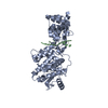
| Title | Structure of ring-stacked Mumps virus nucleocapsid filament | |||||||||
 Map data Map data | ring-stacked MuV nucleocapsid | |||||||||
 Sample Sample |
| |||||||||
| Function / homology |  Function and homology information Function and homology informationnegative stranded viral RNA transcription / negative stranded viral RNA replication / helical viral capsid / viral nucleocapsid / host cell cytoplasm / ribonucleoprotein complex / structural molecule activity / RNA binding Similarity search - Function | |||||||||
| Biological species |  | |||||||||
| Method | single particle reconstruction / cryo EM / Resolution: 3.7 Å | |||||||||
 Authors Authors | Shen Q / Shan H / Zhang N / Qin Y | |||||||||
| Funding support |  China, 1 items China, 1 items
| |||||||||
 Citation Citation |  Journal: Commun Biol / Year: 2021 Journal: Commun Biol / Year: 2021Title: Structural plasticity of mumps virus nucleocapsids with cryo-EM structures. Authors: Hong Shan / Xin Su / Tianhao Li / Yuqi Qin / Na Zhang / Liuyan Yang / Linsha Ma / Yun Bai / Lei Qi / Yunhui Liu / Qing-Tao Shen /  Abstract: Mumps virus (MuV) is a highly contagious human pathogen and frequently causes worldwide outbreaks despite available vaccines. Similar to other mononegaviruses such as Ebola and rabies, MuV uses a ...Mumps virus (MuV) is a highly contagious human pathogen and frequently causes worldwide outbreaks despite available vaccines. Similar to other mononegaviruses such as Ebola and rabies, MuV uses a single-stranded negative-sense RNA as its genome, which is enwrapped by viral nucleoproteins into the helical nucleocapsid. The nucleocapsid acts as a scaffold for genome condensation and as a template for RNA replication and transcription. Conformational changes in the MuV nucleocapsid are required to switch between different activities, but the underlying mechanism remains elusive due to the absence of high-resolution structures. Here, we report two MuV nucleoprotein-RNA rings with 13 and 14 protomers, one stacked-ring filament and two nucleocapsids with distinct helical pitches, in dense and hyperdense states, at near-atomic resolutions using cryo-electron microscopy. Structural analysis of these in vitro assemblies indicates that the C-terminal tail of MuV nucleoprotein likely regulates the assembly of helical nucleocapsids, and the C-terminal arm may be relevant for the transition between the dense and hyperdense states of helical nucleocapsids. Our results provide the molecular mechanism for structural plasticity among different MuV nucleocapsids and create a possible link between structural plasticity and genome condensation. | |||||||||
| History |
|
- Structure visualization
| Movie |
 Movie viewer Movie viewer |
|---|---|
| Structure viewer | EM map:  SurfView SurfView Molmil Molmil Jmol/JSmol Jmol/JSmol |
| Supplemental images |
- Downloads & links
-EMDB archive
| Map data |  emd_31370.map.gz emd_31370.map.gz | 159.5 MB |  EMDB map data format EMDB map data format | |
|---|---|---|---|---|
| Header (meta data) |  emd-31370-v30.xml emd-31370-v30.xml emd-31370.xml emd-31370.xml | 9.3 KB 9.3 KB | Display Display |  EMDB header EMDB header |
| FSC (resolution estimation) |  emd_31370_fsc.xml emd_31370_fsc.xml | 17.8 KB | Display |  FSC data file FSC data file |
| Images |  emd_31370.png emd_31370.png | 252 KB | ||
| Archive directory |  http://ftp.pdbj.org/pub/emdb/structures/EMD-31370 http://ftp.pdbj.org/pub/emdb/structures/EMD-31370 ftp://ftp.pdbj.org/pub/emdb/structures/EMD-31370 ftp://ftp.pdbj.org/pub/emdb/structures/EMD-31370 | HTTPS FTP |
-Validation report
| Summary document |  emd_31370_validation.pdf.gz emd_31370_validation.pdf.gz | 407.7 KB | Display |  EMDB validaton report EMDB validaton report |
|---|---|---|---|---|
| Full document |  emd_31370_full_validation.pdf.gz emd_31370_full_validation.pdf.gz | 407.2 KB | Display | |
| Data in XML |  emd_31370_validation.xml.gz emd_31370_validation.xml.gz | 15.5 KB | Display | |
| Data in CIF |  emd_31370_validation.cif.gz emd_31370_validation.cif.gz | 21.2 KB | Display | |
| Arichive directory |  https://ftp.pdbj.org/pub/emdb/validation_reports/EMD-31370 https://ftp.pdbj.org/pub/emdb/validation_reports/EMD-31370 ftp://ftp.pdbj.org/pub/emdb/validation_reports/EMD-31370 ftp://ftp.pdbj.org/pub/emdb/validation_reports/EMD-31370 | HTTPS FTP |
-Related structure data
- Links
Links
| EMDB pages |  EMDB (EBI/PDBe) / EMDB (EBI/PDBe) /  EMDataResource EMDataResource |
|---|---|
| Related items in Molecule of the Month |
- Map
Map
| File |  Download / File: emd_31370.map.gz / Format: CCP4 / Size: 476.8 MB / Type: IMAGE STORED AS FLOATING POINT NUMBER (4 BYTES) Download / File: emd_31370.map.gz / Format: CCP4 / Size: 476.8 MB / Type: IMAGE STORED AS FLOATING POINT NUMBER (4 BYTES) | ||||||||||||||||||||||||||||||||||||||||||||||||||||||||||||||||||||
|---|---|---|---|---|---|---|---|---|---|---|---|---|---|---|---|---|---|---|---|---|---|---|---|---|---|---|---|---|---|---|---|---|---|---|---|---|---|---|---|---|---|---|---|---|---|---|---|---|---|---|---|---|---|---|---|---|---|---|---|---|---|---|---|---|---|---|---|---|---|
| Annotation | ring-stacked MuV nucleocapsid | ||||||||||||||||||||||||||||||||||||||||||||||||||||||||||||||||||||
| Projections & slices | Image control
Images are generated by Spider. | ||||||||||||||||||||||||||||||||||||||||||||||||||||||||||||||||||||
| Voxel size | X=Y=Z: 0.65 Å | ||||||||||||||||||||||||||||||||||||||||||||||||||||||||||||||||||||
| Density |
| ||||||||||||||||||||||||||||||||||||||||||||||||||||||||||||||||||||
| Symmetry | Space group: 1 | ||||||||||||||||||||||||||||||||||||||||||||||||||||||||||||||||||||
| Details | EMDB XML:
CCP4 map header:
| ||||||||||||||||||||||||||||||||||||||||||||||||||||||||||||||||||||
-Supplemental data
- Sample components
-Entire : Mumps virus nucleocapsid
| Entire | Name: Mumps virus nucleocapsid |
|---|---|
| Components |
|
-Supramolecule #1: Mumps virus nucleocapsid
| Supramolecule | Name: Mumps virus nucleocapsid / type: complex / ID: 1 / Parent: 0 / Macromolecule list: all |
|---|---|
| Source (natural) | Organism:  |
| Recombinant expression | Organism:  |
-Macromolecule #1: mumps virus N protein
| Macromolecule | Name: mumps virus N protein / type: protein_or_peptide / ID: 1 / Enantiomer: LEVO |
|---|---|
| Source (natural) | Organism:  |
| Sequence | String: MSSVLKAFER FTIEQELQDR GEEGSIPPET LKSAVKVFVI NTPNPTTRYQ MLNFCLRIIC SQNARASHR VGALITLFSL PSAGMQNHIR LADRSPEAQI ERCEIDGFEP GTYRLIPNAR A NLTANEIA AYALLADDLP PTINNGTPYV HADVEGQPCD EIEQFLDRCY ...String: MSSVLKAFER FTIEQELQDR GEEGSIPPET LKSAVKVFVI NTPNPTTRYQ MLNFCLRIIC SQNARASHR VGALITLFSL PSAGMQNHIR LADRSPEAQI ERCEIDGFEP GTYRLIPNAR A NLTANEIA AYALLADDLP PTINNGTPYV HADVEGQPCD EIEQFLDRCY SVLIQAWVMV CK CMTAYDQ PAGSADRRFA KYQQQGRLEA RYMLQPEAQR LIQTAIRKSL VVRQYLTFEL QLA RRQGLL SNRYYAMVGD IGKYIENSGL TAFFLTLKYA LGTKWSPLSL AAFTGELTKL RSLM MLYRG LGEQARYLAL LEAPQIMDFA PGGYPLIFSY AMGVGTVLDV QMRNYTYARP FLNGY YFQI GVETARRQQG TVDNRVADDL GLTPEQRTEV TQLVDRLARG RGAGIPGGPV NPFVPP VQQ QQPAAVYEDI PALEESDDDG DEDGGAGFQN GVQLPAVRQG GQTDFRAQPL QDPIQAQ LF MPLYPQVSNM PNNQNHQINR IGGLEHQDLL RYNENGDSQQ DARGEHVNTF PNNPNQNA Q LQVGDWDE |
-Experimental details
-Structure determination
| Method | cryo EM |
|---|---|
 Processing Processing | single particle reconstruction |
| Aggregation state | filament |
- Sample preparation
| Concentration | 1 mg/mL | |||||||||
|---|---|---|---|---|---|---|---|---|---|---|
| Buffer | pH: 7.4 Component:
| |||||||||
| Vitrification | Cryogen name: ETHANE / Chamber humidity: 100 % / Chamber temperature: 277 K / Instrument: FEI VITROBOT MARK IV |
- Electron microscopy
| Microscope | FEI TITAN KRIOS |
|---|---|
| Image recording | Film or detector model: GATAN K2 SUMMIT (4k x 4k) / Detector mode: SUPER-RESOLUTION / Average electron dose: 40.0 e/Å2 |
| Electron beam | Acceleration voltage: 300 kV / Electron source:  FIELD EMISSION GUN FIELD EMISSION GUN |
| Electron optics | Illumination mode: FLOOD BEAM / Imaging mode: BRIGHT FIELD |
| Experimental equipment |  Model: Titan Krios / Image courtesy: FEI Company |
 Movie
Movie Controller


 Z (Sec.)
 Y (Row.)
 X (Col.)


