 Movie
Movie Controller


+ Open data

- Basic information
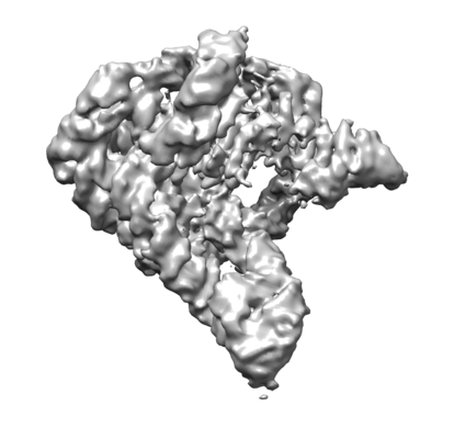
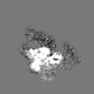
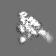
| Entry | Database: EMDB / ID: EMD-30541 | |||||||||
|---|---|---|---|---|---|---|---|---|---|---|
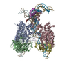
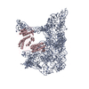
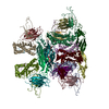
| Title | cryo-EM structure of a pre-catalytic group II intron complex | |||||||||
 Map data Map data | ||||||||||
 Sample Sample |
| |||||||||
| Function / homology |  Function and homology information Function and homology informationintron homing / mRNA processing / RNA-directed DNA polymerase / RNA-directed DNA polymerase activity / endonuclease activity / Hydrolases; Acting on ester bonds Similarity search - Function | |||||||||
| Biological species |  Lactococcus lactis subsp. cremoris (lactic acid bacteria) Lactococcus lactis subsp. cremoris (lactic acid bacteria) | |||||||||
| Method | single particle reconstruction / cryo EM / Resolution: 5.1 Å | |||||||||
 Authors Authors | Liu N / Dong XL / Hu CX / Wang J / Wang HW / Belfort M | |||||||||
| Funding support |  China, China,  United States, 2 items United States, 2 items
| |||||||||
 Citation Citation | Journal: Nucleic Acids Res / Year: 2020 Title: Exon and protein positioning in a pre-catalytic group II intron RNP primed for splicing. Authors: Nan Liu / Xiaolong Dong / Cuixia Hu / Jianwei Zeng / Jiawei Wang / Jia Wang / Hong-Wei Wang / Marlene Belfort / Abstract: Group II introns are the putative progenitors of nuclear spliceosomal introns and use the same two-step splicing pathway. In the cell, the intron RNA forms a ribonucleoprotein (RNP) complex with the ...Group II introns are the putative progenitors of nuclear spliceosomal introns and use the same two-step splicing pathway. In the cell, the intron RNA forms a ribonucleoprotein (RNP) complex with the intron-encoded protein (IEP), which is essential for splicing. Although structures of spliced group II intron RNAs and RNP complexes have been characterized, structural insights into the splicing process remain enigmatic due to lack of pre-catalytic structural models. Here, we report two cryo-EM structures of endogenously produced group II intron RNPs trapped in their pre-catalytic state. Comparison of the catalytically activated precursor RNP to its previously reported spliced counterpart allowed identification of key structural rearrangements accompanying splicing, including a remodeled active site and engagement of the exons. Importantly, altered RNA-protein interactions were observed upon splicing among the RNP complexes. Furthermore, analysis of the catalytically inert precursor RNP demonstrated the structural impact of the formation of the active site on RNP architecture. Taken together, our results not only fill a gap in understanding the structural basis of IEP-assisted group II intron splicing, but also provide parallels to evolutionarily related spliceosomal splicing. | |||||||||
| History |
|
- Structure visualization
| Movie |
Movie viewer |
|---|---|
| Structure viewer | EM map: SurfViewMolmilJmol/JSmol |
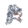
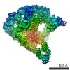
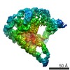
| Supplemental images |
- Downloads & links
-EMDB archive
| Map data | emd_30541.map.gz | 4.2 MB | EMDB map data format | |
|---|---|---|---|---|
| Header (meta data) | emd-30541-v30.xmlemd-30541.xml | 9.8 KB 9.8 KB | Display Display | EMDB header |
| Images |  emd_30541.png emd_30541.png | 58.2 KB | ||
| Archive directory |  http://ftp.pdbj.org/pub/emdb/structures/EMD-30541ftp://ftp.pdbj.org/pub/emdb/structures/EMD-30541 http://ftp.pdbj.org/pub/emdb/structures/EMD-30541ftp://ftp.pdbj.org/pub/emdb/structures/EMD-30541 | HTTPS FTP |
-Validation report
| Summary document | emd_30541_validation.pdf.gz | 78.2 KB | Display | EMDB validaton report |
|---|---|---|---|---|
| Full document | emd_30541_full_validation.pdf.gz | 77.3 KB | Display | |
| Data in XML | emd_30541_validation.xml.gz | 493 B | Display | |
| Arichive directory | https://ftp.pdbj.org/pub/emdb/validation_reports/EMD-30541ftp://ftp.pdbj.org/pub/emdb/validation_reports/EMD-30541 | HTTPS FTP |
-Related structure data


-Links
| EMDB pages | EMDB (EBI/PDBe) / EMDataResource |
|---|
-Map
| File | Download / File: emd_30541.map.gz / Format: CCP4 / Size: 23.8 MB / Type: IMAGE STORED AS FLOATING POINT NUMBER (4 BYTES) | ||||||||||||||||||||||||||||||||||||||||||||||||||||||||||||||||||||
|---|---|---|---|---|---|---|---|---|---|---|---|---|---|---|---|---|---|---|---|---|---|---|---|---|---|---|---|---|---|---|---|---|---|---|---|---|---|---|---|---|---|---|---|---|---|---|---|---|---|---|---|---|---|---|---|---|---|---|---|---|---|---|---|---|---|---|---|---|---|
| Projections & slices | Image control
Images are generated by Spider. | ||||||||||||||||||||||||||||||||||||||||||||||||||||||||||||||||||||
| Voxel size | X=Y=Z: 1.30654 Å | ||||||||||||||||||||||||||||||||||||||||||||||||||||||||||||||||||||
| Density |
| ||||||||||||||||||||||||||||||||||||||||||||||||||||||||||||||||||||
| Symmetry | Space group: 1 | ||||||||||||||||||||||||||||||||||||||||||||||||||||||||||||||||||||
| Details | EMDB XML:
CCP4 map header:
| ||||||||||||||||||||||||||||||||||||||||||||||||||||||||||||||||||||
 Z (Sec.)
 Y (Row.)
 X (Col.)


-Supplemental data
- Sample components
-Entire : a group II intron --LtrB complex with its reverse transcriptase
| Entire | Name: a group II intron --LtrB complex with its reverse transcriptase |
|---|---|
| Components |
|
-Supramolecule #1: a group II intron --LtrB complex with its reverse transcriptase
| Supramolecule | Name: a group II intron --LtrB complex with its reverse transcriptase type: complex / ID: 1 / Parent: 0 / Macromolecule list: all |
|---|---|
| Source (natural) | Organism: Lactococcus lactis subsp. cremoris (lactic acid bacteria) |
| Recombinant expression | Organism: Lactococcus lactis subsp. cremoris (lactic acid bacteria) Recombinant strain: IL1403 |
-Macromolecule #1: group II intron- LtrB
| Macromolecule | Name: group II intron- LtrB / type: rna / ID: 1 |
|---|---|
| Source (natural) | Organism: Lactococcus lactis subsp. cremoris (lactic acid bacteria) |
| Sequence | String: cacauccaua acgugcgccc agauagggug uuaagucaag uaguuuaagg uacuacucug uaagauaaca cagaaaacag ccaaccuaac cgaaaagcga aagcugauac gggaacagag cacgguugga aagcgaugag uuaccuaaag acaaucgggu acgacugagu ...String: cacauccaua acgugcgccc agauagggug uuaagucaag uaguuuaagg uacuacucug uaagauaaca cagaaaacag ccaaccuaac cgaaaagcga aagcugauac gggaacagag cacgguugga aagcgaugag uuaccuaaag acaaucgggu acgacugagu cgcaauguua aucagauaua agguauaagu uguguuuacu gaacgcaagu uucuaauuuc gguuaugugu cgauagagga aagugucuga aaccucuagu acaaagaaag guaaguuaug guuguggacu uaucuguuau caccacauuu guacaaucug uaggagaacc uaugggaacg aaacgaaagc gaugccgaga aucugaauuu accaagacuu aacacuaacu ggggauaccc uaaacaagaa ugccuaauag aaaggaggaa aaaggcuaua gcacuagagc uugaaaaucu ugcaagggua cggaguacuc guaguagucu gagaagggua acgcccuuua cauggcaaag ggguacaguu auuguguacu aaaauuaaaa auugauuagg gaggaaaacc ucaaaaugaa accaacaaug gcaauuuuag aaagaaucag uaaaaauuca caagaaaaua uagacgaagu uuuuacaaga cuuuaucguu aucuuuuacg uccagauauu uauuacgugg cgacgcguug ggaaauggca augauagcga aacaacguaa aacucuuguu guaugcuuuc auugucaucg ucacgugauu cauaaacaca agugaauuuu uacgaacgaa caauaacaga gccguauacu ccgagagggg uacguacggu ucccgaagag gguggugcaa accagucaca guaaugugaa caaggcggua ccucccucuu cacca |
-Experimental details
-Structure determination
| Method | cryo EM |
|---|---|
 Processing Processing | single particle reconstruction |
| Aggregation state | particle |
-Sample preparation
| Concentration | 0.1 mg/mL |
|---|---|
| Buffer | pH: 7.5 |
| Vitrification | Cryogen name: ETHANE / Chamber humidity: 100 % / Chamber temperature: 285.15 K |
- Electron microscopy
| Microscope | FEI TITAN KRIOS |
|---|---|
| Image recording | #0 - Image recording ID: 1 / #0 - Film or detector model: GATAN K2 SUMMIT (4k x 4k) / #0 - Detector mode: SUPER-RESOLUTION / #0 - Average electron dose: 50.0 e/Å2 / #1 - Image recording ID: 2 / #1 - Film or detector model: GATAN K2 SUMMIT (4k x 4k) / #1 - Average electron dose: 50.0 e/Å2 |
| Electron beam | Acceleration voltage: 300 kV / Electron source:  FIELD EMISSION GUN FIELD EMISSION GUN |
| Electron optics | Illumination mode: FLOOD BEAM / Imaging mode: BRIGHT FIELD |
| Experimental equipment |  Model: Titan Krios / Image courtesy: FEI Company |
-Image processing
| Image recording ID | 1 |
|---|---|
| Final reconstruction | Resolution.type: BY AUTHOR / Resolution: 5.1 Å / Resolution method: FSC 0.143 CUT-OFF / Number images used: 231724 |
| Initial angle assignment | Type: PROJECTION MATCHING |
| Final angle assignment | Type: MAXIMUM LIKELIHOOD |
