+ Open data
- Basic information
| Entry |  | ||||||||||||
|---|---|---|---|---|---|---|---|---|---|---|---|---|---|
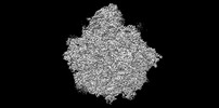
| Title | 60S subunit of the Giardia lamblia 80S ribosome | ||||||||||||
 Map data Map data | |||||||||||||
 Sample Sample |
| ||||||||||||
 Keywords Keywords | Giardia lamblia / Ribosome structure / Translation / parasite / RIBOSOME | ||||||||||||
| Function / homology |  Function and homology information Function and homology informationmaturation of LSU-rRNA / maturation of LSU-rRNA from tricistronic rRNA transcript (SSU-rRNA, 5.8S rRNA, LSU-rRNA) / ribosomal large subunit biogenesis / modification-dependent protein catabolic process / protein tag activity / large ribosomal subunit / ribosome biogenesis / 5S rRNA binding / large ribosomal subunit rRNA binding / ribosomal large subunit assembly ...maturation of LSU-rRNA / maturation of LSU-rRNA from tricistronic rRNA transcript (SSU-rRNA, 5.8S rRNA, LSU-rRNA) / ribosomal large subunit biogenesis / modification-dependent protein catabolic process / protein tag activity / large ribosomal subunit / ribosome biogenesis / 5S rRNA binding / large ribosomal subunit rRNA binding / ribosomal large subunit assembly / cytoplasmic translation / cytosolic large ribosomal subunit / negative regulation of translation / rRNA binding / ribosome / protein ubiquitination / structural constituent of ribosome / ribonucleoprotein complex / translation / mRNA binding / ubiquitin protein ligase binding / nucleolus / RNA binding / metal ion binding Similarity search - Function | ||||||||||||
| Biological species |  Giardia intestinalis assemblage A (eukaryote) Giardia intestinalis assemblage A (eukaryote) | ||||||||||||
| Method | single particle reconstruction / cryo EM / Resolution: 2.49 Å | ||||||||||||
 Authors Authors | Eiler DR / Wimberly BT / Bilodeau DY / Rissland OS / Kieft JS | ||||||||||||
| Funding support |  United States, 3 items United States, 3 items
| ||||||||||||
 Citation Citation |  Journal: Structure / Year: 2024 Journal: Structure / Year: 2024Title: The Giardia lamblia ribosome structure reveals divergence in several biological pathways and the mode of emetine function. Authors: Daniel R Eiler / Brian T Wimberly / Danielle Y Bilodeau / J Matthew Taliaferro / Philip Reigan / Olivia S Rissland / Jeffrey S Kieft /  Abstract: Giardia lamblia is a deeply branching protist and a human pathogen. Its unusual biology presents the opportunity to explore conserved and fundamental molecular mechanisms. We determined the structure ...Giardia lamblia is a deeply branching protist and a human pathogen. Its unusual biology presents the opportunity to explore conserved and fundamental molecular mechanisms. We determined the structure of the G. lamblia 80S ribosome bound to tRNA, mRNA, and the antibiotic emetine by cryo-electron microscopy, to an overall resolution of 2.49 Å. The structure reveals rapidly evolving protein and nucleotide regions, differences in the peptide exit tunnel, and likely altered ribosome quality control pathways. Examination of translation initiation factor binding sites suggests these interactions are conserved despite a divergent initiation mechanism. Highlighting the potential of G. lamblia to resolve conserved biological principles; our structure reveals the interactions of the translation inhibitor emetine with the ribosome and mRNA, thus providing insight into the mechanism of action for this widely used antibiotic. Our work defines key questions in G. lamblia and motivates future experiments to explore the diversity of eukaryotic gene regulation. | ||||||||||||
| History |
|
- Structure visualization
| Supplemental images |
|---|
- Downloads & links
-EMDB archive
| Map data |  emd_29407.map.gz emd_29407.map.gz | 321 MB |  EMDB map data format EMDB map data format | |
|---|---|---|---|---|
| Header (meta data) |  emd-29407-v30.xml emd-29407-v30.xml emd-29407.xml emd-29407.xml | 60.6 KB 60.6 KB | Display Display |  EMDB header EMDB header |
| FSC (resolution estimation) |  emd_29407_fsc.xml emd_29407_fsc.xml | 24.3 KB | Display |  FSC data file FSC data file |
| Images |  emd_29407.png emd_29407.png | 51.4 KB | ||
| Filedesc metadata |  emd-29407.cif.gz emd-29407.cif.gz | 12.5 KB | ||
| Others |  emd_29407_half_map_1.map.gz emd_29407_half_map_1.map.gz emd_29407_half_map_2.map.gz emd_29407_half_map_2.map.gz | 1.2 GB 1.2 GB | ||
| Archive directory |  http://ftp.pdbj.org/pub/emdb/structures/EMD-29407 http://ftp.pdbj.org/pub/emdb/structures/EMD-29407 ftp://ftp.pdbj.org/pub/emdb/structures/EMD-29407 ftp://ftp.pdbj.org/pub/emdb/structures/EMD-29407 | HTTPS FTP |
-Validation report
| Summary document |  emd_29407_validation.pdf.gz emd_29407_validation.pdf.gz | 1.1 MB | Display |  EMDB validaton report EMDB validaton report |
|---|---|---|---|---|
| Full document |  emd_29407_full_validation.pdf.gz emd_29407_full_validation.pdf.gz | 1.1 MB | Display | |
| Data in XML |  emd_29407_validation.xml.gz emd_29407_validation.xml.gz | 33 KB | Display | |
| Data in CIF |  emd_29407_validation.cif.gz emd_29407_validation.cif.gz | 42.3 KB | Display | |
| Arichive directory |  https://ftp.pdbj.org/pub/emdb/validation_reports/EMD-29407 https://ftp.pdbj.org/pub/emdb/validation_reports/EMD-29407 ftp://ftp.pdbj.org/pub/emdb/validation_reports/EMD-29407 ftp://ftp.pdbj.org/pub/emdb/validation_reports/EMD-29407 | HTTPS FTP |
-Related structure data
| Related structure data |  8fruMC  8fvyC  8g4iC  8g4sC M: atomic model generated by this map C: citing same article ( |
|---|---|
| Similar structure data | Similarity search - Function & homology  F&H Search F&H Search |
- Links
Links
| EMDB pages |  EMDB (EBI/PDBe) / EMDB (EBI/PDBe) /  EMDataResource EMDataResource |
|---|---|
| Related items in Molecule of the Month |
- Map
Map
| File |  Download / File: emd_29407.map.gz / Format: CCP4 / Size: 347.6 MB / Type: IMAGE STORED AS FLOATING POINT NUMBER (4 BYTES) Download / File: emd_29407.map.gz / Format: CCP4 / Size: 347.6 MB / Type: IMAGE STORED AS FLOATING POINT NUMBER (4 BYTES) | ||||||||||||||||||||||||||||||||||||
|---|---|---|---|---|---|---|---|---|---|---|---|---|---|---|---|---|---|---|---|---|---|---|---|---|---|---|---|---|---|---|---|---|---|---|---|---|---|
| Projections & slices | Image control
Images are generated by Spider. | ||||||||||||||||||||||||||||||||||||
| Voxel size | X=Y=Z: 0.8211 Å | ||||||||||||||||||||||||||||||||||||
| Density |
| ||||||||||||||||||||||||||||||||||||
| Symmetry | Space group: 1 | ||||||||||||||||||||||||||||||||||||
| Details | EMDB XML:
|
-Supplemental data
-Half map: #1
| File | emd_29407_half_map_1.map | ||||||||||||
|---|---|---|---|---|---|---|---|---|---|---|---|---|---|
| Projections & Slices |
| ||||||||||||
| Density Histograms |
-Half map: #2
| File | emd_29407_half_map_2.map | ||||||||||||
|---|---|---|---|---|---|---|---|---|---|---|---|---|---|
| Projections & Slices |
| ||||||||||||
| Density Histograms |
- Sample components
+Entire : 60S ribosomal subunit of the 80S Giardia intestinalis assemblage ...
+Supramolecule #1: 60S ribosomal subunit of the 80S Giardia intestinalis assemblage ...
+Macromolecule #1: 60S ribosomal protein uL3
+Macromolecule #2: 60S ribosomal protein uL15
+Macromolecule #3: 60S ribosomal protein uL4
+Macromolecule #4: 60S ribosomal protein uL16
+Macromolecule #5: 60S ribosomal protein eL43
+Macromolecule #6: 60S ribosomal protein uL18
+Macromolecule #7: 60S ribosomal protein uL6
+Macromolecule #8: 60S ribosomal protein eL8
+Macromolecule #9: 60S ribosomal protein uL5
+Macromolecule #10: 60S ribosomal protein uL13
+Macromolecule #11: 60S ribosomal protein eL13
+Macromolecule #12: 60S ribosomal protein eL14
+Macromolecule #13: 60S ribosomal protein eL15
+Macromolecule #14: 60S ribosomal protein eL18
+Macromolecule #15: 60S ribosomal protein eL20
+Macromolecule #16: 60S ribosomal protein eL19
+Macromolecule #17: 60S ribosomal protein eL21
+Macromolecule #18: 60S ribosomal protein uL14
+Macromolecule #19: 60S ribosomal protein uL22
+Macromolecule #20: 60S ribosomal protein uL23
+Macromolecule #21: 60S ribosomal protein uL24
+Macromolecule #22: 60S ribosomal protein eL24
+Macromolecule #23: 60S ribosomal protein eL27
+Macromolecule #24: 60S ribosomal protein eL29
+Macromolecule #25: 60S ribosomal protein uL29
+Macromolecule #26: 60S ribosomal protein uL30
+Macromolecule #27: 60S ribosomal protein eL31
+Macromolecule #28: 60S ribosomal protein eL32
+Macromolecule #29: 60S ribosomal protein eL34
+Macromolecule #30: 60S ribosomal protein eL33
+Macromolecule #31: 60S ribosomal protein eL36
+Macromolecule #32: 60S ribosomal protein eL38
+Macromolecule #33: 60S ribosomal protein eL42
+Macromolecule #34: 60S ribosomal protein eL22
+Macromolecule #35: 60S ribosomal protein eL30
+Macromolecule #36: 60S ribosomal protein eL37
+Macromolecule #37: 60S ribosomal protein eL39
+Macromolecule #38: 60S ribosomal protein eL40
+Macromolecule #39: 60S ribosomal protein uL2
+Macromolecule #40: 60S ribosomal protein eL41
+Macromolecule #41: 5S rRNA
+Macromolecule #42: 5.8S rRNA
+Macromolecule #43: 28S rRNA
+Macromolecule #44: POTASSIUM ION
+Macromolecule #45: MAGNESIUM ION
+Macromolecule #46: ZINC ION
+Macromolecule #47: SODIUM ION
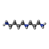
+Macromolecule #48: SPERMIDINE
+Macromolecule #49: water
-Experimental details
-Structure determination
| Method | cryo EM |
|---|---|
 Processing Processing | single particle reconstruction |
| Aggregation state | particle |
- Sample preparation
| Buffer | pH: 7.4 |
|---|---|
| Vitrification | Cryogen name: ETHANE |
- Electron microscopy
| Microscope | TFS KRIOS |
|---|---|
| Image recording | Film or detector model: GATAN K3 (6k x 4k) / Average electron dose: 72.26 e/Å2 |
| Electron beam | Acceleration voltage: 300 kV / Electron source:  FIELD EMISSION GUN FIELD EMISSION GUN |
| Electron optics | Illumination mode: FLOOD BEAM / Imaging mode: BRIGHT FIELD / Nominal defocus max: 1.9000000000000001 µm / Nominal defocus min: 0.5 µm |
| Experimental equipment |  Model: Titan Krios / Image courtesy: FEI Company |
 Movie
Movie Controller


 Z (Sec.)
 Y (Row.)
 X (Col.)


