[English] 日本語
 Yorodumi
- EMDB-28996: Nodavirus RNA replication crown from BSRT7/5 cells expressing vir... -
+ Open data
- Basic information
| Entry |  | |||||||||
|---|---|---|---|---|---|---|---|---|---|---|
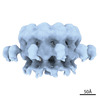
| Title | Nodavirus RNA replication crown from BSRT7/5 cells expressing viral protein A and fsRNA1 template in presence of B2 | |||||||||
 Map data Map data | Nodavirus RNA replication crown from BSRT7/5 cells expressing viral protein A and fsRNA1 template in presence of B2 | |||||||||
 Sample Sample |
| |||||||||
 Keywords Keywords | Nodavirus RNA replication and RNA capping complex / Dodecamer ring / Outer mitochondrial membrane protein complex / VIRAL PROTEIN | |||||||||
| Biological species |  Flock House virus Flock House virus | |||||||||
| Method | subtomogram averaging / cryo EM / Resolution: 26.7 Å | |||||||||
 Authors Authors | den Boon J / Zhan H / Unchwaniwala N / Horswill M / Slavik K / Pennington J / Navine A / Ahlquist P | |||||||||
| Funding support |  United States, 2 items United States, 2 items
| |||||||||
 Citation Citation |  Journal: Viruses / Year: 2022 Journal: Viruses / Year: 2022Title: Multifunctional Protein A Is the Only Viral Protein Required for Nodavirus RNA Replication Crown Formation. Authors: Johan A den Boon / Hong Zhan / Nuruddin Unchwaniwala / Mark Horswill / Kailey Slavik / Janice Pennington / Amanda Navine / Paul Ahlquist /  Abstract: Positive-strand RNA virus RNA genome replication occurs in membrane-associated RNA replication complexes (RCs). Nodavirus RCs are outer mitochondrial membrane invaginations whose necked openings to ...Positive-strand RNA virus RNA genome replication occurs in membrane-associated RNA replication complexes (RCs). Nodavirus RCs are outer mitochondrial membrane invaginations whose necked openings to the cytosol are "crowned" by a 12-fold symmetrical proteinaceous ring that functions as the main engine of RNA replication. Similar protein crowns recently visualized at the openings of alphavirus and coronavirus RCs highlight their broad conservation and functional importance. Using cryo-EM tomography, we earlier showed that the major nodavirus crown constituent is viral protein A, whose polymerase, RNA capping, membrane interaction and multimerization domains drive RC formation and function. Other viral proteins are strong candidates for unassigned EM density in the crown. RNA-binding RNAi inhibitor protein B2 co-immunoprecipitates with protein A and could form crown subdomains that protect nascent viral RNA and dsRNA templates. Capsid protein may interact with the crown since nodavirus virion assembly has spatial and other links to RNA replication. Using cryoelectron tomography and complementary approaches, we show that, even when formed in mammalian cells, nodavirus RC crowns generated without B2 and capsid proteins are functional and structurally indistinguishable from mature crowns in infected cells expressing all viral proteins. Thus, the only nodaviral factors essential to form functional RCs and crowns are RNA replication protein A and an RNA template. We also resolve apparent conflicts in prior results on B2 localization in infected cells, revealing at least two distinguishable pools of B2. The results have significant implications for crown structure, assembly, function and control as an antiviral target. | |||||||||
| History |
|
- Structure visualization
| Supplemental images |
|---|
- Downloads & links
-EMDB archive
| Map data |  emd_28996.map.gz emd_28996.map.gz | 1.8 MB |  EMDB map data format EMDB map data format | |
|---|---|---|---|---|
| Header (meta data) |  emd-28996-v30.xml emd-28996-v30.xml emd-28996.xml emd-28996.xml | 16.4 KB 16.4 KB | Display Display |  EMDB header EMDB header |
| FSC (resolution estimation) |  emd_28996_fsc.xml emd_28996_fsc.xml | 3.8 KB | Display |  FSC data file FSC data file |
| Images |  emd_28996.png emd_28996.png | 77.9 KB | ||
| Filedesc metadata |  emd-28996.cif.gz emd-28996.cif.gz | 5.4 KB | ||
| Others |  emd_28996_half_map_1.map.gz emd_28996_half_map_1.map.gz emd_28996_half_map_2.map.gz emd_28996_half_map_2.map.gz | 1.8 MB 1.8 MB | ||
| Archive directory |  http://ftp.pdbj.org/pub/emdb/structures/EMD-28996 http://ftp.pdbj.org/pub/emdb/structures/EMD-28996 ftp://ftp.pdbj.org/pub/emdb/structures/EMD-28996 ftp://ftp.pdbj.org/pub/emdb/structures/EMD-28996 | HTTPS FTP |
-Validation report
| Summary document |  emd_28996_validation.pdf.gz emd_28996_validation.pdf.gz | 774.2 KB | Display |  EMDB validaton report EMDB validaton report |
|---|---|---|---|---|
| Full document |  emd_28996_full_validation.pdf.gz emd_28996_full_validation.pdf.gz | 773.8 KB | Display | |
| Data in XML |  emd_28996_validation.xml.gz emd_28996_validation.xml.gz | 9.1 KB | Display | |
| Data in CIF |  emd_28996_validation.cif.gz emd_28996_validation.cif.gz | 11.7 KB | Display | |
| Arichive directory |  https://ftp.pdbj.org/pub/emdb/validation_reports/EMD-28996 https://ftp.pdbj.org/pub/emdb/validation_reports/EMD-28996 ftp://ftp.pdbj.org/pub/emdb/validation_reports/EMD-28996 ftp://ftp.pdbj.org/pub/emdb/validation_reports/EMD-28996 | HTTPS FTP |
-Related structure data
- Links
Links
| EMDB pages |  EMDB (EBI/PDBe) / EMDB (EBI/PDBe) /  EMDataResource EMDataResource |
|---|
- Map
Map
| File |  Download / File: emd_28996.map.gz / Format: CCP4 / Size: 2 MB / Type: IMAGE STORED AS FLOATING POINT NUMBER (4 BYTES) Download / File: emd_28996.map.gz / Format: CCP4 / Size: 2 MB / Type: IMAGE STORED AS FLOATING POINT NUMBER (4 BYTES) | ||||||||||||||||||||||||||||||||||||
|---|---|---|---|---|---|---|---|---|---|---|---|---|---|---|---|---|---|---|---|---|---|---|---|---|---|---|---|---|---|---|---|---|---|---|---|---|---|
| Annotation | Nodavirus RNA replication crown from BSRT7/5 cells expressing viral protein A and fsRNA1 template in presence of B2 | ||||||||||||||||||||||||||||||||||||
| Projections & slices | Image control
Images are generated by Spider. | ||||||||||||||||||||||||||||||||||||
| Voxel size | X=Y=Z: 5.4 Å | ||||||||||||||||||||||||||||||||||||
| Density |
| ||||||||||||||||||||||||||||||||||||
| Symmetry | Space group: 1 | ||||||||||||||||||||||||||||||||||||
| Details | EMDB XML:
|
-Supplemental data
-Half map: Nodavirus RNA replication crown from BSRT7/5 cells expressing...
| File | emd_28996_half_map_1.map | ||||||||||||
|---|---|---|---|---|---|---|---|---|---|---|---|---|---|
| Annotation | Nodavirus RNA replication crown from BSRT7/5 cells expressing viral protein A and fsRNA1 template in presence of B2, half map1. | ||||||||||||
| Projections & Slices |
| ||||||||||||
| Density Histograms |
-Half map: Nodavirus RNA replication crown from BSRT7/5 cells expressing...
| File | emd_28996_half_map_2.map | ||||||||||||
|---|---|---|---|---|---|---|---|---|---|---|---|---|---|
| Annotation | Nodavirus RNA replication crown from BSRT7/5 cells expressing viral protein A and fsRNA1 template in presence of B2, half map2. | ||||||||||||
| Projections & Slices |
| ||||||||||||
| Density Histograms |
- Sample components
-Entire : Flock house nodavirus protein A
| Entire | Name: Flock house nodavirus protein A |
|---|---|
| Components |
|
-Supramolecule #1: Flock house nodavirus protein A
| Supramolecule | Name: Flock house nodavirus protein A / type: complex / ID: 1 / Parent: 0 / Macromolecule list: all |
|---|---|
| Source (natural) | Organism:  Flock House virus / Tissue: BSRT7/5 cells / Organelle: mitochondria / Location in cell: outer mitochondrial membrane Flock House virus / Tissue: BSRT7/5 cells / Organelle: mitochondria / Location in cell: outer mitochondrial membrane |
-Macromolecule #1: Flock House nodavirus protein A
| Macromolecule | Name: Flock House nodavirus protein A / type: protein_or_peptide / ID: 1 / Enantiomer: LEVO |
|---|---|
| Sequence | String: MTLKVILGEH QITRTELLVG IATVSGCGAV VYCISKFWGY GAIAPYPQSG GNRVTRALQR AVIDKTKTPI ETRFYPLDSL RTVTPKRVAD NGHAVSGAVR DAARRLIDES ITAVGGSKFE VNPNPNSSTG LRNHFHFAVG DLAQDFRNDT PADDAFIVGV DVDYYVTEPD ...String: MTLKVILGEH QITRTELLVG IATVSGCGAV VYCISKFWGY GAIAPYPQSG GNRVTRALQR AVIDKTKTPI ETRFYPLDSL RTVTPKRVAD NGHAVSGAVR DAARRLIDES ITAVGGSKFE VNPNPNSSTG LRNHFHFAVG DLAQDFRNDT PADDAFIVGV DVDYYVTEPD VLLEHMRPVV LHTFNPKKVS GFDADSPFTI KNNLVEYKVS GGAAWVHPVW DWCEAGEFIA SRVRTSWKEW FLQLPLRMIG LEKVGYHKIH HCRPWTDCPD RALVYTIPQY VIWRFNWIDT ELHVRKLKRI EYQDETKPGW NRLEYVTDKN ELLVSIGREG EHAQITIEKE KLDMLSGLSA TQSVNARLIG MGHKDPQYTS MIVQYYTGKK VVSPISPTVY KPTMPRVHWP VTSDADVPEV SARQYTLPIV SDCMMMPMIK RWETMSESIE RRVTFVANDK KPSDRIAKIA ETFVKLMNGP FKDLDPLSIE ETIERLNKPS QQLQLRAVFE MIGVKPRQLI ESFNKNEPGM KSSRIISGFP DILFILKVSR YTLAYSDIVL HAEHNEHWYY PGRNPTEIAD GVCEFVSDCD AEVIETDFSN LDGRVSSWMQ RNIAQKAMVQ AFRPEYRDEI ISFMDTIINC PAKAKRFGFR YEPGVGVKSG SPTTTPHNTQ YNGCVEFTAL TFEHPDAEPE DLFRLIGPKC GDDGLSRAII QKSINRAAKC FGLELKVERY NPEIGLCFLS RVFVDPLATT TTIQDPLRTL RKLHLTTRDP TIPLADAACD RVEGYLCTDA LTPLISDYCK MVLRLYGPTA STEQVRNQRR SRNKEKPYWL TCDGSWPQHP QDAHLMKQVL IKRTAIDEDQ VDALIGRFAA MKDVWEKITH DSEESAAACT FDEDGVAPNS VDESLPLLND AKQTRANPGT SRPHSNGGGS SHGNELPRRT EQRAQGPRQP ARLPKQGKTN GKSDGNITAG ETQRGGIPRG KGPRGGKTNT RRTPPKAGAQ PQPSNNRK |
-Experimental details
-Structure determination
| Method | cryo EM |
|---|---|
 Processing Processing | subtomogram averaging |
| Aggregation state | particle |
- Sample preparation
| Buffer | pH: 7.4 |
|---|---|
| Grid | Model: Quantifoil / Material: COPPER / Mesh: 200 / Support film - Material: CARBON / Support film - topology: HOLEY / Pretreatment - Type: GLOW DISCHARGE / Pretreatment - Time: 60 sec. |
| Vitrification | Cryogen name: ETHANE / Chamber humidity: 100 % / Chamber temperature: 295 K / Instrument: FEI VITROBOT MARK III |
- Electron microscopy
| Microscope | FEI TITAN KRIOS |
|---|---|
| Image recording | Film or detector model: GATAN K2 SUMMIT (4k x 4k) / Detector mode: COUNTING / Average electron dose: 4.86 e/Å2 / Details: Grid ID: N56 |
| Electron beam | Acceleration voltage: 300 kV / Electron source:  FIELD EMISSION GUN FIELD EMISSION GUN |
| Electron optics | C2 aperture diameter: 100.0 µm / Calibrated magnification: 19500 / Illumination mode: FLOOD BEAM / Imaging mode: BRIGHT FIELD / Cs: 0.01 mm / Nominal defocus max: 3.5 µm / Nominal defocus min: 3.5 µm |
| Experimental equipment |  Model: Titan Krios / Image courtesy: FEI Company |
 Movie
Movie Controller


 Z (Sec.)
 Y (Row.)
 X (Col.)


