[English] 日本語
 Yorodumi
- EMDB-2897: Cryo-EM structure of helical ANTH and ENTH tubules on PI(4,5)P2-c... -
+ Open data
- Basic information
| Entry | Database: EMDB / ID: EMD-2897 | |||||||||
|---|---|---|---|---|---|---|---|---|---|---|
| Title | Cryo-EM structure of helical ANTH and ENTH tubules on PI(4,5)P2-containing membranes | |||||||||
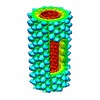
 Map data Map data | Reconstruction of the one-start helix, of pitch 63.2 Angstrom with 9.96 subunits per turn, formed by ANTH-ENTH assembly on PIP2 containing GUVs. | |||||||||
 Sample Sample |
| |||||||||
 Keywords Keywords | epsin / Hip1R / ENTH / ANTH / clathrin adaptors / endocytosis / PI(4 / 5)P2 | |||||||||
| Function / homology |  Function and homology information Function and homology informationCargo recognition for clathrin-mediated endocytosis / actin cortical patch assembly / clathrin vesicle coat / clathrin light chain binding / incipient cellular bud site / negative regulation of Arp2/3 complex-mediated actin nucleation / cellular bud tip / actin cortical patch / clathrin coat assembly / clathrin adaptor activity ...Cargo recognition for clathrin-mediated endocytosis / actin cortical patch assembly / clathrin vesicle coat / clathrin light chain binding / incipient cellular bud site / negative regulation of Arp2/3 complex-mediated actin nucleation / cellular bud tip / actin cortical patch / clathrin coat assembly / clathrin adaptor activity / cellular bud neck / mating projection tip / phosphatidylinositol-3,4-bisphosphate binding / phosphatidylinositol-3,5-bisphosphate binding / clathrin-coated vesicle / clathrin binding / K63-linked polyubiquitin modification-dependent protein binding / cortical actin cytoskeleton / ubiquitin binding / actin filament organization / phospholipid binding / endocytosis / actin filament binding / early endosome / endosome / plasma membrane / cytoplasm Similarity search - Function | |||||||||
| Biological species |  | |||||||||
| Method | single particle reconstruction / cryo EM / Resolution: 18.3 Å | |||||||||
 Authors Authors | Skruzny M / Desfosses A / Prinz S / Dodonova SO / Gieras A / Uetrecht C / Jakobi AJ / Abella M / Hagen WJH / Schulz J ...Skruzny M / Desfosses A / Prinz S / Dodonova SO / Gieras A / Uetrecht C / Jakobi AJ / Abella M / Hagen WJH / Schulz J / Meijers R / Rybin V / Briggs JAG / Sachse C / Kaksonen M | |||||||||
 Citation Citation |  Journal: Dev Cell / Year: 2015 Journal: Dev Cell / Year: 2015Title: An organized co-assembly of clathrin adaptors is essential for endocytosis. Authors: Michal Skruzny / Ambroise Desfosses / Simone Prinz / Svetlana O Dodonova / Anna Gieras / Charlotte Uetrecht / Arjen J Jakobi / Marc Abella / Wim J H Hagen / Joachim Schulz / Rob Meijers / ...Authors: Michal Skruzny / Ambroise Desfosses / Simone Prinz / Svetlana O Dodonova / Anna Gieras / Charlotte Uetrecht / Arjen J Jakobi / Marc Abella / Wim J H Hagen / Joachim Schulz / Rob Meijers / Vladimir Rybin / John A G Briggs / Carsten Sachse / Marko Kaksonen /  Abstract: Clathrin-mediated endocytosis, the main trafficking route from the plasma membrane to the cytoplasm, is critical to many fundamental cellular processes. Clathrin, coupled to the membrane by adaptor ...Clathrin-mediated endocytosis, the main trafficking route from the plasma membrane to the cytoplasm, is critical to many fundamental cellular processes. Clathrin, coupled to the membrane by adaptor proteins, is thought to play a major structural role in endocytosis by self-assembling into a cage-like lattice around the forming vesicle. Although clathrin adaptors are essential for endocytosis, little is known about their structural role in this process. Here we show that the membrane-binding domains of two conserved clathrin adaptors, Sla2 and Ent1, co-assemble in a PI(4,5)P2-dependent manner to form organized lattices on membranes. We determined the structure of the co-assembled lattice by electron cryo-microscopy and designed mutations that specifically impair the lattice formation in vitro. We show that these mutations block endocytosis in vivo. We suggest that clathrin adaptors not only link the polymerized clathrin to the membrane but also form an oligomeric structure, which is essential for membrane remodeling during endocytosis. | |||||||||
| History |
|
- Structure visualization
| Movie |
 Movie viewer Movie viewer |
|---|---|
| Structure viewer | EM map:  SurfView SurfView Molmil Molmil Jmol/JSmol Jmol/JSmol |
| Supplemental images |
- Downloads & links
-EMDB archive
| Map data |  emd_2897.map.gz emd_2897.map.gz | 52.5 MB |  EMDB map data format EMDB map data format | |
|---|---|---|---|---|
| Header (meta data) |  emd-2897-v30.xml emd-2897-v30.xml emd-2897.xml emd-2897.xml | 11.7 KB 11.7 KB | Display Display |  EMDB header EMDB header |
| Images |  EMD-2897_preview.png EMD-2897_preview.png | 158.5 KB | ||
| Archive directory |  http://ftp.pdbj.org/pub/emdb/structures/EMD-2897 http://ftp.pdbj.org/pub/emdb/structures/EMD-2897 ftp://ftp.pdbj.org/pub/emdb/structures/EMD-2897 ftp://ftp.pdbj.org/pub/emdb/structures/EMD-2897 | HTTPS FTP |
-Validation report
| Summary document |  emd_2897_validation.pdf.gz emd_2897_validation.pdf.gz | 224.8 KB | Display |  EMDB validaton report EMDB validaton report |
|---|---|---|---|---|
| Full document |  emd_2897_full_validation.pdf.gz emd_2897_full_validation.pdf.gz | 224 KB | Display | |
| Data in XML |  emd_2897_validation.xml.gz emd_2897_validation.xml.gz | 4.8 KB | Display | |
| Arichive directory |  https://ftp.pdbj.org/pub/emdb/validation_reports/EMD-2897 https://ftp.pdbj.org/pub/emdb/validation_reports/EMD-2897 ftp://ftp.pdbj.org/pub/emdb/validation_reports/EMD-2897 ftp://ftp.pdbj.org/pub/emdb/validation_reports/EMD-2897 | HTTPS FTP |
-Related structure data
- Links
Links
| EMDB pages |  EMDB (EBI/PDBe) / EMDB (EBI/PDBe) /  EMDataResource EMDataResource |
|---|---|
| Related items in Molecule of the Month |
- Map
Map
| File |  Download / File: emd_2897.map.gz / Format: CCP4 / Size: 73 MB / Type: IMAGE STORED AS FLOATING POINT NUMBER (4 BYTES) Download / File: emd_2897.map.gz / Format: CCP4 / Size: 73 MB / Type: IMAGE STORED AS FLOATING POINT NUMBER (4 BYTES) | ||||||||||||||||||||||||||||||||||||||||||||||||||||||||||||
|---|---|---|---|---|---|---|---|---|---|---|---|---|---|---|---|---|---|---|---|---|---|---|---|---|---|---|---|---|---|---|---|---|---|---|---|---|---|---|---|---|---|---|---|---|---|---|---|---|---|---|---|---|---|---|---|---|---|---|---|---|---|
| Annotation | Reconstruction of the one-start helix, of pitch 63.2 Angstrom with 9.96 subunits per turn, formed by ANTH-ENTH assembly on PIP2 containing GUVs. | ||||||||||||||||||||||||||||||||||||||||||||||||||||||||||||
| Projections & slices | Image control
Images are generated by Spider. generated in cubic-lattice coordinate | ||||||||||||||||||||||||||||||||||||||||||||||||||||||||||||
| Voxel size | X=Y=Z: 1.78 Å | ||||||||||||||||||||||||||||||||||||||||||||||||||||||||||||
| Density |
| ||||||||||||||||||||||||||||||||||||||||||||||||||||||||||||
| Symmetry | Space group: 1 | ||||||||||||||||||||||||||||||||||||||||||||||||||||||||||||
| Details | EMDB XML:
CCP4 map header:
| ||||||||||||||||||||||||||||||||||||||||||||||||||||||||||||
-Supplemental data
- Sample components
-Entire : ANTH and ENTH domains of the clathrin adaptors Sla2 and Ent1, res...
| Entire | Name: ANTH and ENTH domains of the clathrin adaptors Sla2 and Ent1, respectively. |
|---|---|
| Components |
|
-Supramolecule #1000: ANTH and ENTH domains of the clathrin adaptors Sla2 and Ent1, res...
| Supramolecule | Name: ANTH and ENTH domains of the clathrin adaptors Sla2 and Ent1, respectively. type: sample / ID: 1000 / Number unique components: 2 |
|---|
-Macromolecule #1: ANTH domain of endocytic adaptor Sla2
| Macromolecule | Name: ANTH domain of endocytic adaptor Sla2 / type: protein_or_peptide / ID: 1 / Recombinant expression: Yes |
|---|---|
| Source (natural) | Organism:  |
| Molecular weight | Experimental: 33.215 KDa / Theoretical: 33.21 KDa |
| Recombinant expression | Organism:  |
| Sequence | UniProtKB: Protein SLA2 / InterPro: AP180 N-terminal homology (ANTH) domain |
-Macromolecule #2: ENTH domain of epsin Ent1
| Macromolecule | Name: ENTH domain of epsin Ent1 / type: protein_or_peptide / ID: 2 / Recombinant expression: Yes |
|---|---|
| Source (natural) | Organism:  |
| Molecular weight | Experimental: 18.87 KDa / Theoretical: 18.847 KDa |
| Recombinant expression | Organism:  |
| Sequence | UniProtKB: Epsin-1 / InterPro: ENTH domain |
-Experimental details
-Structure determination
| Method | cryo EM |
|---|---|
 Processing Processing | single particle reconstruction |
| Aggregation state | helical array |
- Sample preparation
| Buffer | pH: 7.5 / Details: 20 mM HEPES, pH 7.5; 100 mM KCl |
|---|---|
| Grid | Details: on C-flat holey carbon coated grids (Protochips) |
| Vitrification | Cryogen name: ETHANE / Chamber humidity: 80 % / Instrument: HOMEMADE PLUNGER Method: applied on C-flat holey carbon coated grids (Protochips) and vitrified |
- Electron microscopy
| Microscope | FEI TITAN KRIOS |
|---|---|
| Date | Mar 1, 2013 |
| Image recording | Category: CCD / Film or detector model: GATAN ULTRASCAN 4000 (4k x 4k) / Number real images: 1419 / Average electron dose: 10 e/Å2 |
| Electron beam | Acceleration voltage: 120 kV / Electron source:  FIELD EMISSION GUN FIELD EMISSION GUN |
| Electron optics | Calibrated magnification: 59000 / Illumination mode: FLOOD BEAM / Imaging mode: BRIGHT FIELD / Cs: 2.7 mm / Nominal defocus max: 5.0 µm / Nominal defocus min: 0.5 µm / Nominal magnification: 59000 |
| Sample stage | Specimen holder model: FEI TITAN KRIOS AUTOGRID HOLDER |
| Experimental equipment |  Model: Titan Krios / Image courtesy: FEI Company |
- Image processing
| Details | Processing done with SPRING |
|---|---|
| CTF correction | Details: Each Particle |
| Final reconstruction | Applied symmetry - Helical parameters - Δz: 6.345 Å Applied symmetry - Helical parameters - Δ&Phi: 36.145 ° Applied symmetry - Helical parameters - Axial symmetry: C1 (asymmetric) Resolution.type: BY AUTHOR / Resolution: 18.3 Å / Resolution method: OTHER / Software - Name: SPRING / Number images used: 154432 |
 Movie
Movie Controller


 Z (Sec.)
 Y (Row.)
 X (Col.)


