Movie
Movie Controller


+ Open data

- Basic information
| Entry |  | ||||||||||||
|---|---|---|---|---|---|---|---|---|---|---|---|---|---|
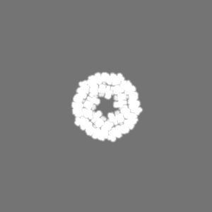
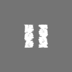
| Title | Cryo-EM structure of the Agrobacterium T-pilus | ||||||||||||
 Map data Map data | |||||||||||||
 Sample Sample |
| ||||||||||||
 Keywords Keywords | VirB/D4 T4SS / Bacterial conjugation / T-pilus / helical filament / PROTEIN FIBRIL | ||||||||||||
| Function / homology | Conjugal transfer TrbC/type IV secretion VirB2 / TrbC/VIRB2 pilin / type IV secretion system complex / protein secretion by the type IV secretion system / cell outer membrane / Type IV secretion system pilin protein VirB2 Function and homology information Function and homology information | ||||||||||||
| Biological species |  Agrobacterium tumefaciens (bacteria) Agrobacterium tumefaciens (bacteria) | ||||||||||||
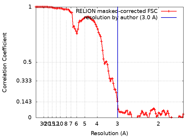
| Method | helical reconstruction / cryo EM / Resolution: 3.0 Å | ||||||||||||
 Authors Authors | Kreida S / Narita A / Johnson MD / Tocheva EI / Das A / Jensen GJ / Ghosal D | ||||||||||||
| Funding support |  United States, United States,  Australia, Australia,  Canada, 3 items Canada, 3 items
| ||||||||||||
 Citation Citation | Journal: Structure / Year: 2023 Title: Cryo-EM structure of the Agrobacterium tumefaciens T4SS-associated T-pilus reveals stoichiometric protein-phospholipid assembly. Authors: Stefan Kreida / Akihiro Narita / Matthew D Johnson / Elitza I Tocheva / Anath Das / Debnath Ghosal / Grant J Jensen /   Abstract: Agrobacterium tumefaciens causes crown gall disease in plants by the horizontal transfer of oncogenic DNA. The conjugation is mediated by the VirB/D4 type 4 secretion system (T4SS) that assembles an ...Agrobacterium tumefaciens causes crown gall disease in plants by the horizontal transfer of oncogenic DNA. The conjugation is mediated by the VirB/D4 type 4 secretion system (T4SS) that assembles an extracellular filament, the T-pilus, and is involved in mating pair formation between A. tumefaciens and the recipient plant cell. Here, we present a 3 Å cryoelectron microscopy (cryo-EM) structure of the T-pilus solved by helical reconstruction. Our structure reveals that the T-pilus is a stoichiometric assembly of the VirB2 major pilin and phosphatidylglycerol (PG) phospholipid with 5-start helical symmetry. We show that PG head groups and the positively charged Arg 91 residues of VirB2 protomers form extensive electrostatic interactions in the lumen of the T-pilus. Mutagenesis of Arg 91 abolished pilus formation. While our T-pilus structure is architecturally similar to previously published conjugative pili structures, the T-pilus lumen is narrower and positively charged, raising questions of whether the T-pilus is a conduit for ssDNA transfer. | ||||||||||||
| History |
|
- Structure visualization
| Supplemental images |
|---|

- Downloads & links
-EMDB archive
| Map data | emd_28957.map.gz | 7.4 MB | EMDB map data format | |
|---|---|---|---|---|
| Header (meta data) | emd-28957-v30.xmlemd-28957.xml | 18 KB 18 KB | Display Display | EMDB header |
| FSC (resolution estimation) | emd_28957_fsc.xml | 10.7 KB | Display | FSC data file |
| Images | 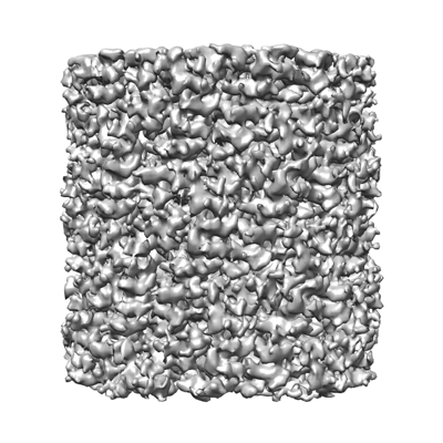 emd_28957.png emd_28957.png | 143.5 KB | ||
| Masks | emd_28957_msk_1.map | 103 MB | Mask map | |
| Filedesc metadata | emd-28957.cif.gz | 5.8 KB | ||
| Others | emd_28957_half_map_1.map.gzemd_28957_half_map_2.map.gz | 80.7 MB 80.9 MB | ||
| Archive directory |  http://ftp.pdbj.org/pub/emdb/structures/EMD-28957ftp://ftp.pdbj.org/pub/emdb/structures/EMD-28957 http://ftp.pdbj.org/pub/emdb/structures/EMD-28957ftp://ftp.pdbj.org/pub/emdb/structures/EMD-28957 | HTTPS FTP |
-Related structure data
| Related structure data |  8faiMC M: atomic model generated by this map C: citing same article ( |
|---|---|
| Similar structure data |
-Links
| EMDB pages | EMDB (EBI/PDBe) / EMDataResource |
|---|
-Map
| File | Download / File: emd_28957.map.gz / Format: CCP4 / Size: 103 MB / Type: IMAGE STORED AS FLOATING POINT NUMBER (4 BYTES) | ||||||||||||||||||||||||||||||||||||
|---|---|---|---|---|---|---|---|---|---|---|---|---|---|---|---|---|---|---|---|---|---|---|---|---|---|---|---|---|---|---|---|---|---|---|---|---|---|
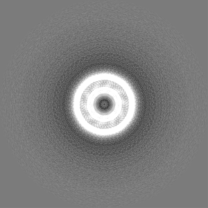
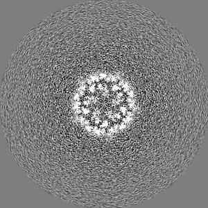
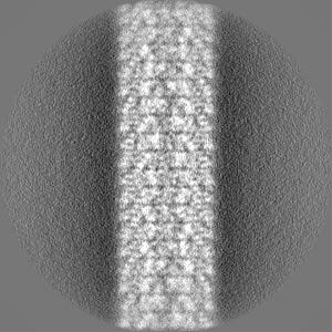
| Projections & slices | Image control
Images are generated by Spider. | ||||||||||||||||||||||||||||||||||||
| Voxel size | X=Y=Z: 0.832 Å | ||||||||||||||||||||||||||||||||||||
| Density |
| ||||||||||||||||||||||||||||||||||||
| Symmetry | Space group: 1 | ||||||||||||||||||||||||||||||||||||
| Details | EMDB XML:
|
 Z (Sec.)
 Y (Row.)
 X (Col.)


-Supplemental data
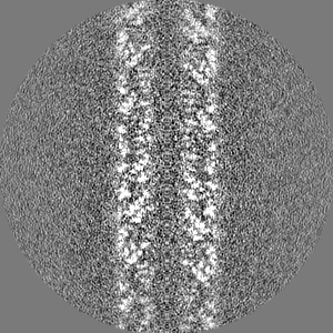
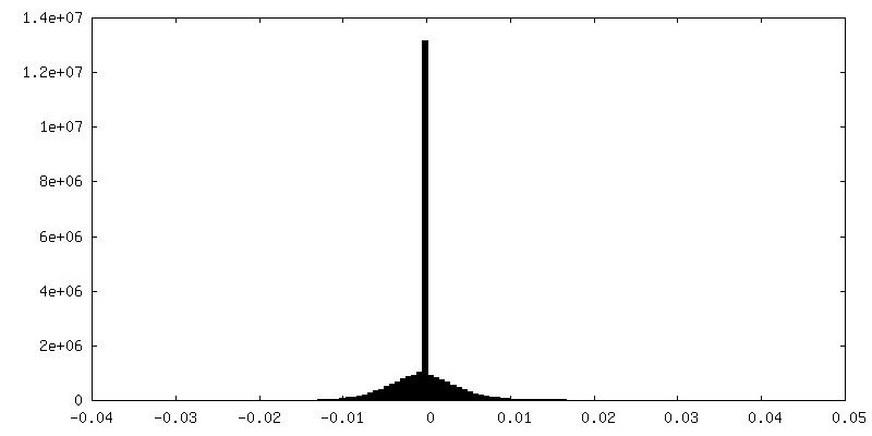
-Mask #1
| File | emd_28957_msk_1.map | ||||||||||||
|---|---|---|---|---|---|---|---|---|---|---|---|---|---|
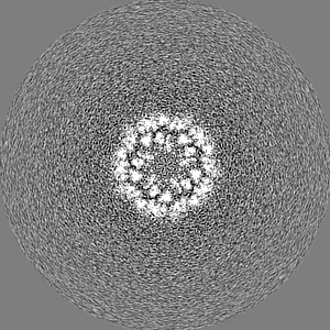
| Projections & Slices |
| ||||||||||||
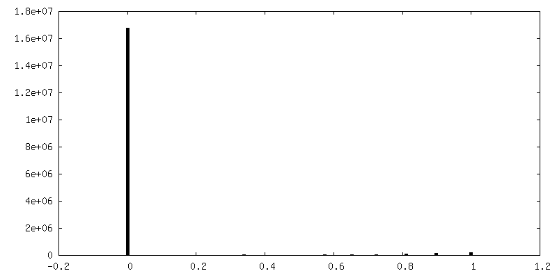
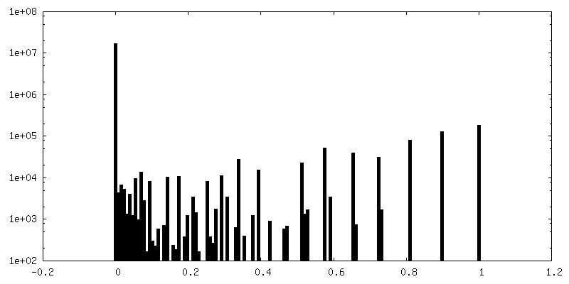
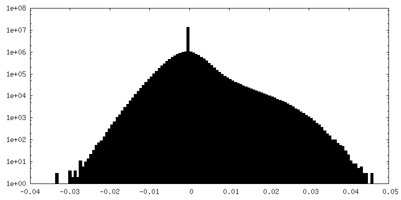
| Density Histograms |


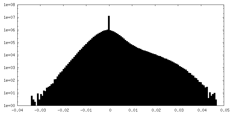
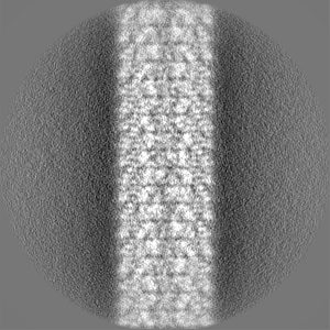
-Half map: #2
| File | emd_28957_half_map_1.map | ||||||||||||
|---|---|---|---|---|---|---|---|---|---|---|---|---|---|
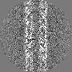
| Projections & Slices |
| ||||||||||||
| Density Histograms |


-Half map: #1
| File | emd_28957_half_map_2.map | ||||||||||||
|---|---|---|---|---|---|---|---|---|---|---|---|---|---|
| Projections & Slices |
| ||||||||||||
| Density Histograms |


- Sample components
-Entire : T-pilus
| Entire | Name: T-pilus |
|---|---|
| Components |
|
-Supramolecule #1: T-pilus
| Supramolecule | Name: T-pilus / type: complex / ID: 1 / Parent: 0 / Macromolecule list: #1 |
|---|---|
| Source (natural) | Organism: Agrobacterium tumefaciens (bacteria) / Strain: NT1REB(pJK270) |
| Molecular weight | Theoretical: 2.787 kDa/nm |
-Supramolecule #2: VirB2
| Supramolecule | Name: VirB2 / type: organelle_or_cellular_component / ID: 2 / Parent: 1 / Macromolecule list: #1 |
|---|---|
| Source (natural) | Organism: Agrobacterium tumefaciens (bacteria) / Strain: NT1REB(pJK270) |
-Supramolecule #3: Phosphatidylglycerol 16:1/18:1
| Supramolecule | Name: Phosphatidylglycerol 16:1/18:1 / type: organelle_or_cellular_component / ID: 3 / Parent: 1 |
|---|---|
| Source (natural) | Organism: Agrobacterium tumefaciens (bacteria) / Strain: NT1REB(pJK270) |
-Macromolecule #1: Protein virB2
| Macromolecule | Name: Protein virB2 / type: protein_or_peptide / ID: 1 / Number of copies: 15 / Enantiomer: LEVO |
|---|---|
| Source (natural) | Organism: Agrobacterium tumefaciens (bacteria) / Strain: NT1REB(pJK270) |
| Molecular weight | Theoretical: 12.326439 KDa |
| Sequence | String: MRCFERYRVH LNRLSLSNAV MRMVSGYAPS VVGAMGWSIF SSGPAAAQSA GGGTDPATMV NNICTFILGP FGQSLAVLGI VAIGISWMF GRASLGLVAG VVGGIVIMFG ASFLGKTLTG GG UniProtKB: Type IV secretion system pilin protein VirB2 |
-Macromolecule #2: (7Z,19R,22S,25R)-22,25,26-trihydroxy-16,22-dioxo-17,21,23-trioxa-...
| Macromolecule | Name: (7Z,19R,22S,25R)-22,25,26-trihydroxy-16,22-dioxo-17,21,23-trioxa-22lambda~5~-phosphahexacos-7-en-19-yl (9Z)-octadec-9-enoate type: ligand / ID: 2 / Number of copies: 15 / Formula: XL0 |
|---|---|
| Molecular weight | Theoretical: 746.991 Da |
-Experimental details
-Structure determination
| Method | cryo EM |
|---|---|
 Processing Processing | helical reconstruction |
| Aggregation state | filament |
-Sample preparation
| Concentration | 1 mg/mL |
|---|---|
| Buffer | pH: 5.5 / Component - Concentration: 50.0 mM / Component - Formula: MES / Component - Name: 2-(N-morpholino)ethanesulfonic acid |
| Grid | Model: UltrAuFoil R1.2/1.3 / Material: GOLD / Mesh: 300 / Pretreatment - Type: GLOW DISCHARGE / Pretreatment - Time: 60 sec. / Pretreatment - Atmosphere: AIR |
| Vitrification | Cryogen name: ETHANE-PROPANE / Chamber humidity: 100 % / Chamber temperature: 295 K / Instrument: FEI VITROBOT MARK IV |
- Electron microscopy
| Microscope | FEI TITAN KRIOS |
|---|---|
| Image recording | Film or detector model: GATAN K3 (6k x 4k) / Average electron dose: 60.0 e/Å2 |
| Electron beam | Acceleration voltage: 300 kV / Electron source:  FIELD EMISSION GUN FIELD EMISSION GUN |
| Electron optics | C2 aperture diameter: 100.0 µm / Illumination mode: FLOOD BEAM / Imaging mode: BRIGHT FIELD / Cs: 2.7 mm / Nominal defocus max: 2.0 µm / Nominal defocus min: 0.7000000000000001 µm / Nominal magnification: 105000 |
| Experimental equipment |  Model: Titan Krios / Image courtesy: FEI Company |