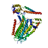
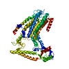
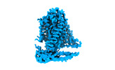
+ Open data
- Basic information
| Entry |  | |||||||||
|---|---|---|---|---|---|---|---|---|---|---|
| Title | cryo-EM structure of TMEM63B in LMNG | |||||||||
 Map data Map data | ||||||||||
 Sample Sample |
| |||||||||
 Keywords Keywords | ion channel / mechanosensitive / monomeric / MEMBRANE PROTEIN | |||||||||
| Function / homology |  Function and homology information Function and homology informationalveolar lamellar body membrane / surfactant secretion / osmolarity-sensing monoatomic cation channel activity / mechanosensitive monoatomic cation channel activity / mechanosensitive monoatomic ion channel activity / calcium-activated cation channel activity / exocytosis / sensory perception of sound / actin cytoskeleton / early endosome membrane / plasma membrane Similarity search - Function | |||||||||
| Biological species |  Homo sapiens (human) Homo sapiens (human) | |||||||||
| Method | single particle reconstruction / cryo EM / Resolution: 3.6 Å | |||||||||
 Authors Authors | Zheng W / Fu TM / Holt JR | |||||||||
| Funding support |  United States, 1 items United States, 1 items
| |||||||||
 Citation Citation |  Journal: Neuron / Year: 2023 Journal: Neuron / Year: 2023Title: TMEM63 proteins function as monomeric high-threshold mechanosensitive ion channels. Authors: Wang Zheng / Shaun Rawson / Zhangfei Shen / Elakkiya Tamilselvan / Harper E Smith / Julia Halford / Chen Shen / Swetha E Murthy / Maximilian H Ulbrich / Marcos Sotomayor / Tian-Min Fu / Jeffrey R Holt /   Abstract: OSCA/TMEM63s form mechanically activated (MA) ion channels in plants and animals, respectively. OSCAs and related TMEM16s and transmembrane channel-like (TMC) proteins form homodimers with two pores. ...OSCA/TMEM63s form mechanically activated (MA) ion channels in plants and animals, respectively. OSCAs and related TMEM16s and transmembrane channel-like (TMC) proteins form homodimers with two pores. Here, we uncover an unanticipated monomeric configuration of TMEM63 proteins. Structures of TMEM63A and TMEM63B (referred to as TMEM63s) revealed a single highly restricted pore. Functional analyses demonstrated that TMEM63s are bona fide mechanosensitive ion channels, characterized by small conductance and high thresholds. TMEM63s possess evolutionary variations in the intracellular linker IL2, which mediates dimerization in OSCAs. Replacement of OSCA1.2 IL2 with TMEM63A IL2 or mutations to key variable residues resulted in monomeric OSCA1.2 and MA currents with significantly higher thresholds. Structural analyses revealed substantial conformational differences in the mechano-sensing domain IL2 and gating helix TM6 between TMEM63s and OSCA1.2. Our studies reveal that mechanosensitivity in OSCA/TMEM63 channels is affected by oligomerization and suggest gating mechanisms that may be shared by OSCA/TMEM63, TMEM16, and TMC channels. | |||||||||
| History |
|
- Structure visualization
| Supplemental images |
|---|
- Downloads & links
-EMDB archive
| Map data |  emd_28154.map.gz emd_28154.map.gz | 97.1 MB |  EMDB map data format EMDB map data format | |
|---|---|---|---|---|
| Header (meta data) |  emd-28154-v30.xml emd-28154-v30.xml emd-28154.xml emd-28154.xml | 17.3 KB 17.3 KB | Display Display |  EMDB header EMDB header |
| Images |  emd_28154.png emd_28154.png | 43.6 KB | ||
| Filedesc metadata |  emd-28154.cif.gz emd-28154.cif.gz | 6.1 KB | ||
| Others |  emd_28154_half_map_1.map.gz emd_28154_half_map_1.map.gz emd_28154_half_map_2.map.gz emd_28154_half_map_2.map.gz | 95.5 MB 95.5 MB | ||
| Archive directory |  http://ftp.pdbj.org/pub/emdb/structures/EMD-28154 http://ftp.pdbj.org/pub/emdb/structures/EMD-28154 ftp://ftp.pdbj.org/pub/emdb/structures/EMD-28154 ftp://ftp.pdbj.org/pub/emdb/structures/EMD-28154 | HTTPS FTP |
-Validation report
| Summary document |  emd_28154_validation.pdf.gz emd_28154_validation.pdf.gz | 1 MB | Display |  EMDB validaton report EMDB validaton report |
|---|---|---|---|---|
| Full document |  emd_28154_full_validation.pdf.gz emd_28154_full_validation.pdf.gz | 1 MB | Display | |
| Data in XML |  emd_28154_validation.xml.gz emd_28154_validation.xml.gz | 13.2 KB | Display | |
| Data in CIF |  emd_28154_validation.cif.gz emd_28154_validation.cif.gz | 15.6 KB | Display | |
| Arichive directory |  https://ftp.pdbj.org/pub/emdb/validation_reports/EMD-28154 https://ftp.pdbj.org/pub/emdb/validation_reports/EMD-28154 ftp://ftp.pdbj.org/pub/emdb/validation_reports/EMD-28154 ftp://ftp.pdbj.org/pub/emdb/validation_reports/EMD-28154 | HTTPS FTP |
-Related structure data
| Related structure data |  8ehxMC  8ehwC M: atomic model generated by this map C: citing same article ( |
|---|---|
| Similar structure data | Similarity search - Function & homology  F&H Search F&H Search |
- Links
Links
| EMDB pages |  EMDB (EBI/PDBe) / EMDB (EBI/PDBe) /  EMDataResource EMDataResource |
|---|
- Map
Map
| File |  Download / File: emd_28154.map.gz / Format: CCP4 / Size: 103 MB / Type: IMAGE STORED AS FLOATING POINT NUMBER (4 BYTES) Download / File: emd_28154.map.gz / Format: CCP4 / Size: 103 MB / Type: IMAGE STORED AS FLOATING POINT NUMBER (4 BYTES) | ||||||||||||||||||||||||||||||||||||
|---|---|---|---|---|---|---|---|---|---|---|---|---|---|---|---|---|---|---|---|---|---|---|---|---|---|---|---|---|---|---|---|---|---|---|---|---|---|
| Projections & slices | Image control
Images are generated by Spider. | ||||||||||||||||||||||||||||||||||||
| Voxel size | X=Y=Z: 0.825 Å | ||||||||||||||||||||||||||||||||||||
| Density |
| ||||||||||||||||||||||||||||||||||||
| Symmetry | Space group: 1 | ||||||||||||||||||||||||||||||||||||
| Details | EMDB XML:
|
-Supplemental data
-Half map: #1
| File | emd_28154_half_map_1.map | ||||||||||||
|---|---|---|---|---|---|---|---|---|---|---|---|---|---|
| Projections & Slices |
| ||||||||||||
| Density Histograms |
-Half map: #2
| File | emd_28154_half_map_2.map | ||||||||||||
|---|---|---|---|---|---|---|---|---|---|---|---|---|---|
| Projections & Slices |
| ||||||||||||
| Density Histograms |
- Sample components
-Entire : TMEM63B purified protein in LMNG
| Entire | Name: TMEM63B purified protein in LMNG |
|---|---|
| Components |
|
-Supramolecule #1: TMEM63B purified protein in LMNG
| Supramolecule | Name: TMEM63B purified protein in LMNG / type: complex / ID: 1 / Parent: 0 / Macromolecule list: all |
|---|---|
| Source (natural) | Organism:  Homo sapiens (human) Homo sapiens (human) |
| Molecular weight | Theoretical: 110 KDa |
-Macromolecule #1: CSC1-like protein 2
| Macromolecule | Name: CSC1-like protein 2 / type: protein_or_peptide / ID: 1 / Number of copies: 1 / Enantiomer: LEVO |
|---|---|
| Source (natural) | Organism:  Homo sapiens (human) Homo sapiens (human) |
| Molecular weight | Theoretical: 96.051203 KDa |
| Recombinant expression | Organism:  Homo sapiens (human) Homo sapiens (human) |
| Sequence | String: MLPFLLATLG TTALNNSNPK DYCYSARIRS TVLQGLPFGG VPTVLALDFM CFLALLFLFS ILRKVAWDYG RLALVTDADR LRRQERDRV EQEYVASAMH GDSHDRYERL TSVSSSVDFD QRDNGFCSWL TAIFRIKDDE IRDKCGGDAV HYLSFQRHII G LLVVVGVL ...String: MLPFLLATLG TTALNNSNPK DYCYSARIRS TVLQGLPFGG VPTVLALDFM CFLALLFLFS ILRKVAWDYG RLALVTDADR LRRQERDRV EQEYVASAMH GDSHDRYERL TSVSSSVDFD QRDNGFCSWL TAIFRIKDDE IRDKCGGDAV HYLSFQRHII G LLVVVGVL SVGIVLPVNF SGDLLENNAY SFGRTTIANL KSGNNLLWLH TSFAFLYLLL TVYSMRRHTS KMRYKEDDLV KR TLFINGI SKYAESEKIK KHFEEAYPNC TVLEARPCYN VARLMFLDAE RKKAERGKLY FTNLQSKENV PTMINPKPCG HLC CCVVRG CEQVEAIEYY TKLEQKLKED YKREKEKVNE KPLGMAFVTF HNETITAIIL KDFNVCKCQG CTCRGEPRPS SCSE SLHIS NWTVSYAPDP QNIYWEHLSI RGFIWWLRCL VINVVLFILL FFLTTPAIII TTMDKFNVTK PVEYLNNPII TQFFP TLLL WCFSALLPTI VYYSAFFEAH WTRSGENRTT MHKCYTFLIF MVLLLPSLGL SSLDLFFRWL FDKKFLAEAA IRFECV FLP DNGAFFVNYV IASAFIGNAM DLLRIPGLLM YMIRLCLARS AAERRNVKRH QAYEFQFGAA YAWMMCVFTV VMTYSIT CP IIVPFGLMYM LLKHLVDRYN LYYAYLPAKL DKKIHSGAVN QVVAAPILCL FWLLFFSTMR TGFLAPTSMF TFVVLVIT I VICLCHVCFG HFKYLSAHNY KIEHTETDTV DPRSNGRPPT AAAVPKSAKY IAQVLQDSEV DGDGDGAPGS SGDEPPSSS SQDEELLMPP DALTDTDFQS CEDSLIENEI HQTRLEVLFQ UniProtKB: CSC1-like protein 2 |
-Experimental details
-Structure determination
| Method | cryo EM |
|---|---|
 Processing Processing | single particle reconstruction |
| Aggregation state | particle |
- Sample preparation
| Concentration | 2.5 mg/mL | |||||||||
|---|---|---|---|---|---|---|---|---|---|---|
| Buffer | pH: 7.4 Component:
| |||||||||
| Grid | Model: Quantifoil R1.2/1.3 / Material: GOLD / Mesh: 400 / Support film - Material: CARBON / Support film - topology: HOLEY / Pretreatment - Type: GLOW DISCHARGE / Pretreatment - Time: 60 sec. | |||||||||
| Vitrification | Cryogen name: ETHANE / Chamber humidity: 100 % / Chamber temperature: 279 K / Instrument: FEI VITROBOT MARK IV |
- Electron microscopy
| Microscope | FEI TITAN KRIOS |
|---|---|
| Specialist optics | Energy filter - Slit width: 20 eV |
| Image recording | Film or detector model: GATAN K3 (6k x 4k) / Average exposure time: 2.8 sec. / Average electron dose: 53.0 e/Å2 |
| Electron beam | Acceleration voltage: 300 kV / Electron source:  FIELD EMISSION GUN FIELD EMISSION GUN |
| Electron optics | C2 aperture diameter: 50.0 µm / Illumination mode: FLOOD BEAM / Imaging mode: BRIGHT FIELD / Cs: 2.7 mm / Nominal defocus max: 2.1 µm / Nominal defocus min: 0.8 µm / Nominal magnification: 105000 |
| Sample stage | Specimen holder model: FEI TITAN KRIOS AUTOGRID HOLDER / Cooling holder cryogen: NITROGEN |
| Experimental equipment |  Model: Titan Krios / Image courtesy: FEI Company |
- Image processing
| Startup model | Type of model: NONE |
|---|---|
| Final reconstruction | Applied symmetry - Point group: C1 (asymmetric) / Algorithm: BACK PROJECTION / Resolution.type: BY AUTHOR / Resolution: 3.6 Å / Resolution method: FSC 0.143 CUT-OFF / Software - Name: cryoSPARC / Number images used: 198144 |
| Initial angle assignment | Type: MAXIMUM LIKELIHOOD / Software - Name: cryoSPARC (ver. 3.3.2) |
| Final angle assignment | Type: MAXIMUM LIKELIHOOD / Software - Name: cryoSPARC (ver. 3.3.2) |
-Atomic model buiding 1
| Refinement | Space: REAL / Protocol: AB INITIO MODEL |
|---|---|
| Output model |  PDB-8ehx: |
 Movie
Movie Controller


 Z (Sec.)
 Y (Row.)
 X (Col.)


