 Movie
Movie Controller


+ Open data

- Basic information
| Entry |  | |||||||||
|---|---|---|---|---|---|---|---|---|---|---|
| Title | Klebsiella pneumoniae adenosine monophosphate nucleosidase | |||||||||
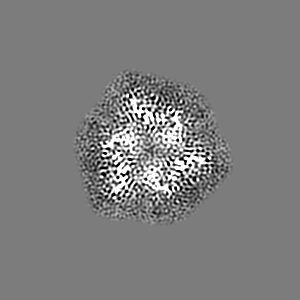
 Map data Map data | Sharpened map for building | |||||||||
 Sample Sample |
| |||||||||
 Keywords Keywords | nucleosidase / AMP / salvage / HYDROLASE | |||||||||
| Function / homology |  Function and homology information Function and homology informationAMP nucleosidase / AMP nucleosidase activity / nucleoside metabolic process / AMP salvage / cytosol Similarity search - Function | |||||||||
| Biological species |  Klebsiella pneumoniae (bacteria) Klebsiella pneumoniae (bacteria) | |||||||||
| Method | single particle reconstruction / cryo EM / Resolution: 3.05 Å | |||||||||
 Authors Authors | Richardson BC / French JB | |||||||||
| Funding support |  United States, 1 items United States, 1 items
| |||||||||
 Citation Citation | Journal: PLoS One / Year: 2022 Title: Structure of Klebsiella pneumoniae adenosine monophosphate nucleosidase. Authors: Brian C Richardson / Roger Shek / Wesley C Van Voorhis / Jarrod B French / Abstract: Klebsiella pneumoniae is a bacterial pathogen that is increasingly responsible for hospital-acquired pneumonia and sepsis. Progressive development of antibiotic resistance has led to higher mortality ...Klebsiella pneumoniae is a bacterial pathogen that is increasingly responsible for hospital-acquired pneumonia and sepsis. Progressive development of antibiotic resistance has led to higher mortality rates and creates a need for novel treatments. Because of the essential role that nucleotides play in many bacterial processes, enzymes involved in purine and pyrimidine metabolism and transport are ideal targets for the development of novel antibiotics. Herein we describe the structure of K. pneumoniae adenosine monophosphate nucleosidase (KpAmn), a purine salvage enzyme unique to bacteria, as determined by cryoelectron microscopy. The data detail a well conserved fold with a hexameric overall structure and clear density for the putative active site residues. Comparison to the crystal structures of homologous prokaryotic proteins confirms the presence of many of the conserved structural features of this protein yet reveals differences in distal loops in the absence of crystal contacts. This first cryo-EM structure of an Amn enzyme provides a basis for future structure-guided drug development and extends the accuracy of structural characterization of this family of proteins beyond this clinically relevant organism. | |||||||||
| History |
|
- Structure visualization
| Supplemental images |
|---|

- Downloads & links
-EMDB archive
| Map data | emd_26838.map.gz | 17.4 MB | EMDB map data format | |
|---|---|---|---|---|
| Header (meta data) | emd-26838-v30.xmlemd-26838.xml | 17.7 KB 17.7 KB | Display Display | EMDB header |
| FSC (resolution estimation) | emd_26838_fsc.xml | 11.2 KB | Display | FSC data file |
| Images |  emd_26838.png emd_26838.png | 132.7 KB | ||
| Masks | emd_26838_msk_1.map | 103 MB | Mask map | |
| Filedesc metadata | emd-26838.cif.gz | 6.3 KB | ||
| Others | emd_26838_half_map_1.map.gzemd_26838_half_map_2.map.gz | 95.6 MB 95.6 MB | ||
| Archive directory |  http://ftp.pdbj.org/pub/emdb/structures/EMD-26838ftp://ftp.pdbj.org/pub/emdb/structures/EMD-26838 http://ftp.pdbj.org/pub/emdb/structures/EMD-26838ftp://ftp.pdbj.org/pub/emdb/structures/EMD-26838 | HTTPS FTP |
-Validation report
| Summary document | emd_26838_validation.pdf.gz | 830.7 KB | Display | EMDB validaton report |
|---|---|---|---|---|
| Full document | emd_26838_full_validation.pdf.gz | 830.2 KB | Display | |
| Data in XML | emd_26838_validation.xml.gz | 18 KB | Display | |
| Data in CIF | emd_26838_validation.cif.gz | 22.7 KB | Display | |
| Arichive directory | https://ftp.pdbj.org/pub/emdb/validation_reports/EMD-26838ftp://ftp.pdbj.org/pub/emdb/validation_reports/EMD-26838 | HTTPS FTP |
-Related structure data
| Related structure data |  7uwqMC M: atomic model generated by this map C: citing same article ( |
|---|---|
| Similar structure data |
-Links
| EMDB pages | EMDB (EBI/PDBe) / EMDataResource |
|---|
-Map
| File | Download / File: emd_26838.map.gz / Format: CCP4 / Size: 103 MB / Type: IMAGE STORED AS FLOATING POINT NUMBER (4 BYTES) | ||||||||||||||||||||||||||||||||||||
|---|---|---|---|---|---|---|---|---|---|---|---|---|---|---|---|---|---|---|---|---|---|---|---|---|---|---|---|---|---|---|---|---|---|---|---|---|---|
| Annotation | Sharpened map for building | ||||||||||||||||||||||||||||||||||||
| Projections & slices | Image control
Images are generated by Spider. | ||||||||||||||||||||||||||||||||||||
| Voxel size | X=Y=Z: 0.8891 Å | ||||||||||||||||||||||||||||||||||||
| Density |
| ||||||||||||||||||||||||||||||||||||
| Symmetry | Space group: 1 | ||||||||||||||||||||||||||||||||||||
| Details | EMDB XML:
|
 Z (Sec.)
 Y (Row.)
 X (Col.)


-Supplemental data
-Mask #1
| File | emd_26838_msk_1.map | ||||||||||||
|---|---|---|---|---|---|---|---|---|---|---|---|---|---|
| Projections & Slices |
| ||||||||||||
| Density Histograms |


-Half map: #2
| File | emd_26838_half_map_1.map | ||||||||||||
|---|---|---|---|---|---|---|---|---|---|---|---|---|---|
| Projections & Slices |
| ||||||||||||
| Density Histograms |


-Half map: #1
| File | emd_26838_half_map_2.map | ||||||||||||
|---|---|---|---|---|---|---|---|---|---|---|---|---|---|
| Projections & Slices |
| ||||||||||||
| Density Histograms |


- Sample components
-Entire : Adenosine monophosphate nucleosidase
| Entire | Name: Adenosine monophosphate nucleosidase |
|---|---|
| Components |
|
-Supramolecule #1: Adenosine monophosphate nucleosidase
| Supramolecule | Name: Adenosine monophosphate nucleosidase / type: complex / ID: 1 / Parent: 0 / Macromolecule list: all |
|---|---|
| Source (natural) | Organism: Klebsiella pneumoniae (bacteria) |
-Macromolecule #1: AMP nucleosidase
| Macromolecule | Name: AMP nucleosidase / type: protein_or_peptide / ID: 1 / Number of copies: 6 / Enantiomer: LEVO / EC number: AMP nucleosidase |
|---|---|
| Source (natural) | Organism: Klebsiella pneumoniae (bacteria) |
| Molecular weight | Theoretical: 55.326336 KDa |
| Recombinant expression | Organism: |
| Sequence | String: MAHHHHHHMN IKAASLTPEQ ALAELEARYE ASVTALRKAI GDYIDHNTLP DTEARAEGLF VYPQLSVSWD GADHKALKTR AWGRFTHAG CYTTTITNPK LFRNYLLEQL TLLYQDYGAH ISVELSQHEI PYPYVIDGST LTLDRSMSAG LTRYFPTTEL S QIGDETAD ...String: MAHHHHHHMN IKAASLTPEQ ALAELEARYE ASVTALRKAI GDYIDHNTLP DTEARAEGLF VYPQLSVSWD GADHKALKTR AWGRFTHAG CYTTTITNPK LFRNYLLEQL TLLYQDYGAH ISVELSQHEI PYPYVIDGST LTLDRSMSAG LTRYFPTTEL S QIGDETAD GLFHPTEFYP LSHFDARRVD FSLARLRHYT GTPAEHFQPY VLFTNYTRYV DEFVSWGCSQ ILDPDSPYIA LS CAGGIWI TAETEAPEQA ISDLAWKKHQ MPAWHLITHD GKGITLINIG VGPANAKTIC DHLAVLRPDV WLMIGHCGGL RES QAIGDY VLAHAYLRDD HVLDAVLPPD IPIPSIAEVQ RALYDATKQV SGMPGEEVKQ RLRTGTVVTT DDRNWELRYS ASAL RFNLS RAVAIDMESA TIAAQGYRFR VPYGTLLCVS DKPLHGEIKL PGQANRFYEG AISEHLQIGI RAIDLLRAEG DHMHS RKLR TFNEPPFR UniProtKB: AMP nucleosidase |
-Experimental details
-Structure determination
| Method | cryo EM |
|---|---|
 Processing Processing | single particle reconstruction |
| Aggregation state | particle |
-Sample preparation
| Concentration | 2 mg/mL | ||||||||||||||||||
|---|---|---|---|---|---|---|---|---|---|---|---|---|---|---|---|---|---|---|---|
| Buffer | pH: 7 Component:
| ||||||||||||||||||
| Grid | Model: C-flat-1.2/1.3 / Material: GOLD / Pretreatment - Type: GLOW DISCHARGE / Pretreatment - Time: 60 sec. | ||||||||||||||||||
| Vitrification | Cryogen name: ETHANE / Chamber humidity: 96 % / Chamber temperature: 298 K / Instrument: FEI VITROBOT MARK IV / Details: 9 second blot. |
- Electron microscopy
| Microscope | FEI TITAN KRIOS |
|---|---|
| Image recording | Film or detector model: FEI FALCON III (4k x 4k) / Digitization - Dimensions - Width: 4096 pixel / Digitization - Dimensions - Height: 4096 pixel / Number grids imaged: 1 / Number real images: 679 / Average electron dose: 60.0 e/Å2 |
| Electron beam | Acceleration voltage: 300 kV / Electron source:  FIELD EMISSION GUN FIELD EMISSION GUN |
| Electron optics | Illumination mode: FLOOD BEAM / Imaging mode: BRIGHT FIELD / Nominal defocus max: 2.6 µm / Nominal defocus min: 1.0 µm / Nominal magnification: 96000 |
| Experimental equipment |  Model: Titan Krios / Image courtesy: FEI Company |


