National Institutes of Health/National Institute of General Medical Sciences (NIH/NIGMS)
R01GM136936
United States
Robert A. Welch Foundation
H-2032-20200401
United States
Citation
Journal: Nat Commun / Year: 2022 Title: Compact IF2 allows initiator tRNA accommodation into the P site and gates the ribosome to elongation. Authors: Ritwika S Basu / Michael B Sherman / Matthieu G Gagnon / Abstract: During translation initiation, initiation factor 2 (IF2) holds initiator transfer RNA (fMet-tRNA) in a specific orientation in the peptidyl (P) site of the ribosome. Upon subunit joining IF2 ...During translation initiation, initiation factor 2 (IF2) holds initiator transfer RNA (fMet-tRNA) in a specific orientation in the peptidyl (P) site of the ribosome. Upon subunit joining IF2 hydrolyzes GTP and, concomitant with inorganic phosphate (P) release, changes conformation facilitating fMet-tRNA accommodation into the P site and transition of the 70 S ribosome initiation complex (70S-IC) to an elongation-competent ribosome. The mechanism by which IF2 separates from initiator tRNA at the end of translation initiation remains elusive. Here, we report cryo-electron microscopy (cryo-EM) structures of the 70S-IC from Pseudomonas aeruginosa bound to compact IF2-GDP and initiator tRNA. Relative to GTP-bound IF2, rotation of the switch 2 α-helix in the G-domain bound to GDP unlocks a cascade of large-domain movements in IF2 that propagate to the distal tRNA-binding domain C2. The C2-domain relocates 35 angstroms away from tRNA, explaining how IF2 makes way for fMet-tRNA accommodation into the P site. Our findings provide the basis by which IF2 gates the ribosome to the elongation phase.
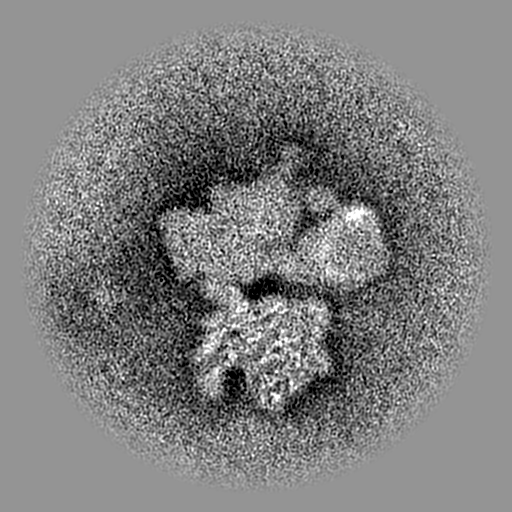
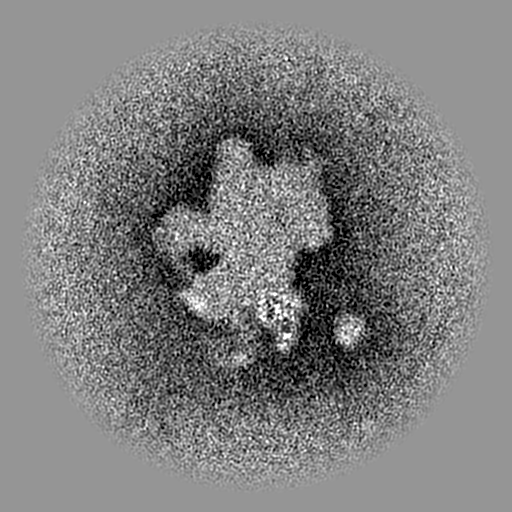
EMPIAR-11012 (Title: Pseudomonas aeruginosa 70S ribosome initiation complex bound to compact IF2 Data size: 1.7 TB Data #1: Unaligned multiframe micrographs of the Pseudomonas aeruginosa 70S ribosome initiation complex bound to IF2 collected on Gatan K3 in Super Resolution [micrographs - multiframe])
Entire : Focused map of the N2 sub-domain of IF2 bound to the 30S subunit ...
Entire
Name: Focused map of the N2 sub-domain of IF2 bound to the 30S subunit in the 70S ribosome initiation complex
Components
Complex: Focused map of the N2 sub-domain of IF2 bound to the 30S subunit in the 70S ribosome initiation complex
Protein or peptide: Translation initiation factor IF-2
-
Supramolecule #1: Focused map of the N2 sub-domain of IF2 bound to the 30S subunit ...
Supramolecule
Name: Focused map of the N2 sub-domain of IF2 bound to the 30S subunit in the 70S ribosome initiation complex type: complex / ID: 1 / Parent: 0 / Macromolecule list: all
Cryogen name: ETHANE / Chamber humidity: 85 % / Chamber temperature: 295 K / Instrument: LEICA EM GP
-
Electron microscopy
Microscope
TFS KRIOS
Image recording
Film or detector model: GATAN K3 BIOQUANTUM (6k x 4k) / Number grids imaged: 1 / Number real images: 8056 / Average exposure time: 1.0 sec. / Average electron dose: 31.0 e/Å2
Electron beam
Acceleration voltage: 300 kV / Electron source: FIELD EMISSION GUN
Applied symmetry - Point group: C1 (asymmetric) / Resolution.type: BY AUTHOR / Resolution: 2.8 Å / Resolution method: FSC 0.143 CUT-OFF / Software - Name: cryoSPARC (ver. 3.1.0) / Number images used: 123146
Initial angle assignment
Type: MAXIMUM LIKELIHOOD / Software - Name: cryoSPARC (ver. 3.1.0)
Final angle assignment
Type: MAXIMUM LIKELIHOOD / Software - Name: cryoSPARC (ver. 3.1.0)
Final 3D classification
Software - Name: cryoSPARC (ver. 3.1.0)
-
Atomic model buiding 1
Refinement
Space: REAL / Protocol: RIGID BODY FIT
Output model
PDB-7uiu: N2 sub-domain of IF2 bound to the 30S subunit in the Pseudomonas aeruginosa 70S ribosome initiation complex (focused classification and refinement)
+
About Yorodumi
-
News
-
Feb 9, 2022. New format data for meta-information of EMDB entries
New format data for meta-information of EMDB entries
Version 3 of the EMDB header file is now the official format.
The previous official version 1.9 will be removed from the archive.
In the structure databanks used in Yorodumi, some data are registered as the other names, "COVID-19 virus" and "2019-nCoV". Here are the details of the virus and the list of structure data.
Jan 31, 2019. EMDB accession codes are about to change! (news from PDBe EMDB page)
EMDB accession codes are about to change! (news from PDBe EMDB page)
The allocation of 4 digits for EMDB accession codes will soon come to an end. Whilst these codes will remain in use, new EMDB accession codes will include an additional digit and will expand incrementally as the available range of codes is exhausted. The current 4-digit format prefixed with “EMD-” (i.e. EMD-XXXX) will advance to a 5-digit format (i.e. EMD-XXXXX), and so on. It is currently estimated that the 4-digit codes will be depleted around Spring 2019, at which point the 5-digit format will come into force.
The EM Navigator/Yorodumi systems omit the EMD- prefix.
Related info.:Q: What is EMD? / ID/Accession-code notation in Yorodumi/EM Navigator
Yorodumi is a browser for structure data from EMDB, PDB, SASBDB, etc.
This page is also the successor to EM Navigator detail page, and also detail information page/front-end page for Omokage search.
The word "yorodu" (or yorozu) is an old Japanese word meaning "ten thousand". "mi" (miru) is to see.
Related info.:EMDB / PDB / SASBDB / Comparison of 3 databanks / Yorodumi Search / Aug 31, 2016. New EM Navigator & Yorodumi / Yorodumi Papers / Jmol/JSmol / Function and homology information / Changes in new EM Navigator and Yorodumi
 Movie
Movie Controller

 Yorodumi
 Open data
 Basic information
 Map data
 Sample
 Keywords
 Function and homology information
 Pseudomonas aeruginosa PAO1 (bacteria)
 Authors
 United States, 2 items
  Citation
 Structure visualization
 Downloads & links
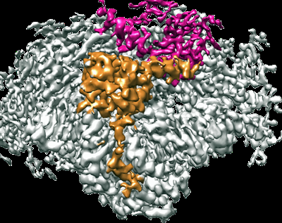 emd_26553.png
 http://ftp.pdbj.org/pub/emdb/structures/EMD-26553


 Z (Sec.)
 Y (Row.)
 X (Col.)


 Sample components
 Processing
 Electron microscopy
 FIELD EMISSION GUN
