 Movie
Movie Controller

[English] 日本語
 Yorodumi
- EMDB-2628: Structural similarity of secretins from Type II and Type III Secr... -
+ Open data

- Basic information
| Entry | Database: EMDB / ID: EMD-2628 | |||||||||
|---|---|---|---|---|---|---|---|---|---|---|
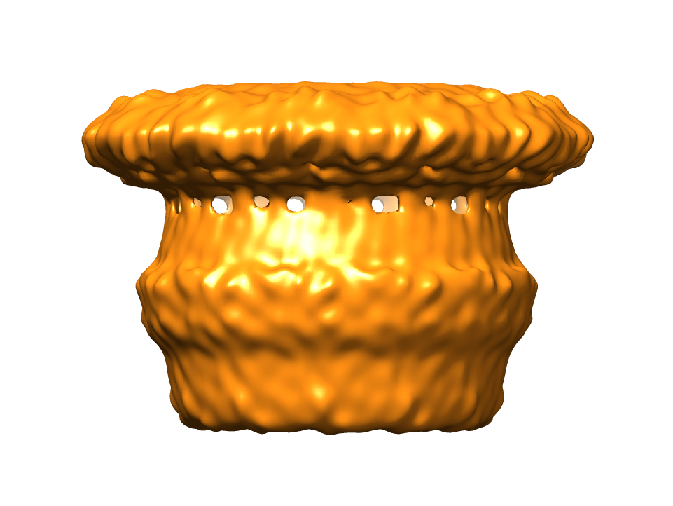
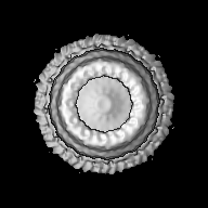
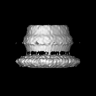
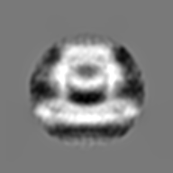
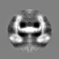
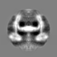
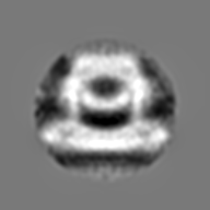
| Title | Structural similarity of secretins from Type II and Type III Secretion Systems | |||||||||
 Map data Map data | PulD | |||||||||
 Sample Sample |
| |||||||||
 Keywords Keywords | bacterial secretion system / membrane protein / electron microscopy / Single Particle Analysis / secretin | |||||||||
| Biological species |  Klebsiella oxytoca (bacteria) Klebsiella oxytoca (bacteria) | |||||||||
| Method | single particle reconstruction / cryo EM / Resolution: 8.2 Å | |||||||||
 Authors Authors | Tosi T / Estrozi LF / Job V / Guilvout I / Pugsley AP / Schoehn G / Dessen A | |||||||||
 Citation Citation | Journal: Structure / Year: 2014 Title: Structural similarity of secretins from type II and type III secretion systems. Authors: Tommaso Tosi / Leandro F Estrozi / Viviana Job / Ingrid Guilvout / Anthony P Pugsley / Guy Schoehn / Andréa Dessen /   Abstract: Secretins, the outer membrane components of several secretion systems in Gram-negative bacteria, assemble into channels that allow exoproteins to traverse the membrane. The membrane-inserted, ...Secretins, the outer membrane components of several secretion systems in Gram-negative bacteria, assemble into channels that allow exoproteins to traverse the membrane. The membrane-inserted, multimeric regions of PscC, the Pseudomonas aeruginosa type III secretion system secretin, and PulD, the Klebsiella oxytoca type II secretion system secretin, were purified after cell-free synthesis and their structures analyzed by single particle cryoelectron microscopy. Both homomultimeric, barrel-like structures display a "cup and saucer" architecture. The "saucer" region of both secretins is composed of two distinct rings, with that of PulD being less segmented than that of PscC. Both secretins have a central chamber that is occluded by a plug linked to the chamber walls through hairpin-like structures. Comparisons with published structures from other bacterial systems reveal that secretins have regions of local structural flexibility, probably reflecting their evolved functions in protein secretion and needle assembly. | |||||||||
| History |
|
- Structure visualization
| Movie |
 Movie viewer Movie viewer |
|---|---|
| Structure viewer | EM map: SurfViewMolmilJmol/JSmol |
| Supplemental images |

- Downloads & links
-EMDB archive
| Map data | emd_2628.map.gz | 21.7 MB | EMDB map data format | |
|---|---|---|---|---|
| Header (meta data) | emd-2628-v30.xmlemd-2628.xml | 8.1 KB 8.1 KB | Display Display | EMDB header |
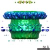
| Images |  EMD-2628.png EMD-2628.png puld_final.png puld_final.png | 255.1 KB 255.1 KB | ||
| Archive directory |  http://ftp.pdbj.org/pub/emdb/structures/EMD-2628ftp://ftp.pdbj.org/pub/emdb/structures/EMD-2628 http://ftp.pdbj.org/pub/emdb/structures/EMD-2628ftp://ftp.pdbj.org/pub/emdb/structures/EMD-2628 | HTTPS FTP |
-Validation report
| Summary document | emd_2628_validation.pdf.gz | 208.6 KB | Display | EMDB validaton report |
|---|---|---|---|---|
| Full document | emd_2628_full_validation.pdf.gz | 207.7 KB | Display | |
| Data in XML | emd_2628_validation.xml.gz | 6.1 KB | Display | |
| Arichive directory | https://ftp.pdbj.org/pub/emdb/validation_reports/EMD-2628ftp://ftp.pdbj.org/pub/emdb/validation_reports/EMD-2628 | HTTPS FTP |
-Related structure data


-Links
| EMDB pages | EMDB (EBI/PDBe) / EMDataResource |
|---|
-Map
| File | Download / File: emd_2628.map.gz / Format: CCP4 / Size: 26.4 MB / Type: IMAGE STORED AS FLOATING POINT NUMBER (4 BYTES) | ||||||||||||||||||||||||||||||||||||||||||||||||||||||||||||||||||||
|---|---|---|---|---|---|---|---|---|---|---|---|---|---|---|---|---|---|---|---|---|---|---|---|---|---|---|---|---|---|---|---|---|---|---|---|---|---|---|---|---|---|---|---|---|---|---|---|---|---|---|---|---|---|---|---|---|---|---|---|---|---|---|---|---|---|---|---|---|---|
| Annotation | PulD | ||||||||||||||||||||||||||||||||||||||||||||||||||||||||||||||||||||
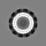
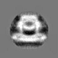
| Projections & slices | Image control
Images are generated by Spider. | ||||||||||||||||||||||||||||||||||||||||||||||||||||||||||||||||||||
| Voxel size | X=Y=Z: 1.3355 Å | ||||||||||||||||||||||||||||||||||||||||||||||||||||||||||||||||||||
| Density |
| ||||||||||||||||||||||||||||||||||||||||||||||||||||||||||||||||||||
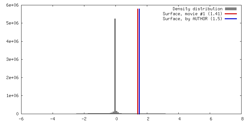
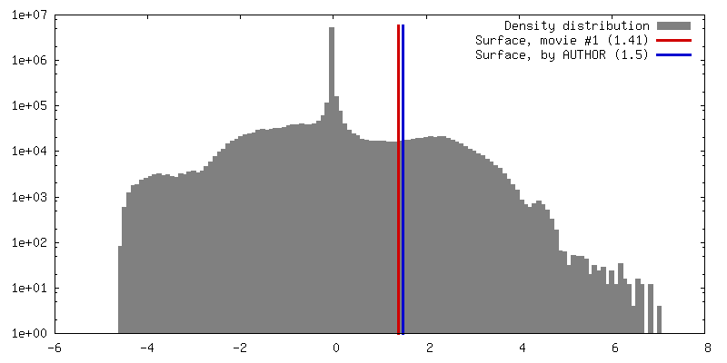
| Symmetry | Space group: 1 | ||||||||||||||||||||||||||||||||||||||||||||||||||||||||||||||||||||
| Details | EMDB XML:
CCP4 map header:
| ||||||||||||||||||||||||||||||||||||||||||||||||||||||||||||||||||||
 Z (Sec.)
 Y (Row.)
 X (Col.)


-Supplemental data
- Sample components
-Entire : PulD
| Entire | Name: PulD |
|---|---|
| Components |
|
-Supramolecule #1000: PulD
| Supramolecule | Name: PulD / type: sample / ID: 1000 / Oligomeric state: dodecamer / Number unique components: 1 |
|---|---|
| Molecular weight | Experimental: 468.6 KDa / Theoretical: 468.6 KDa |
-Macromolecule #1: PulD
| Macromolecule | Name: PulD / type: protein_or_peptide / ID: 1 / Number of copies: 1 / Oligomeric state: dodecamer / Recombinant expression: Yes |
|---|---|
| Source (natural) | Organism: Klebsiella oxytoca (bacteria) |
| Molecular weight | Experimental: 468.6 KDa / Theoretical: 468.6 KDa |
| Recombinant expression | Organism: Cell-free |
-Experimental details
-Structure determination
| Method | cryo EM |
|---|---|
 Processing Processing | single particle reconstruction |
| Aggregation state | particle |
-Sample preparation
| Concentration | 1.0 mg/mL |
|---|---|
| Grid | Details: 200 mesh coper quantifoil grid with thin carbon support |
| Vitrification | Cryogen name: ETHANE / Instrument: FEI VITROBOT MARK IV |
- Electron microscopy
| Microscope | FEI POLARA 300 |
|---|---|
| Date | Jan 1, 2012 |
| Image recording | Category: CCD / Film or detector model: GATAN ULTRASCAN 4000 (4k x 4k) / Number real images: 1060 / Average electron dose: 20 e/Å2 / Bits/pixel: 8 |
| Electron beam | Acceleration voltage: 300 kV / Electron source:  FIELD EMISSION GUN FIELD EMISSION GUN |
| Electron optics | Illumination mode: FLOOD BEAM / Imaging mode: BRIGHT FIELD / Cs: 2.0 mm / Nominal magnification: 59000 |
| Sample stage | Specimen holder model: GATAN LIQUID NITROGEN |
| Experimental equipment |  Model: Tecnai Polara / Image courtesy: FEI Company |
-Image processing
| Details | The particles were selected using an automatic selection program. |
|---|---|
| CTF correction | Details: Phase-flipping |
| Final reconstruction | Applied symmetry - Point group: C12 (12 fold cyclic) / Algorithm: OTHER / Resolution.type: BY AUTHOR / Resolution: 8.2 Å / Resolution method: OTHER / Software - Name: Imagic, RIco, bsoft, ctffind3, FPM / Number images used: 63500 |
