National Institutes of Health/National Institute Of Allergy and Infectious Diseases (NIH/NIAID)
R01AI089728
United States
National Institutes of Health/National Institute Of Allergy and Infectious Diseases (NIH/NIAID)
R01AI110700
United States
Citation
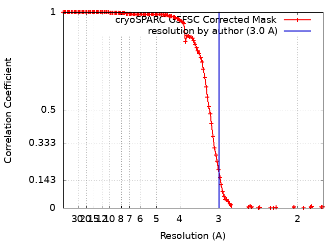
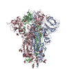
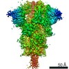
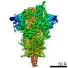
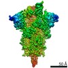
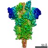
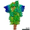
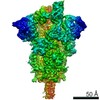
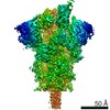
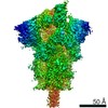
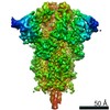
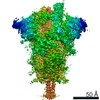
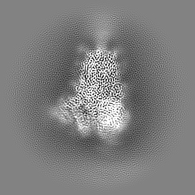
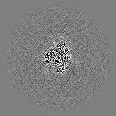
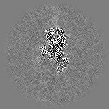
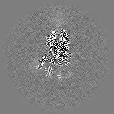
Journal: Nat Commun / Year: 2022 Title: Cryo-EM structure of a SARS-CoV-2 omicron spike protein ectodomain. Authors: Gang Ye / Bin Liu / Fang Li / Abstract: The omicron variant of SARS-CoV-2 has been spreading rapidly across the globe. The virus-surface spike protein plays a critical role in the cell entry and immune evasion of SARS-CoV-2. Here we ...The omicron variant of SARS-CoV-2 has been spreading rapidly across the globe. The virus-surface spike protein plays a critical role in the cell entry and immune evasion of SARS-CoV-2. Here we determined the 3.0 Å cryo-EM structure of the omicron spike protein ectodomain. In contrast to the original strain of SARS-CoV-2 where the receptor-binding domain (RBD) of the spike protein takes a mixture of open ("standing up") and closed ("lying down") conformations, the omicron spike molecules are predominantly in the open conformation, with one upright RBD ready for receptor binding. The open conformation of the omicron spike is stabilized by enhanced inter-domain and inter-subunit packing, which involves new mutations in the omicron strain. Moreover, the omicron spike has undergone extensive mutations in RBD regions where known neutralizing antibodies target, allowing the omicron variant to escape immune surveillance aimed at the original viral strain. The stable open conformation of the omicron spike sheds light on the cell entry and immune evasion mechanisms of the omicron variant.
History
Deposition
Jan 9, 2022
-
Header (metadata) release
Mar 9, 2022
-
Map release
Mar 9, 2022
-
Update
Nov 20, 2024
-
Current status
Nov 20, 2024
Processing site: RCSB / Status: Released
-
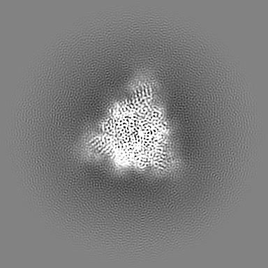
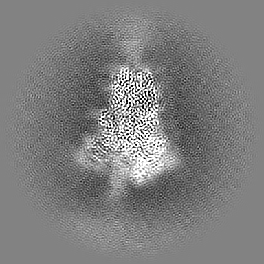
Structure visualization
Movie
Surface view with section colored by density value
In the structure databanks used in Yorodumi, some data are registered as the other names, "COVID-19 virus" and "2019-nCoV". Here are the details of the virus and the list of structure data.
Jan 31, 2019. EMDB accession codes are about to change! (news from PDBe EMDB page)
EMDB accession codes are about to change! (news from PDBe EMDB page)
The allocation of 4 digits for EMDB accession codes will soon come to an end. Whilst these codes will remain in use, new EMDB accession codes will include an additional digit and will expand incrementally as the available range of codes is exhausted. The current 4-digit format prefixed with “EMD-” (i.e. EMD-XXXX) will advance to a 5-digit format (i.e. EMD-XXXXX), and so on. It is currently estimated that the 4-digit codes will be depleted around Spring 2019, at which point the 5-digit format will come into force.
The EM Navigator/Yorodumi systems omit the EMD- prefix.
Related info.:Q: What is EMD? / ID/Accession-code notation in Yorodumi/EM Navigator
Yorodumi is a browser for structure data from EMDB, PDB, SASBDB, etc.
This page is also the successor to EM Navigator detail page, and also detail information page/front-end page for Omokage search.
The word "yorodu" (or yorozu) is an old Japanese word meaning "ten thousand". "mi" (miru) is to see.
Related info.:EMDB / PDB / SASBDB / Comparison of 3 databanks / Yorodumi Search / Aug 31, 2016. New EM Navigator & Yorodumi / Yorodumi Papers / Jmol/JSmol / Function and homology information / Changes in new EM Navigator and Yorodumi
 Movie
Movie Controller


 Open data
 Basic information
 Map data
 Sample
 Keywords
 Function and homology information
 Severe acute respiratory syndrome coronavirus 2
 Authors
 United States, 2 items
  Citation
 Structure visualization
 Downloads & links
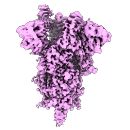 emd_25887.png
 http://ftp.pdbj.org/pub/emdb/structures/EMD-25887


 Z (Sec.)
 Y (Row.)
 X (Col.)


 Sample components
 Homo sapiens (human)
 Processing
 Electron microscopy
 FIELD EMISSION GUN