 Movie
Movie Controller


+ Open data

- Basic information
| Entry |  | |||||||||
|---|---|---|---|---|---|---|---|---|---|---|
| Title | Cryo-EM of the OmcE nanowires from Geobacter sulfurreducens | |||||||||
 Map data Map data | Cryo-EM of the OmcE nanowires from Geobacter sulfurreducens | |||||||||
 Sample Sample |
| |||||||||
| Function / homology | Doubled CXXCH motif / Doubled CXXCH motif (Paired_CXXCH_1) / Multiheme cytochrome superfamily / Cytochrome c Function and homology information Function and homology information | |||||||||
| Biological species |  Geobacter sulfurreducens (bacteria) Geobacter sulfurreducens (bacteria) | |||||||||
| Method | helical reconstruction / cryo EM / Resolution: 4.3 Å | |||||||||
 Authors Authors | Wang F / Mustafa K / Chan CH / Joshi K / Hochbaum AI / Bond DR / Egelman EH | |||||||||
| Funding support |  United States, 2 items United States, 2 items
| |||||||||
 Citation Citation | Journal: Nat Microbiol / Year: 2022 Title: Cryo-EM structure of an extracellular Geobacter OmcE cytochrome filament reveals tetrahaem packing. Authors: Fengbin Wang / Khawla Mustafa / Victor Suciu / Komal Joshi / Chi H Chan / Sol Choi / Zhangli Su / Dong Si / Allon I Hochbaum / Edward H Egelman / Daniel R Bond / Abstract: Electrically conductive appendages from the anaerobic bacterium Geobacter sulfurreducens were first observed two decades ago, with genetic and biochemical data suggesting that conductive fibres were ...Electrically conductive appendages from the anaerobic bacterium Geobacter sulfurreducens were first observed two decades ago, with genetic and biochemical data suggesting that conductive fibres were type IV pili. Recently, an extracellular conductive filament of G. sulfurreducens was found to contain polymerized c-type cytochrome OmcS subunits, not pilin subunits. Here we report that G. sulfurreducens also produces a second, thinner appendage comprised of cytochrome OmcE subunits and solve its structure using cryo-electron microscopy at ~4.3 Å resolution. Although OmcE and OmcS subunits have no overall sequence or structural similarities, upon polymerization both form filaments that share a conserved haem packing arrangement in which haems are coordinated by histidines in adjacent subunits. Unlike OmcS filaments, OmcE filaments are highly glycosylated. In extracellular fractions from G. sulfurreducens, we detected type IV pili comprising PilA-N and -C chains, along with abundant B-DNA. OmcE is the second cytochrome filament to be characterized using structural and biophysical methods. We propose that there is a broad class of conductive bacterial appendages with conserved haem packing (rather than sequence homology) that enable long-distance electron transport to chemicals or other microbial cells. | |||||||||
| History |
|
- Structure visualization
| Supplemental images |
|---|

- Downloads & links
-EMDB archive
| Map data | emd_25879.map.gz | 5.2 MB | EMDB map data format | |
|---|---|---|---|---|
| Header (meta data) | emd-25879-v30.xmlemd-25879.xml | 10.4 KB 10.4 KB | Display Display | EMDB header |
| Images | 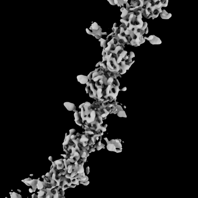 emd_25879.png emd_25879.png | 63.4 KB | ||
| Archive directory |  http://ftp.pdbj.org/pub/emdb/structures/EMD-25879ftp://ftp.pdbj.org/pub/emdb/structures/EMD-25879 http://ftp.pdbj.org/pub/emdb/structures/EMD-25879ftp://ftp.pdbj.org/pub/emdb/structures/EMD-25879 | HTTPS FTP |
-Validation report
| Summary document | emd_25879_validation.pdf.gz | 304.3 KB | Display | EMDB validaton report |
|---|---|---|---|---|
| Full document | emd_25879_full_validation.pdf.gz | 303.9 KB | Display | |
| Data in XML | emd_25879_validation.xml.gz | 6.6 KB | Display | |
| Data in CIF | emd_25879_validation.cif.gz | 7.6 KB | Display | |
| Arichive directory | https://ftp.pdbj.org/pub/emdb/validation_reports/EMD-25879ftp://ftp.pdbj.org/pub/emdb/validation_reports/EMD-25879 | HTTPS FTP |
-Related structure data
| Related structure data |  7tfsMC  7tggC M: atomic model generated by this map C: citing same article ( |
|---|---|
| Similar structure data |

-Links
| EMDB pages | EMDB (EBI/PDBe) / EMDataResource |
|---|---|
| Related items in Molecule of the Month |

-Map
| File | Download / File: emd_25879.map.gz / Format: CCP4 / Size: 125 MB / Type: IMAGE STORED AS FLOATING POINT NUMBER (4 BYTES) | ||||||||||||||||||||
|---|---|---|---|---|---|---|---|---|---|---|---|---|---|---|---|---|---|---|---|---|---|
| Annotation | Cryo-EM of the OmcE nanowires from Geobacter sulfurreducens | ||||||||||||||||||||
| Voxel size | X=Y=Z: 1.08 Å | ||||||||||||||||||||
| Density |
| ||||||||||||||||||||
| Symmetry | Space group: 1 | ||||||||||||||||||||
| Details | EMDB XML:
|
-Supplemental data
- Sample components
-Entire : Filament of OmcE protein
| Entire | Name: Filament of OmcE protein |
|---|---|
| Components |
|
-Supramolecule #1: Filament of OmcE protein
| Supramolecule | Name: Filament of OmcE protein / type: complex / Chimera: Yes / ID: 1 / Parent: 0 / Macromolecule list: #1 |
|---|---|
| Source (natural) | Organism: Geobacter sulfurreducens (bacteria) / Strain: ATCC 51573 / DSM 12127 / PCA |
-Macromolecule #1: Cytochrome c
| Macromolecule | Name: Cytochrome c / type: protein_or_peptide / ID: 1 / Number of copies: 1 / Enantiomer: LEVO |
|---|---|
| Source (natural) | Organism: Geobacter sulfurreducens (bacteria) / Strain: ATCC 51573 / DSM 12127 / PCA |
| Molecular weight | Theoretical: 23.839742 KDa |
| Sequence | String: MRSEVKIGLA LTALLVAVTA AGAASIKNTK HDLSSGSTGA TFKATNTDQI CVFCHTPHNA QQDIPLWNRG NPTASTFTLY SSSSMNNVP VKQGFTADSI SLFCMSCHDG ATGLGGAVHN DPNGAAIAMV GGNDLITGEA NLGTDLSNDH PVNFEVTPAG I AADGNLGA ...String: MRSEVKIGLA LTALLVAVTA AGAASIKNTK HDLSSGSTGA TFKATNTDQI CVFCHTPHNA QQDIPLWNRG NPTASTFTLY SSSSMNNVP VKQGFTADSI SLFCMSCHDG ATGLGGAVHN DPNGAAIAMV GGNDLITGEA NLGTDLSNDH PVNFEVTPAG I AADGNLGA LDTGTNPPTM KTGDVTNGLP LFKSARGATT LECGSCHKVH DNTDAPFLRT TMAGSKLCLG CHKK |
-Macromolecule #2: HEME C
| Macromolecule | Name: HEME C / type: ligand / ID: 2 / Number of copies: 4 / Formula: HEC |
|---|---|
| Molecular weight | Theoretical: 618.503 Da |
| Chemical component information |  ChemComp-HEC: |
-Experimental details
-Structure determination
| Method | cryo EM |
|---|---|
 Processing Processing | helical reconstruction |
| Aggregation state | filament |
-Sample preparation
| Buffer | pH: 6 |
|---|---|
| Vitrification | Cryogen name: ETHANE |
- Electron microscopy
| Microscope | FEI TITAN KRIOS |
|---|---|
| Image recording | Film or detector model: GATAN K3 (6k x 4k) / Average electron dose: 50.0 e/Å2 |
| Electron beam | Acceleration voltage: 300 kV / Electron source:  FIELD EMISSION GUN FIELD EMISSION GUN |
| Electron optics | Illumination mode: FLOOD BEAM / Imaging mode: BRIGHT FIELD / Nominal defocus max: 3.0 µm / Nominal defocus min: 1.0 µm |
| Experimental equipment |  Model: Titan Krios / Image courtesy: FEI Company |
-Image processing
| Final reconstruction | Applied symmetry - Helical parameters - Δz: 33.9 Å Applied symmetry - Helical parameters - Δ&Phi: 58.8 ° Applied symmetry - Helical parameters - Axial symmetry: C1 (asymmetric) Resolution.type: BY AUTHOR / Resolution: 4.3 Å / Resolution method: FSC 0.143 CUT-OFF / Number images used: 346826 |
|---|---|
| Final angle assignment | Type: NOT APPLICABLE |
