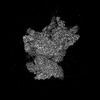
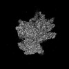
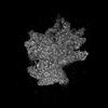
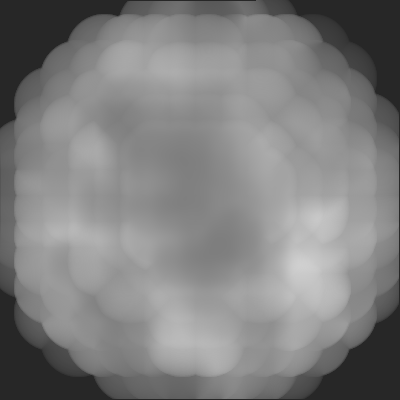
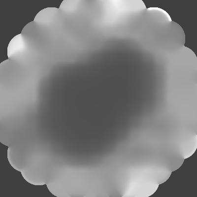
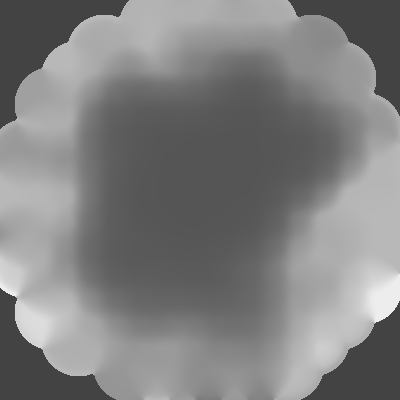
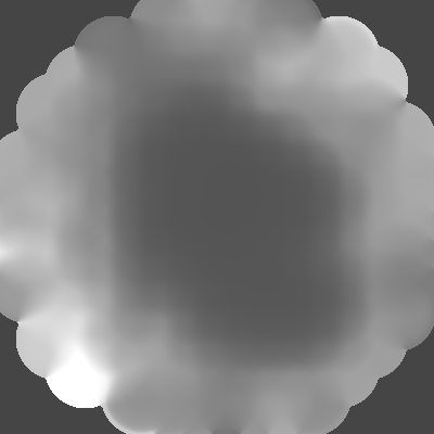
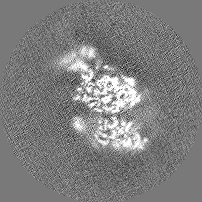
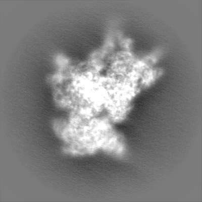
- EMDB-25534: Structure of the HCV IRES bound to the 40S ribosomal subunit, hea... -
+
Open data
ID or keywords:
Loading...
-
Basic information
Entry
Database: EMDB / ID: EMD-25534
Title
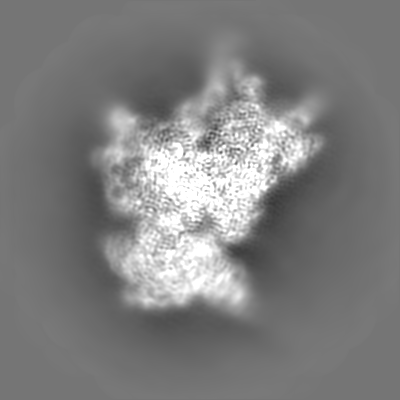
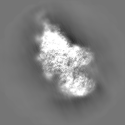
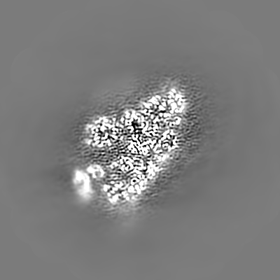
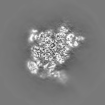
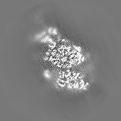
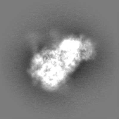
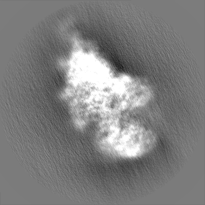
Structure of the HCV IRES bound to the 40S ribosomal subunit, head opening. Structure 8(delta dII)
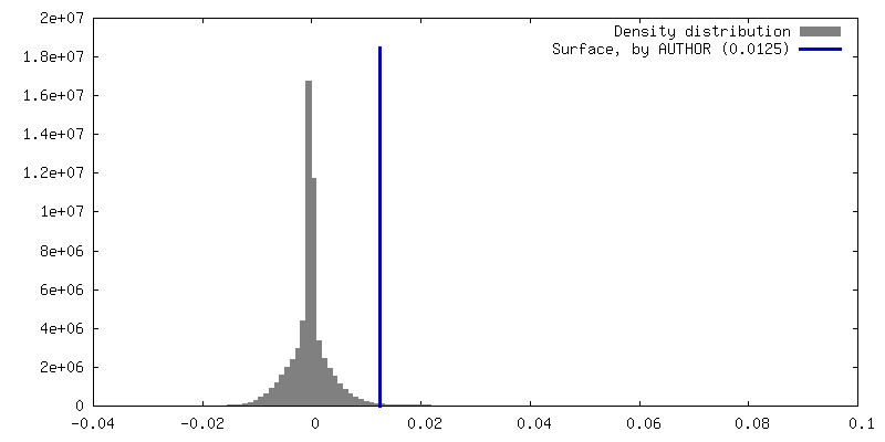
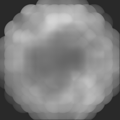
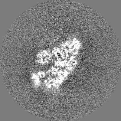
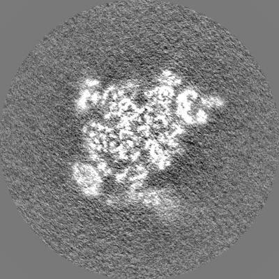
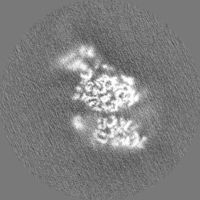
Map data
Post processed map
Sample
Complex: 40S ribosomal small subunit with HCV IRES
RNA: x 2 types
Protein or peptide: x 33 types
Protein or peptide: x 1 types
Ligand: x 1 types
Keywords
HCV / IRES / 40S / RIBOSOME
Function / homology
Function and homology information
laminin receptor activity / 90S preribosome / ubiquitin ligase inhibitor activity / positive regulation of signal transduction by p53 class mediator / phagocytic cup / translation regulator activity / rough endoplasmic reticulum / ribosomal small subunit export from nucleus / laminin binding / gastrulation ...laminin receptor activity / 90S preribosome / ubiquitin ligase inhibitor activity / positive regulation of signal transduction by p53 class mediator / phagocytic cup / translation regulator activity / rough endoplasmic reticulum / ribosomal small subunit export from nucleus / laminin binding / gastrulation / MDM2/MDM4 family protein binding / cytosolic ribosome / class I DNA-(apurinic or apyrimidinic site) endonuclease activity / DNA-(apurinic or apyrimidinic site) lyase / maturation of LSU-rRNA from tricistronic rRNA transcript (SSU-rRNA, 5.8S rRNA, LSU-rRNA) / positive regulation of apoptotic signaling pathway / maturation of SSU-rRNA from tricistronic rRNA transcript (SSU-rRNA, 5.8S rRNA, LSU-rRNA) / maturation of SSU-rRNA / small-subunit processome / spindle / rRNA processing / regulation of translation / rhythmic process / positive regulation of canonical Wnt signaling pathway / ribosomal small subunit assembly / virus receptor activity / ribosome binding / ribosomal small subunit biogenesis / small ribosomal subunit / small ribosomal subunit rRNA binding / cytosolic small ribosomal subunit / cytosolic large ribosomal subunit / perikaryon / cell differentiation / cytoplasmic translation / mitochondrial inner membrane / postsynaptic density / rRNA binding / structural constituent of ribosome / ribosome / translation / ribonucleoprotein complex / cell division / DNA repair / mRNA binding / apoptotic process / centrosome / synapse / dendrite / nucleolus / perinuclear region of cytoplasm / Golgi apparatus / DNA binding / RNA binding / zinc ion binding / membrane / nucleus / plasma membrane / cytoplasm Similarity search - Function
40S ribosomal protein SA / 40S ribosomal protein SA, C-terminal domain / 40S ribosomal protein SA C-terminus / Ubiquitin-like protein FUBI / : / Ribosomal protein S26e signature. / Ribosomal protein L41 / Ribosomal protein L41 / Ribosomal protein S21e, conserved site / Ribosomal protein S21e signature. ...40S ribosomal protein SA / 40S ribosomal protein SA, C-terminal domain / 40S ribosomal protein SA C-terminus / Ubiquitin-like protein FUBI / : / Ribosomal protein S26e signature. / Ribosomal protein L41 / Ribosomal protein L41 / Ribosomal protein S21e, conserved site / Ribosomal protein S21e signature. / : / Ribosomal protein S12e signature. / Ribosomal protein S26e / Ribosomal protein S26e superfamily / Ribosomal protein S26e / Ribosomal protein S12e / Small (40S) ribosomal subunit Asc1/RACK1 / Ribosomal protein S21e / Ribosomal protein S21e superfamily / Ribosomal protein S21e / Ribosomal protein S19e, conserved site / Ribosomal protein S19e signature. / Ribosomal protein S2, eukaryotic / 40S Ribosomal protein S10 / S27a-like superfamily / Plectin/S10, N-terminal / Plectin/S10 domain / Ribosomal protein S30 / Ribosomal protein S30 / Ribosomal protein S10, eukaryotic/archaeal / : / Ribosomal protein S7e signature. / Ribosomal protein S8e subdomain, eukaryotes / Ribosomal protein S25 / S25 ribosomal protein / Ribosomal protein S17e, conserved site / Ribosomal protein S17e signature. / Ribosomal protein S2, eukaryotic/archaeal / Ribosomal protein S27a / Ribosomal protein S27a / Ribosomal protein S27a / Ribosomal protein S3Ae, conserved site / Ribosomal protein S3Ae signature. / 40S ribosomal protein S29/30S ribosomal protein S14 type Z / Ribosomal protein S3, eukaryotic/archaeal / 40S ribosomal protein S4, C-terminal domain / 40S ribosomal protein S4 C-terminus / Ribosomal protein L7, eukaryotic / Ribosomal protein L30, N-terminal / Ribosomal L30 N-terminal domain / Ribosomal protein S4e, N-terminal, conserved site / Ribosomal protein S4e signature. / Ribosomal protein S8e, conserved site / Ribosomal protein S8e signature. / Ribosomal protein S19A/S15e / Ribosomal protein S27e signature. / Ribosomal protein S6, eukaryotic / Ribosomal protein S19e / Ribosomal protein S19e / Ribosomal_S19e / Ribosomal protein S17e / Ribosomal protein S17e-like superfamily / Ribosomal S17 / 40S ribosomal protein S1/3, eukaryotes / 40S ribosomal protein S11, N-terminal / Ribosomal_S17 N-terminal / Ribosomal protein S7e / Ribosomal protein S7e / : / Ribosomal S24e conserved site / Ribosomal protein S24e signature. / Ribosomal protein S4e, N-terminal / RS4NT (NUC023) domain / Ribosomal protein S4, KOW domain / Ribosomal protein S4e / Ribosomal protein S4e, central region / Ribosomal protein S4e, central domain superfamily / Ribosomal family S4e / Ribosomal protein S28e conserved site / Ribosomal protein S28e signature. / Ribosomal protein S6/S6e/A/B/2, conserved site / Ribosomal protein S17, archaeal/eukaryotic / Ribosomal protein S6e signature. / Ribosomal protein S23, eukaryotic/archaeal / Ribosomal protein S24e / Ribosomal protein S24e / Ribosomal protein S8e / Ribosomal protein S27 / Ribosomal protein S27, zinc-binding domain superfamily / Ribosomal protein S27 / Ribosomal protein S3Ae / Ribosomal S3Ae family / Ribosomal S3Ae family / Ribosomal protein S28e / Ribosomal protein S28e / Ribosomal protein S6e / Ribosomal protein S6e / Ribosomal protein S5/S7, eukaryotic/archaeal / Ribosomal protein S6e / Ribosomal protein S13/S15, N-terminal Similarity search - Domain/homology
Small ribosomal subunit protein eS32 / Small ribosomal subunit protein uS4 / Small ribosomal subunit protein eS12 / Small ribosomal subunit protein uS9 / Small ribosomal subunit protein uS10 / Small ribosomal subunit protein RACK1 / Ubiquitin-ribosomal protein eS31 fusion protein / Small ribosomal subunit protein uS15 / Small ribosomal subunit protein eS1 / Small ribosomal subunit protein eS7 ...Small ribosomal subunit protein eS32 / Small ribosomal subunit protein uS4 / Small ribosomal subunit protein eS12 / Small ribosomal subunit protein uS9 / Small ribosomal subunit protein uS10 / Small ribosomal subunit protein RACK1 / Ubiquitin-ribosomal protein eS31 fusion protein / Small ribosomal subunit protein uS15 / Small ribosomal subunit protein eS1 / Small ribosomal subunit protein eS7 / Small ribosomal subunit protein uS12 / 40S ribosomal protein S24 / Ubiquitin-like FUBI-ribosomal protein eS30 fusion protein / Small ribosomal subunit protein eS25 / Small ribosomal subunit protein eS26 / Small ribosomal subunit protein uS7 / Small ribosomal subunit protein uS8 / Small ribosomal subunit protein eS28 / Small ribosomal subunit protein eS8 / Small ribosomal subunit protein eS4 / Small ribosomal subunit protein uS2 / Small ribosomal subunit protein eS6 / Small ribosomal subunit protein eS21 / Small ribosomal subunit protein eS19 / Small ribosomal subunit protein uS3 / Small ribosomal subunit protein uS13 / Small ribosomal subunit protein eS10 / Small ribosomal subunit protein uS17 / Small ribosomal subunit protein eS17 / Large ribosomal subunit protein uL30 / Small ribosomal subunit protein eS27 / Small ribosomal subunit protein uS19 / Small ribosomal subunit protein uS11 / Small ribosomal subunit protein uS14 Similarity search - Component
Biological species
Oryctolagus cuniculus (rabbit) / Hepatitis C virus (isolate 1)
Method
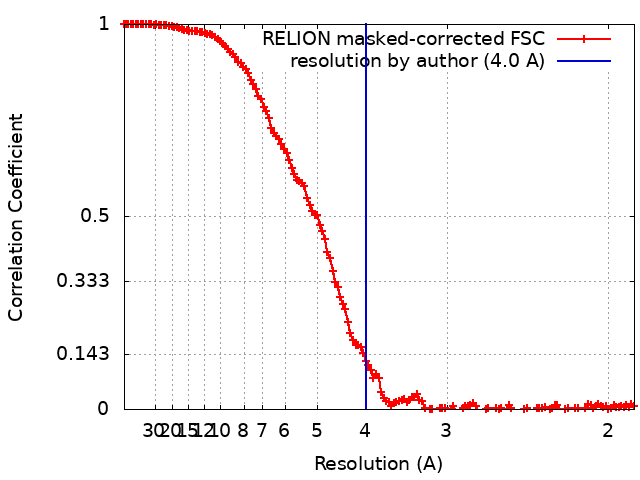
single particle reconstruction / cryo EM / Resolution: 4.0 Å
Name: eL41 / type: protein_or_peptide / ID: 35 / Number of copies: 1 / Enantiomer: LEVO
Source (natural)
Organism: Oryctolagus cuniculus (rabbit)
Molecular weight
Theoretical: 3.473451 KDa
Sequence
String:
MRAKWRKKRM RRLKRKRRKM RQRSK
UniProtKB: Small ribosomal subunit protein eS32
+
Macromolecule #37: ZINC ION
Macromolecule
Name: ZINC ION / type: ligand / ID: 37 / Number of copies: 2 / Formula: ZN
Molecular weight
Theoretical: 65.409 Da
-
Experimental details
-
Structure determination
Method
cryo EM
Processing
single particle reconstruction
Aggregation state
particle
-
Sample preparation
Concentration
0.000075 mg/mL
Buffer
pH: 7.5
Grid
Model: Quantifoil R0.6/1 / Material: GOLD / Mesh: 300 / Support film - Material: GOLD / Support film - topology: HOLEY / Support film - Film thickness: 50 / Pretreatment - Type: PLASMA CLEANING / Pretreatment - Time: 25 sec. / Pretreatment - Atmosphere: OTHER / Details: H2/O2 mixture for 25 seconds at 25W power
Vitrification
Cryogen name: ETHANE-PROPANE / Chamber humidity: 100 % / Chamber temperature: 277.15 K / Instrument: FEI VITROBOT MARK IV / Details: 4 second blot time, force 3.
-
Electron microscopy
Microscope
FEI TECNAI F30
Image recording
Film or detector model: GATAN K3 (6k x 4k) / Average exposure time: 4.0 sec. / Average electron dose: 70.9 e/Å2
Electron beam
Acceleration voltage: 300 kV / Electron source: FIELD EMISSION GUN
In the structure databanks used in Yorodumi, some data are registered as the other names, "COVID-19 virus" and "2019-nCoV". Here are the details of the virus and the list of structure data.
Jan 31, 2019. EMDB accession codes are about to change! (news from PDBe EMDB page)
EMDB accession codes are about to change! (news from PDBe EMDB page)
The allocation of 4 digits for EMDB accession codes will soon come to an end. Whilst these codes will remain in use, new EMDB accession codes will include an additional digit and will expand incrementally as the available range of codes is exhausted. The current 4-digit format prefixed with “EMD-” (i.e. EMD-XXXX) will advance to a 5-digit format (i.e. EMD-XXXXX), and so on. It is currently estimated that the 4-digit codes will be depleted around Spring 2019, at which point the 5-digit format will come into force.
The EM Navigator/Yorodumi systems omit the EMD- prefix.
Related info.:Q: What is EMD? / ID/Accession-code notation in Yorodumi/EM Navigator
Yorodumi is a browser for structure data from EMDB, PDB, SASBDB, etc.
This page is also the successor to EM Navigator detail page, and also detail information page/front-end page for Omokage search.
The word "yorodu" (or yorozu) is an old Japanese word meaning "ten thousand". "mi" (miru) is to see.
Related info.:EMDB / PDB / SASBDB / Comparison of 3 databanks / Yorodumi Search / Aug 31, 2016. New EM Navigator & Yorodumi / Yorodumi Papers / Jmol/JSmol / Function and homology information / Changes in new EM Navigator and Yorodumi
 Movie
Movie Controller

 Yorodumi
 Open data
 Basic information
 Map data
 Sample
 Keywords
 Function and homology information
 Hepatitis C virus (isolate 1)
 Authors
 United States, 4 items
  Citation
 Structure visualization
 Downloads & links
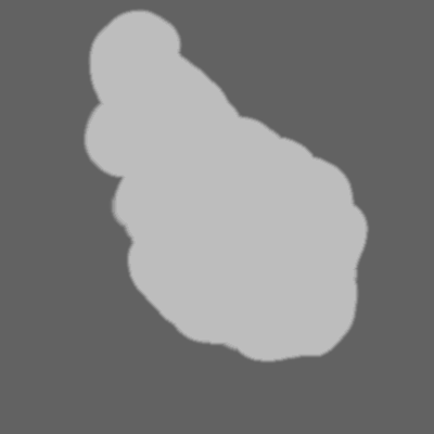
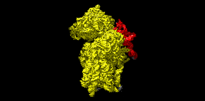 emd_25534.png
 http://ftp.pdbj.org/pub/emdb/structures/EMD-25534


 Z (Sec.)
 Y (Row.)
 X (Col.)


 Sample components
 Processing
 Electron microscopy
 FIELD EMISSION GUN