 Movie
Movie Controller

[English] 日本語
 Yorodumi
- EMDB-2489: Determination of protein structure at 8.5 Angstrom resolution usi... -
+ Open data

- Basic information
| Entry | Database: EMDB / ID: EMD-2489 | |||||||||
|---|---|---|---|---|---|---|---|---|---|---|
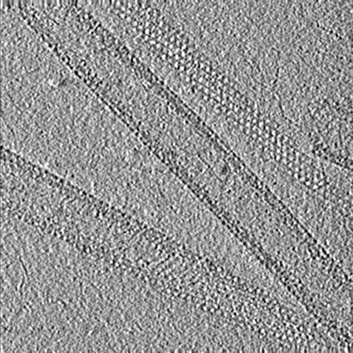
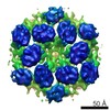
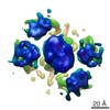
| Title | Determination of protein structure at 8.5 Angstrom resolution using cryo-electron microscopy and subtomogram averaging | |||||||||
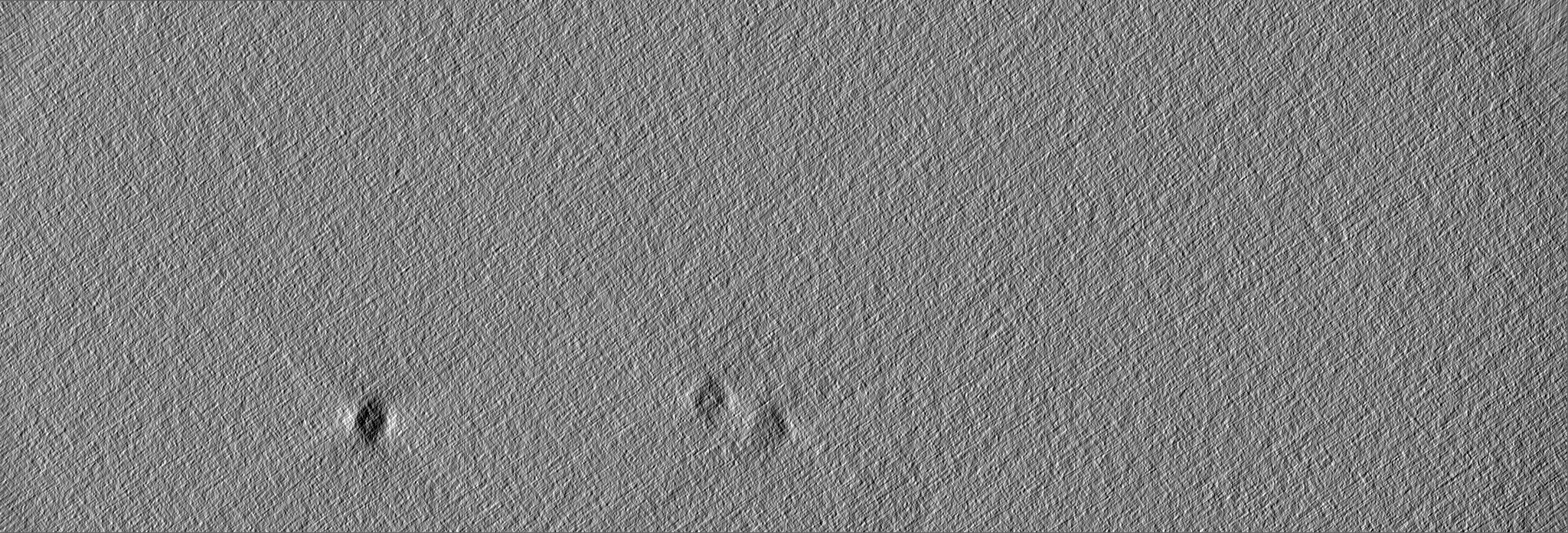
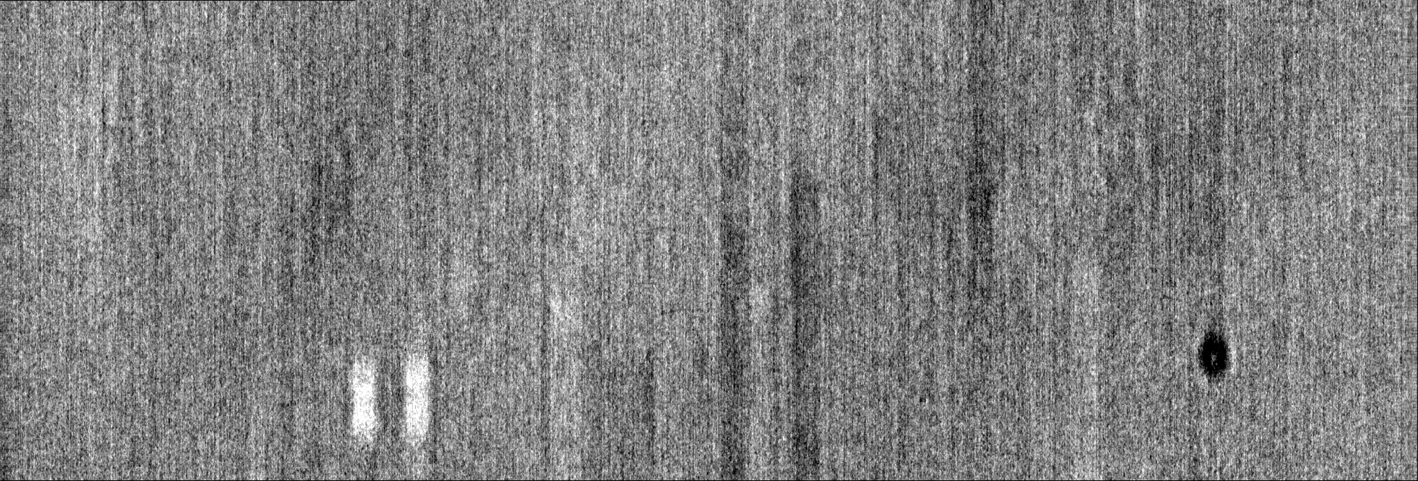
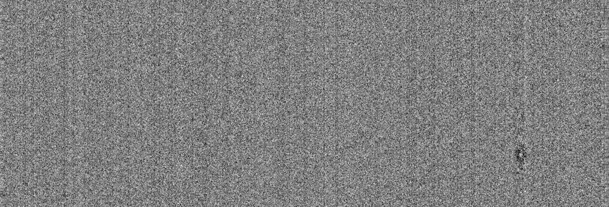
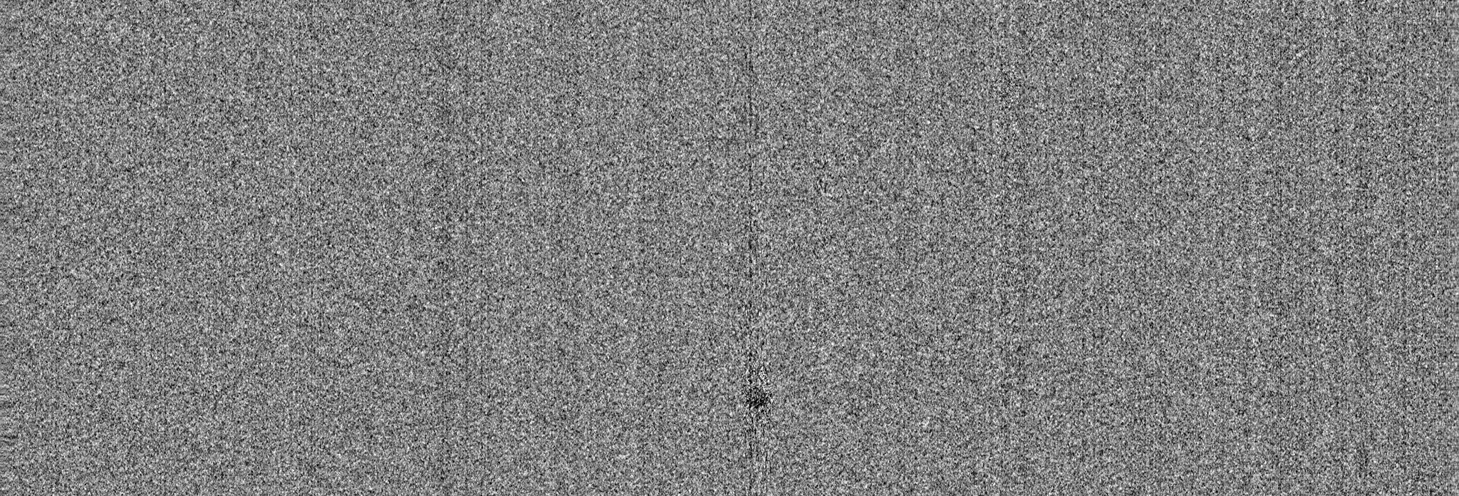
 Map data Map data | Tomogram reconstruction of a tilt-series containing M-MPMV dPro CANC tubes | |||||||||
 Sample Sample |
| |||||||||
 Keywords Keywords | Cryo-electron Tomography / Sub-tomogram Averaging / Retrovirus / Capsid | |||||||||
| Biological species |  Mason-Pfizer monkey virus Mason-Pfizer monkey virus | |||||||||
| Method | electron tomography / cryo EM | |||||||||
 Authors Authors | Schur FKM / Hagen W / de Marco A / Briggs JAG | |||||||||
 Citation Citation | Journal: J Struct Biol / Year: 2013 Title: Determination of protein structure at 8.5Å resolution using cryo-electron tomography and sub-tomogram averaging. Authors: Florian K M Schur / Wim J H Hagen / Alex de Marco / John A G Briggs /  Abstract: Cryo-electron tomography combined with image processing by sub-tomogram averaging is unique in its power to resolve the structures of proteins and macromolecular complexes in situ. Limitations of the ...Cryo-electron tomography combined with image processing by sub-tomogram averaging is unique in its power to resolve the structures of proteins and macromolecular complexes in situ. Limitations of the method, including the low signal to noise ratio within individual images from cryo-tomographic datasets and difficulties in determining the defocus at which the data was collected, mean that to date the very best structures obtained by sub-tomogram averaging are limited to a resolution of approximately 15 Å. Here, by optimizing data collection and defocus determination steps, we have determined the structure of assembled Mason-Pfizer monkey virus Gag protein using sub-tomogram averaging to a resolution of 8.5 Å. At this resolution alpha-helices can be directly and clearly visualized. These data demonstrate for the first time that high-resolution structural information can be obtained from cryo-electron tomograms using sub-tomogram averaging. Sub-tomogram averaging has the potential to allow detailed studies of unsolved and biologically relevant structures under biologically relevant conditions. | |||||||||
| History |
|
- Structure visualization
| Movie |
 Movie viewer Movie viewer |
|---|---|
| Supplemental images |
- Downloads & links
-EMDB archive
| Map data | emd_2489.map.gz | 5.1 GB | EMDB map data format | |
|---|---|---|---|---|
| Header (meta data) | emd-2489-v30.xmlemd-2489.xml | 8.5 KB 8.5 KB | Display Display | EMDB header |
| Images | emd_2489.tif | 800.4 KB | ||
| Archive directory |  http://ftp.pdbj.org/pub/emdb/structures/EMD-2489ftp://ftp.pdbj.org/pub/emdb/structures/EMD-2489 http://ftp.pdbj.org/pub/emdb/structures/EMD-2489ftp://ftp.pdbj.org/pub/emdb/structures/EMD-2489 | HTTPS FTP |
-Validation report
| Summary document | emd_2489_validation.pdf.gz | 141.3 KB | Display | EMDB validaton report |
|---|---|---|---|---|
| Full document | emd_2489_full_validation.pdf.gz | 140.4 KB | Display | |
| Data in XML | emd_2489_validation.xml.gz | 4.1 KB | Display | |
| Arichive directory | https://ftp.pdbj.org/pub/emdb/validation_reports/EMD-2489ftp://ftp.pdbj.org/pub/emdb/validation_reports/EMD-2489 | HTTPS FTP |
-Related structure data
-Links
| EMDB pages | EMDB (EBI/PDBe) / EMDataResource |
|---|
-Map
| File | Download / File: emd_2489.map.gz / Format: CCP4 / Size: 5.3 GB / Type: IMAGE STORED AS SIGNED INTEGER (2 BYTES) | ||||||||||||||||||||||||||||||||||||||||||||||||||||||||||||||||||||
|---|---|---|---|---|---|---|---|---|---|---|---|---|---|---|---|---|---|---|---|---|---|---|---|---|---|---|---|---|---|---|---|---|---|---|---|---|---|---|---|---|---|---|---|---|---|---|---|---|---|---|---|---|---|---|---|---|---|---|---|---|---|---|---|---|---|---|---|---|---|
| Annotation | Tomogram reconstruction of a tilt-series containing M-MPMV dPro CANC tubes | ||||||||||||||||||||||||||||||||||||||||||||||||||||||||||||||||||||
| Projections & slices | Image control
Images are generated by Spider. generated in cubic-lattice coordinate | ||||||||||||||||||||||||||||||||||||||||||||||||||||||||||||||||||||
| Voxel size | X=Y=Z: 2.025 Å | ||||||||||||||||||||||||||||||||||||||||||||||||||||||||||||||||||||
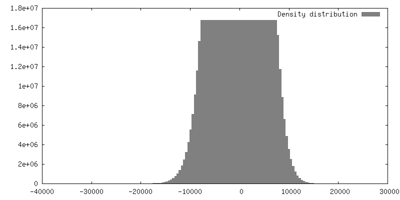
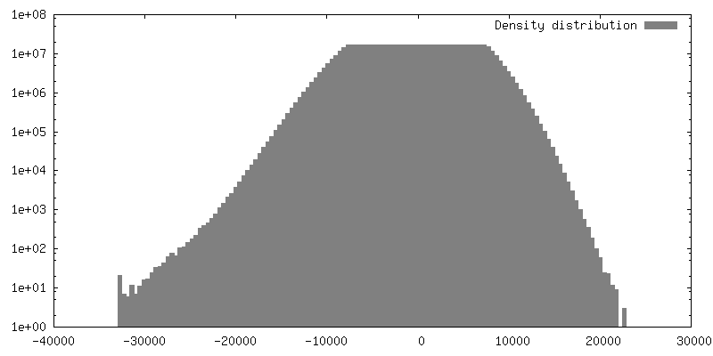
| Density |
| ||||||||||||||||||||||||||||||||||||||||||||||||||||||||||||||||||||
| Symmetry | Space group: 1 | ||||||||||||||||||||||||||||||||||||||||||||||||||||||||||||||||||||
| Details | EMDB XML:
CCP4 map header:
| ||||||||||||||||||||||||||||||||||||||||||||||||||||||||||||||||||||
 Z (Sec.)
 Y (Row.)
 X (Col.)


-Supplemental data
- Sample components
-Entire : M-MPV CANC Gag lattice
| Entire | Name: M-MPV CANC Gag lattice |
|---|---|
| Components |
|
-Supramolecule #1000: M-MPV CANC Gag lattice
| Supramolecule | Name: M-MPV CANC Gag lattice / type: sample / ID: 1000 / Oligomeric state: helical / Number unique components: 1 |
|---|
-Macromolecule #1: M-MPV dPro CANC protein
| Macromolecule | Name: M-MPV dPro CANC protein / type: protein_or_peptide / ID: 1 / Oligomeric state: helical / Recombinant expression: Yes |
|---|---|
| Source (natural) | Organism: Mason-Pfizer monkey virus |
| Recombinant expression | Organism:  |
-Experimental details
-Structure determination
| Method | cryo EM |
|---|---|
 Processing Processing | electron tomography |
| Aggregation state | helical array |
-Sample preparation
| Buffer | pH: 7.7 / Details: 50mM Tris, 100mM NaCl,1uM Zn |
|---|---|
| Grid | Details: 300mesh copper, glow discharged |
| Vitrification | Cryogen name: ETHANE / Instrument: OTHER |
- Electron microscopy
| Microscope | FEI TITAN KRIOS |
|---|---|
| Specialist optics | Energy filter - Name: GIF2002 postcolumn energy filter |
| Date | May 21, 2013 |
| Image recording | Category: CCD / Film or detector model: GENERIC GATAN (2k x 2k) / Number real images: 36 / Average electron dose: 40 e/Å2 |
| Electron beam | Acceleration voltage: 200 kV / Electron source:  FIELD EMISSION GUN FIELD EMISSION GUN |
| Electron optics | Illumination mode: FLOOD BEAM / Imaging mode: BRIGHT FIELD / Cs: 2.7 mm / Nominal defocus max: 3.3 µm / Nominal defocus min: 3.3 µm / Nominal magnification: 42000 |
| Sample stage | Specimen holder: Liquid nitrogen cooled / Specimen holder model: FEI TITAN KRIOS AUTOGRID HOLDER / Tilt series - Axis1 - Min angle: -45 ° / Tilt series - Axis1 - Max angle: 60 ° / Tilt series - Axis1 - Angle increment: 3 ° |
| Experimental equipment |  Model: Titan Krios / Image courtesy: FEI Company |
-Image processing
| Details | Tilt series was reconstructed using IMOD |
|---|---|
| Final reconstruction | Algorithm: OTHER / Software - Name: IMOD / Number images used: 36 |
