 Movie
Movie Controller


+ Open data

- Basic information
| Entry | Database: EMDB / ID: EMD-2444 | |||||||||
|---|---|---|---|---|---|---|---|---|---|---|
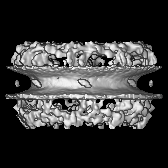
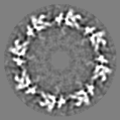
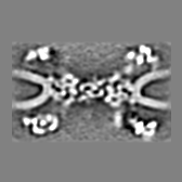
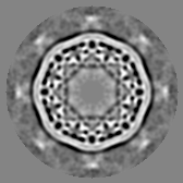
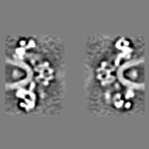
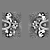
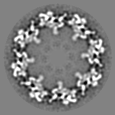
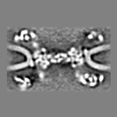
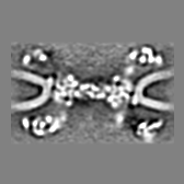
| Title | Cryo-electron tomography of the human nuclear pore complex | |||||||||
 Map data Map data | Reconstruction of the human nuclear pore complex | |||||||||
 Sample Sample |
| |||||||||
 Keywords Keywords | nuclear pore complex | |||||||||
| Biological species |  Homo sapiens (human) Homo sapiens (human) | |||||||||
| Method | subtomogram averaging / Resolution: 35.0 Å | |||||||||
 Authors Authors | Bui KH / von Appen A / DiGuilio AL / Ori A / Sparks L / Mackmull MT / Bock T / Hagen W / Andres-Pons A / Glavy JS / Beck M | |||||||||
 Citation Citation | Journal: Cell / Year: 2013 Title: Integrated structural analysis of the human nuclear pore complex scaffold. Authors: Khanh Huy Bui / Alexander von Appen / Amanda L DiGuilio / Alessandro Ori / Lenore Sparks / Marie-Therese Mackmull / Thomas Bock / Wim Hagen / Amparo Andrés-Pons / Joseph S Glavy / Martin Beck /  Abstract: The nuclear pore complex (NPC) is a fundamental component of all eukaryotic cells that facilitates nucleocytoplasmic exchange of macromolecules. It is assembled from multiple copies of about 30 ...The nuclear pore complex (NPC) is a fundamental component of all eukaryotic cells that facilitates nucleocytoplasmic exchange of macromolecules. It is assembled from multiple copies of about 30 nucleoporins. Due to its size and complex composition, determining the structure of the NPC is an enormous challenge, and the overall architecture of the NPC scaffold remains elusive. In this study, we have used an integrated approach based on electron tomography, single-particle electron microscopy, and crosslinking mass spectrometry to determine the structure of a major scaffold motif of the human NPC, the Nup107 subcomplex, in both isolation and integrated into the NPC. We show that 32 copies of the Nup107 subcomplex assemble into two reticulated rings, one each at the cytoplasmic and nuclear face of the NPC. This arrangement may explain how changes of the diameter are realized that would accommodate transport of huge cargoes. | |||||||||
| History |
|
- Structure visualization
| Movie |
 Movie viewer Movie viewer |
|---|---|
| Structure viewer | EM map: SurfViewMolmilJmol/JSmol |
| Supplemental images |

- Downloads & links
-EMDB archive
| Map data | emd_2444.map.gz | 6.3 MB | EMDB map data format | |
|---|---|---|---|---|
| Header (meta data) | emd-2444-v30.xmlemd-2444.xml | 10.3 KB 10.3 KB | Display Display | EMDB header |
| Images | 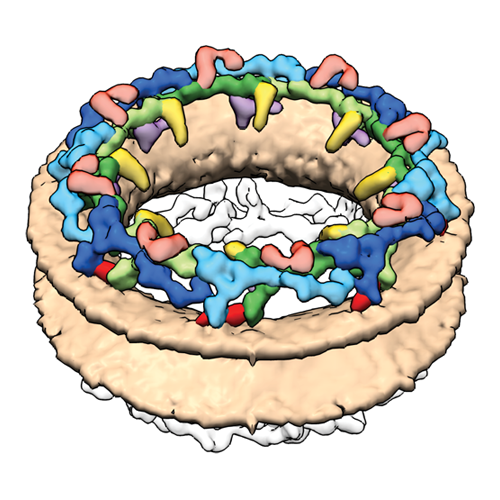 emd_2444.png emd_2444.png | 254.7 KB | ||
| Archive directory |  http://ftp.pdbj.org/pub/emdb/structures/EMD-2444ftp://ftp.pdbj.org/pub/emdb/structures/EMD-2444 http://ftp.pdbj.org/pub/emdb/structures/EMD-2444ftp://ftp.pdbj.org/pub/emdb/structures/EMD-2444 | HTTPS FTP |
-Validation report
| Summary document | emd_2444_validation.pdf.gz | 243.3 KB | Display | EMDB validaton report |
|---|---|---|---|---|
| Full document | emd_2444_full_validation.pdf.gz | 242.4 KB | Display | |
| Data in XML | emd_2444_validation.xml.gz | 6.1 KB | Display | |
| Arichive directory | https://ftp.pdbj.org/pub/emdb/validation_reports/EMD-2444ftp://ftp.pdbj.org/pub/emdb/validation_reports/EMD-2444 | HTTPS FTP |
-Related structure data


-Links
| EMDB pages | EMDB (EBI/PDBe) / EMDataResource |
|---|
-Map
| File | Download / File: emd_2444.map.gz / Format: CCP4 / Size: 17.7 MB / Type: IMAGE STORED AS FLOATING POINT NUMBER (4 BYTES) | ||||||||||||||||||||||||||||||||||||||||||||||||||||||||||||||||||||
|---|---|---|---|---|---|---|---|---|---|---|---|---|---|---|---|---|---|---|---|---|---|---|---|---|---|---|---|---|---|---|---|---|---|---|---|---|---|---|---|---|---|---|---|---|---|---|---|---|---|---|---|---|---|---|---|---|---|---|---|---|---|---|---|---|---|---|---|---|---|
| Annotation | Reconstruction of the human nuclear pore complex | ||||||||||||||||||||||||||||||||||||||||||||||||||||||||||||||||||||
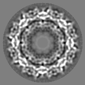
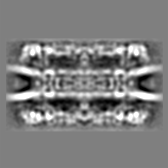
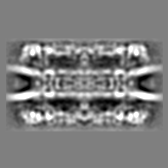
| Projections & slices | Image control
Images are generated by Spider. | ||||||||||||||||||||||||||||||||||||||||||||||||||||||||||||||||||||
| Voxel size | X=Y=Z: 8.6 Å | ||||||||||||||||||||||||||||||||||||||||||||||||||||||||||||||||||||
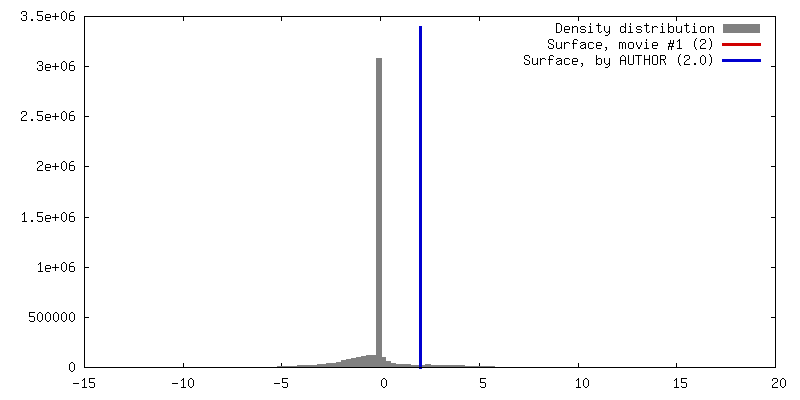
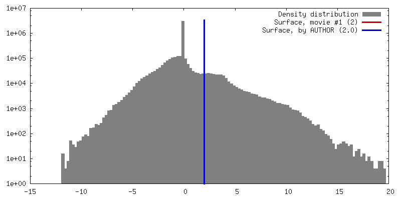
| Density |
| ||||||||||||||||||||||||||||||||||||||||||||||||||||||||||||||||||||
| Symmetry | Space group: 1 | ||||||||||||||||||||||||||||||||||||||||||||||||||||||||||||||||||||
| Details | EMDB XML:
CCP4 map header:
| ||||||||||||||||||||||||||||||||||||||||||||||||||||||||||||||||||||
 Z (Sec.)
 Y (Row.)
 X (Col.)


-Supplemental data
- Sample components
-Entire : Human Nuclear Pore Complex
| Entire | Name: Human Nuclear Pore Complex |
|---|---|
| Components |
|
-Supramolecule #1000: Human Nuclear Pore Complex
| Supramolecule | Name: Human Nuclear Pore Complex / type: sample / ID: 1000 Details: The sample is purified human nuclear envelope containing nuclear pore complex. Number unique components: 1 |
|---|---|
| Molecular weight | Method: Absolute Quantitative Mass Spectrometry |
-Macromolecule #1: Nuclear Pore Complex
| Macromolecule | Name: Nuclear Pore Complex / type: protein_or_peptide / ID: 1 / Number of copies: 1 / Recombinant expression: No / Database: NCBI |
|---|---|
| Source (natural) | Organism: Homo sapiens (human) / synonym: Human / Organelle: Nucleus / Location in cell: Nuclear pore complex |
| Molecular weight | Experimental: 120 MDa / Theoretical: 120 MDa |
-Experimental details
-Structure determination
 Processing Processing | subtomogram averaging |
|---|---|
| Aggregation state | particle |
-Sample preparation
| Buffer | pH: 7.5 / Details: 20mM Tris, 0.2-0.4% Trehalose |
|---|---|
| Grid | Details: 200 mesh Quantifoil Copper Holey Carbon Grid R2/1 |
| Vitrification | Cryogen name: NONE / Instrument: HOMEMADE PLUNGER |
- Electron microscopy
| Microscope | FEI TITAN KRIOS |
|---|---|
| Specialist optics | Energy filter - Name: GIF2002 / Energy filter - Lower energy threshold: 0.0 eV / Energy filter - Upper energy threshold: 20.0 eV |
| Date | Sep 20, 2012 |
| Image recording | Category: CCD / Film or detector model: GATAN MULTISCAN / Average electron dose: 100 e/Å2 / Bits/pixel: 16 |
| Electron beam | Acceleration voltage: 300 kV / Electron source:  FIELD EMISSION GUN FIELD EMISSION GUN |
| Electron optics | Calibrated magnification: 34883 / Illumination mode: FLOOD BEAM / Imaging mode: BRIGHT FIELD / Cs: 2.7 mm / Nominal defocus max: 5.0 µm / Nominal defocus min: 3.0 µm / Nominal magnification: 19500 |
| Sample stage | Specimen holder model: FEI TITAN KRIOS AUTOGRID HOLDER / Tilt series - Axis1 - Min angle: -60 ° / Tilt series - Axis1 - Max angle: 60 ° |
| Experimental equipment |  Model: Titan Krios / Image courtesy: FEI Company |
-Image processing
| Details | The subtomograms were picked manually and further processed iterative symmetry independent averaging. |
|---|---|
| Final reconstruction | Algorithm: OTHER / Resolution.type: BY AUTHOR / Resolution: 35.0 Å / Resolution method: FSC 0.5 CUT-OFF / Software - Name: IMOD, TOM, AV3 Details: Local Resolution. Cytoplasmic Ring 32 Angstrom. Spoke Ring 34 Angstrom. Nuclear Ring 35 Angstrom. Number subtomograms used: 10000 |
