 Movie
Movie Controller

[English] 日本語
 Yorodumi
- EMDB-24417: Microtubule subtomogram average from particles with a uniform sta... -
+ Open data

- Basic information
| Entry | Database: EMDB / ID: EMD-24417 | |||||||||
|---|---|---|---|---|---|---|---|---|---|---|
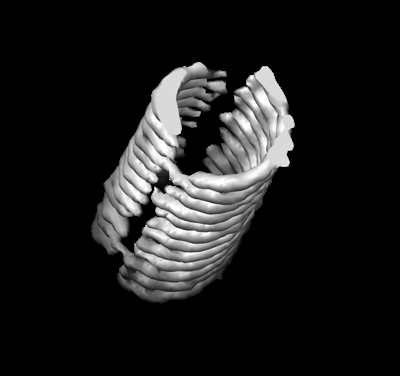
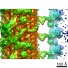
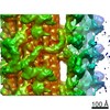
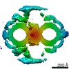
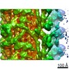
| Title | Microtubule subtomogram average from particles with a uniform starting orientation with respect to the missing wedge | |||||||||
 Map data Map data | ||||||||||
 Sample Sample |
| |||||||||
| Biological species |  Homo sapiens (human) Homo sapiens (human) | |||||||||
| Method | subtomogram averaging / cryo EM / Resolution: 25.0 Å | |||||||||
 Authors Authors | Croxford M / Villa E | |||||||||
| Funding support |  United States, 1 items United States, 1 items
| |||||||||
 Citation Citation | Journal: Proc Natl Acad Sci U S A / Year: 2021 Title: Entropy-regularized deconvolution of cellular cryotransmission electron tomograms. Authors: Matthew Croxford / Michael Elbaum / Muthuvel Arigovindan / Zvi Kam / David Agard / Elizabeth Villa / John Sedat /   Abstract: Cryo-electron tomography (cryo-ET) allows for the high-resolution visualization of biological macromolecules. However, the technique is limited by a low signal-to-noise ratio (SNR) and variance in ...Cryo-electron tomography (cryo-ET) allows for the high-resolution visualization of biological macromolecules. However, the technique is limited by a low signal-to-noise ratio (SNR) and variance in contrast at different frequencies, as well as reduced Z resolution. Here, we applied entropy-regularized deconvolution (ER-DC) to cryo-ET data generated from transmission electron microscopy (TEM) and reconstructed using weighted back projection (WBP). We applied deconvolution to several in situ cryo-ET datasets and assessed the results by Fourier analysis and subtomogram analysis (STA). | |||||||||
| History |
|
- Structure visualization
| Movie |
 Movie viewer Movie viewer |
|---|---|
| Structure viewer | EM map: SurfViewMolmilJmol/JSmol |
| Supplemental images |
- Downloads & links
-EMDB archive
| Map data | emd_24417.map.gz | 778.4 KB | EMDB map data format | |
|---|---|---|---|---|
| Header (meta data) | emd-24417-v30.xmlemd-24417.xml | 13.5 KB 13.5 KB | Display Display | EMDB header |
| Images |  emd_24417.png emd_24417.png | 42.5 KB | ||
| Others | emd_24417_half_map_1.map.gzemd_24417_half_map_2.map.gz | 777.4 KB 778.2 KB | ||
| Archive directory |  http://ftp.pdbj.org/pub/emdb/structures/EMD-24417ftp://ftp.pdbj.org/pub/emdb/structures/EMD-24417 http://ftp.pdbj.org/pub/emdb/structures/EMD-24417ftp://ftp.pdbj.org/pub/emdb/structures/EMD-24417 | HTTPS FTP |
-Validation report
| Summary document | emd_24417_validation.pdf.gz | 467.3 KB | Display | EMDB validaton report |
|---|---|---|---|---|
| Full document | emd_24417_full_validation.pdf.gz | 466.9 KB | Display | |
| Data in XML | emd_24417_validation.xml.gz | 6.7 KB | Display | |
| Data in CIF | emd_24417_validation.cif.gz | 7.8 KB | Display | |
| Arichive directory | https://ftp.pdbj.org/pub/emdb/validation_reports/EMD-24417ftp://ftp.pdbj.org/pub/emdb/validation_reports/EMD-24417 | HTTPS FTP |
-Related structure data
| Related structure data | C: citing same article ( |
|---|---|
| Similar structure data |
-Links
| EMDB pages | EMDB (EBI/PDBe) / EMDataResource |
|---|---|
| Related items in Molecule of the Month |

-Map
| File | Download / File: emd_24417.map.gz / Format: CCP4 / Size: 844.7 KB / Type: IMAGE STORED AS FLOATING POINT NUMBER (4 BYTES) | ||||||||||||||||||||||||||||||||||||||||||||||||||||||||||||||||||||
|---|---|---|---|---|---|---|---|---|---|---|---|---|---|---|---|---|---|---|---|---|---|---|---|---|---|---|---|---|---|---|---|---|---|---|---|---|---|---|---|---|---|---|---|---|---|---|---|---|---|---|---|---|---|---|---|---|---|---|---|---|---|---|---|---|---|---|---|---|---|
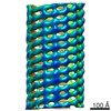
| Projections & slices | Image control
Images are generated by Spider. | ||||||||||||||||||||||||||||||||||||||||||||||||||||||||||||||||||||
| Voxel size | X=Y=Z: 8.984 Å | ||||||||||||||||||||||||||||||||||||||||||||||||||||||||||||||||||||
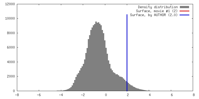
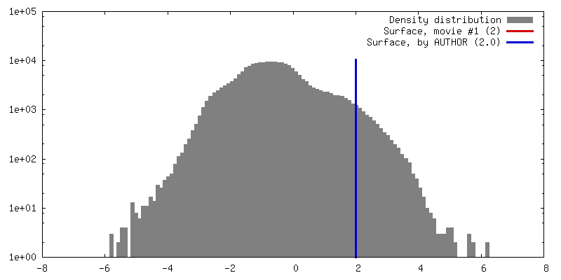
| Density |
| ||||||||||||||||||||||||||||||||||||||||||||||||||||||||||||||||||||
| Symmetry | Space group: 1 | ||||||||||||||||||||||||||||||||||||||||||||||||||||||||||||||||||||
| Details | EMDB XML:
CCP4 map header:
| ||||||||||||||||||||||||||||||||||||||||||||||||||||||||||||||||||||
 Z (Sec.)
 Y (Row.)
 X (Col.)


-Supplemental data
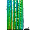
-Half map: #1
| File | emd_24417_half_map_1.map | ||||||||||||
|---|---|---|---|---|---|---|---|---|---|---|---|---|---|
| Projections & Slices |
| ||||||||||||
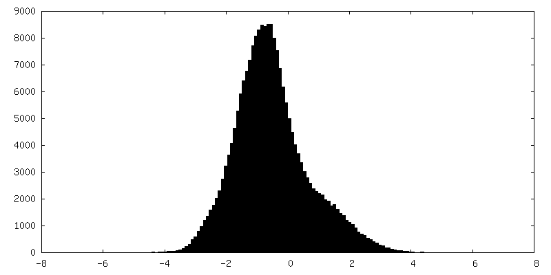
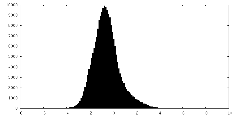
| Density Histograms |


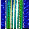
-Half map: #2
| File | emd_24417_half_map_2.map | ||||||||||||
|---|---|---|---|---|---|---|---|---|---|---|---|---|---|
| Projections & Slices |
| ||||||||||||
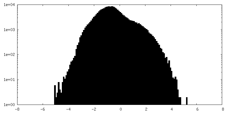
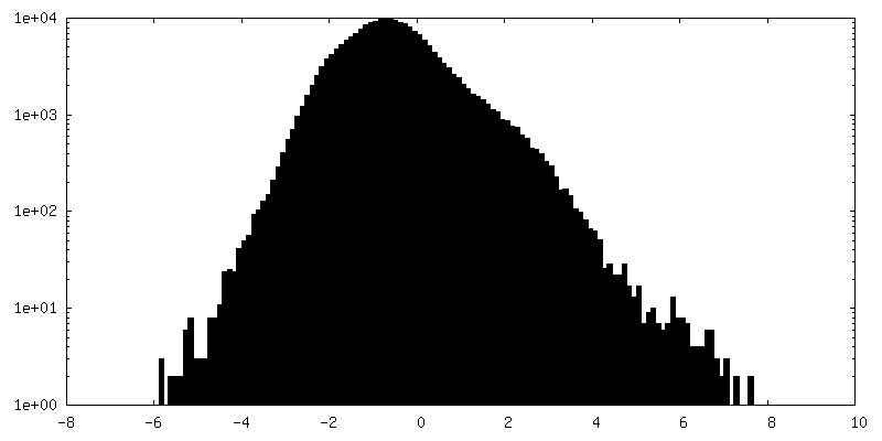
| Density Histograms |


- Sample components
-Entire : Microtubule
| Entire | Name: Microtubule |
|---|---|
| Components |
|
-Supramolecule #1: Microtubule
| Supramolecule | Name: Microtubule / type: organelle_or_cellular_component / ID: 1 / Parent: 0 / Details: Subtomogram average of a polymerized microtubule. |
|---|---|
| Source (natural) | Organism: Homo sapiens (human) / Strain: HEK293 / Organ: Kidney |
-Experimental details
-Structure determination
| Method | cryo EM |
|---|---|
 Processing Processing | subtomogram averaging |
| Aggregation state | filament |
-Sample preparation
| Buffer | pH: 7.4 |
|---|---|
| Grid | Model: Quantifoil / Support film - Material: CARBON |
| Vitrification | Cryogen name: ETHANE-PROPANE |
- Electron microscopy
| Microscope | FEI TITAN KRIOS |
|---|---|
| Image recording | Film or detector model: GATAN K2 SUMMIT (4k x 4k) / Average electron dose: 96.0 e/Å2 |
| Electron beam | Acceleration voltage: 300 kV / Electron source:  FIELD EMISSION GUN FIELD EMISSION GUN |
| Electron optics | Illumination mode: FLOOD BEAM / Imaging mode: BRIGHT FIELD / Cs: 2.7 mm |
| Sample stage | Cooling holder cryogen: NITROGEN |
| Experimental equipment |  Model: Titan Krios / Image courtesy: FEI Company |
-Image processing
| Final reconstruction | Applied symmetry - Point group: C1 (asymmetric) / Resolution.type: BY AUTHOR / Resolution: 25.0 Å / Resolution method: DIFFRACTION PATTERN/LAYERLINES / Software - Name: Dynamo (ver. 11514) / Number subtomograms used: 300 |
|---|---|
| Extraction | Number tomograms: 1 / Number images used: 300 / Method: Filament Tracing / Software - Name: Dynamo (ver. 11514) Details: A single MT filament parallel to the tilt axis was traced and sampled every 4nm. |
| Final 3D classification | Number classes: 1 |
| Final angle assignment | Type: PROJECTION MATCHING / Software - Name: Dynamo (ver. 11514) |
