 Movie
Movie Controller


+ Open data

- Basic information
| Entry | Database: EMDB / ID: EMD-20411 | ||||||||||||
|---|---|---|---|---|---|---|---|---|---|---|---|---|---|
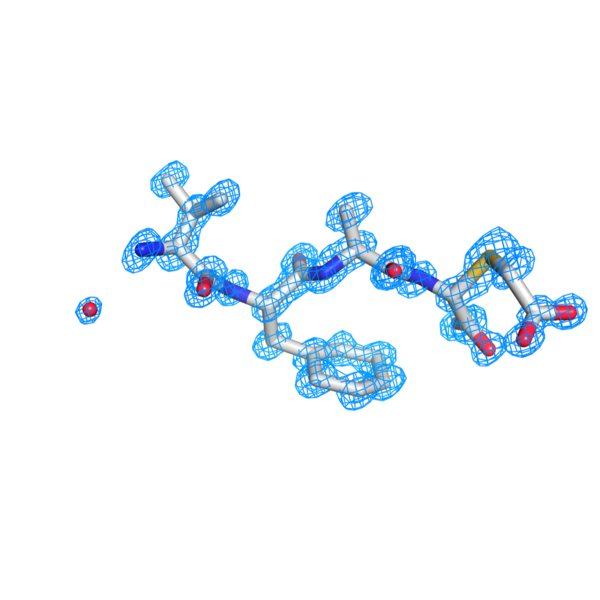
| Title | MicroED Structure of a Natural Product VFAThiaGlu | ||||||||||||
 Map data Map data | Natural Product VFAThiaGlu | ||||||||||||
 Sample Sample |
| ||||||||||||
 Keywords Keywords | Ribosomal synthesized small peptide / MicroED / microcrystal electron diffraction / UNKNOWN FUNCTION | ||||||||||||
| Biological species |  Pseudomonas syringae (bacteria) Pseudomonas syringae (bacteria) | ||||||||||||
| Method | electron crystallography / cryo EM | ||||||||||||
 Authors Authors | Halaby S / Gonen T | ||||||||||||
| Funding support |  United States, 3 items United States, 3 items
| ||||||||||||
 Citation Citation | Journal: Science / Year: 2019 Title: Use of a scaffold peptide in the biosynthesis of amino acid-derived natural products. Authors: Chi P Ting / Michael A Funk / Steve L Halaby / Zhengan Zhang / Tamir Gonen / Wilfred A van der Donk / Abstract: Genome sequencing of environmental bacteria allows identification of biosynthetic gene clusters encoding unusual combinations of enzymes that produce unknown natural products. We identified a pathway ...Genome sequencing of environmental bacteria allows identification of biosynthetic gene clusters encoding unusual combinations of enzymes that produce unknown natural products. We identified a pathway in which a ribosomally synthesized small peptide serves as a scaffold for nonribosomal peptide extension and chemical modification. Amino acids are transferred to the carboxyl terminus of the peptide through adenosine triphosphate and amino acyl-tRNA-dependent chemistry that is independent of the ribosome. Oxidative rearrangement, carboxymethylation, and proteolysis of a terminal cysteine yields an amino acid-derived small molecule. Microcrystal electron diffraction demonstrates that the resulting product is isosteric to glutamate. We show that a similar peptide extension is used during the biosynthesis of the ammosamides, which are cytotoxic pyrroloquinoline alkaloids. These results suggest an alternative paradigm for biosynthesis of amino acid-derived natural products. | ||||||||||||
| History |
|
- Structure visualization
| Movie |
 Movie viewer Movie viewer |
|---|---|
| Structure viewer | EM map: SurfViewMolmilJmol/JSmol |
| Supplemental images |
- Downloads & links
-EMDB archive
| Map data | emd_20411.map.gz | 233.1 KB | EMDB map data format | |
|---|---|---|---|---|
| Header (meta data) | emd-20411-v30.xmlemd-20411.xml | 11.9 KB 11.9 KB | Display Display | EMDB header |
| Images |  emd_20411.png emd_20411.png | 103.2 KB | ||
| Filedesc metadata | emd-20411.cif.gz | 4.6 KB | ||
| Filedesc structureFactors | emd_20411_sf.cif.gz | 50.3 KB | ||
| Archive directory |  http://ftp.pdbj.org/pub/emdb/structures/EMD-20411ftp://ftp.pdbj.org/pub/emdb/structures/EMD-20411 http://ftp.pdbj.org/pub/emdb/structures/EMD-20411ftp://ftp.pdbj.org/pub/emdb/structures/EMD-20411 | HTTPS FTP |
-Related structure data
-Links
| EMDB pages | EMDB (EBI/PDBe) / EMDataResource |
|---|
-Map
| File | Download / File: emd_20411.map.gz / Format: CCP4 / Size: 1004.9 KB / Type: IMAGE STORED AS FLOATING POINT NUMBER (4 BYTES) | ||||||||||||||||||||||||||||||||||||||||||||||||||||||||||||||||||||
|---|---|---|---|---|---|---|---|---|---|---|---|---|---|---|---|---|---|---|---|---|---|---|---|---|---|---|---|---|---|---|---|---|---|---|---|---|---|---|---|---|---|---|---|---|---|---|---|---|---|---|---|---|---|---|---|---|---|---|---|---|---|---|---|---|---|---|---|---|---|
| Annotation | Natural Product VFAThiaGlu | ||||||||||||||||||||||||||||||||||||||||||||||||||||||||||||||||||||
| Voxel size | X: 0.30188 Å / Y: 0.29938 Å / Z: 0.31947 Å | ||||||||||||||||||||||||||||||||||||||||||||||||||||||||||||||||||||
| Density |
| ||||||||||||||||||||||||||||||||||||||||||||||||||||||||||||||||||||
| Symmetry | Space group: 4 | ||||||||||||||||||||||||||||||||||||||||||||||||||||||||||||||||||||
| Details | EMDB XML:
CCP4 map header:
| ||||||||||||||||||||||||||||||||||||||||||||||||||||||||||||||||||||
-Supplemental data
- Sample components
-Entire : VAL-PHE-ALA-ThiaGLU
| Entire | Name: VAL-PHE-ALA-ThiaGLU |
|---|---|
| Components |
|
-Supramolecule #1: VAL-PHE-ALA-ThiaGLU
| Supramolecule | Name: VAL-PHE-ALA-ThiaGLU / type: complex / ID: 1 / Parent: 0 / Macromolecule list: #1 |
|---|---|
| Source (natural) | Organism: Pseudomonas syringae (bacteria) |
-Macromolecule #1: YFAThiaGlu
| Macromolecule | Name: YFAThiaGlu / type: protein_or_peptide / ID: 1 / Number of copies: 1 / Enantiomer: LEVO |
|---|---|
| Source (natural) | Organism: Pseudomonas syringae (bacteria) |
| Molecular weight | Theoretical: 482.55 Da |
| Sequence | String: VFA(OV7) |
-Macromolecule #2: water
| Macromolecule | Name: water / type: ligand / ID: 2 / Number of copies: 1 / Formula: HOH |
|---|---|
| Molecular weight | Theoretical: 18.015 Da |
| Chemical component information |  ChemComp-HOH: |
-Experimental details
-Structure determination
| Method | cryo EM |
|---|---|
 Processing Processing | electron crystallography |
| Aggregation state | 3D array |
-Sample preparation
| Buffer | pH: 7 |
|---|---|
| Grid | Model: Quantifoil R1.2/1.3 / Material: COPPER / Mesh: 300 |
| Vitrification | Cryogen name: NITROGEN / Instrument: HOMEMADE PLUNGER |
- Electron microscopy
| Microscope | FEI TALOS ARCTICA |
|---|---|
| Temperature | Min: 77.0 K / Max: 100.0 K |
| Image recording | Film or detector model: FEI FALCON III (4k x 4k) / Digitization - Dimensions - Width: 2048 pixel / Digitization - Dimensions - Height: 2048 pixel / Number grids imaged: 1 / Average exposure time: 1.0 sec. / Average electron dose: 0.01 e/Å2 |
| Electron beam | Acceleration voltage: 200 kV / Electron source:  FIELD EMISSION GUN FIELD EMISSION GUN |
| Electron optics | Illumination mode: OTHER / Imaging mode: DIFFRACTION / Camera length: 1100 mm |
| Sample stage | Specimen holder model: FEI TITAN KRIOS AUTOGRID HOLDER / Cooling holder cryogen: NITROGEN |
| Experimental equipment |  Model: Talos Arctica / Image courtesy: FEI Company |
-Image processing
| Final reconstruction | Resolution method: DIFFRACTION PATTERN/LAYERLINES |
|---|---|
| Crystallography statistics | Number intensities measured: 7590 / Number structure factors: 3047 / Fourier space coverage: 95.4 / R sym: 23.5 / R merge: 23.5 / Overall phase error: 0 / Overall phase residual: 15 / Phase error rejection criteria: 0 / High resolution: 0.9 Å / Shell - Shell ID: 1 / Shell - High resolution: 0.9 Å / Shell - Low resolution: 0.93 Å / Shell - Number structure factors: 642 / Shell - Phase residual: 15 / Shell - Fourier space coverage: 92.8 / Shell - Multiplicity: 2.25 |
-Atomic model buiding 1
| Refinement | Space: RECIPROCAL / Protocol: AB INITIO MODEL / Overall B value: 3.746 |
|---|---|
| Output model | 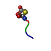 PDB-6po6: |
