Movie
Movie Controller

[English] 日本語
 Yorodumi
- EMDB-20360: MicroED structure of proteinase K from an unpolished, platinum-co... -
+ Open data

- Basic information
| Entry | Database: EMDB / ID: EMD-20360 | |||||||||
|---|---|---|---|---|---|---|---|---|---|---|
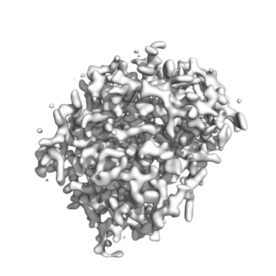
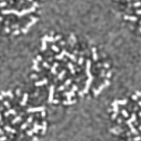
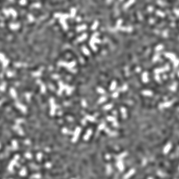
| Title | MicroED structure of proteinase K from an unpolished, platinum-coated, single lamella at 2.08A resolution (#9) | |||||||||
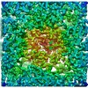
 Map data Map data | MicroED 2Fo-Fc map from a platinum coated, unpolished proteinase K lamella (#9) | |||||||||
 Sample Sample |
| |||||||||
 Keywords Keywords | Hydrolase | |||||||||
| Function / homology |  Function and homology information Function and homology informationpeptidase K / serine-type endopeptidase activity / proteolysis / extracellular region / metal ion binding Similarity search - Function | |||||||||
| Biological species |  Parengyodontium album (fungus) Parengyodontium album (fungus) | |||||||||
| Method | electron crystallography / cryo EM | |||||||||
 Authors Authors | Martynowycz MW / Zhao W | |||||||||
| Funding support |  United States, 2 items United States, 2 items
| |||||||||
 Citation Citation | Journal: Structure / Year: 2019 Title: Qualitative Analyses of Polishing and Precoating FIB Milled Crystals for MicroED. Authors: Michael W Martynowycz / Wei Zhao / Johan Hattne / Grant J Jensen / Tamir Gonen / Abstract: Microcrystal electron diffraction (MicroED) leverages the strong interaction between matter and electrons to determine protein structures from vanishingly small crystals. This strong interaction ...Microcrystal electron diffraction (MicroED) leverages the strong interaction between matter and electrons to determine protein structures from vanishingly small crystals. This strong interaction limits the thickness of crystals that can be investigated by MicroED, mainly due to absorption. Recent studies have demonstrated that focused ion-beam (FIB) milling can thin crystals into ideal-sized lamellae; however, it is not clear how to best apply FIB milling for MicroED. Here, the effects of polishing the lamellae, whereby the last few nanometers are milled away using a low-current gallium beam, are explored in both the platinum-precoated and uncoated samples. Our results suggest that precoating samples with a thin layer of platinum followed by polishing the crystal surfaces prior to data collection consistently led to superior results as indicated by higher signal-to-noise ratio, higher resolution, and better refinement statistics. This study lays the foundation for routine and reproducible methodology for sample preparation in MicroED. | |||||||||
| History |
|
- Structure visualization
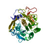
| Movie |
Movie viewer |
|---|---|
| Structure viewer | EM map: SurfViewMolmilJmol/JSmol |
| Supplemental images |
- Downloads & links
-EMDB archive
| Map data | emd_20360.map.gz | 29.6 MB | EMDB map data format | |
|---|---|---|---|---|
| Header (meta data) | emd-20360-v30.xmlemd-20360.xml | 16.3 KB 16.3 KB | Display Display | EMDB header |
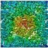
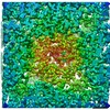
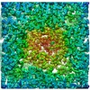
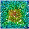
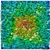
| Images |  emd_20360.png emd_20360.png | 81.9 KB | ||
| Filedesc metadata | emd-20360.cif.gz | 5.6 KB | ||
| Filedesc structureFactors | emd_20360_sf.cif.gz | 405.8 KB | ||
| Archive directory |  http://ftp.pdbj.org/pub/emdb/structures/EMD-20360ftp://ftp.pdbj.org/pub/emdb/structures/EMD-20360 http://ftp.pdbj.org/pub/emdb/structures/EMD-20360ftp://ftp.pdbj.org/pub/emdb/structures/EMD-20360 | HTTPS FTP |
-Related structure data
| Related structure data | 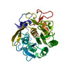 6pknMC 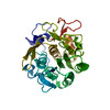 6pkjC 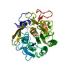 6pkkC 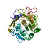 6pklC 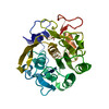 6pkmC 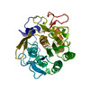 6pkoC 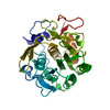 6pkpC 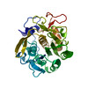 6pkqC  6pkrC  6pksC 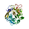 6pktC C: citing same article ( M: atomic model generated by this map |
|---|---|
| Similar structure data |
-Links
| EMDB pages | EMDB (EBI/PDBe) / EMDataResource |
|---|---|
| Related items in Molecule of the Month |


-Map
| File | Download / File: emd_20360.map.gz / Format: CCP4 / Size: 33.4 MB / Type: IMAGE STORED AS FLOATING POINT NUMBER (4 BYTES) | ||||||||||||||||||||||||||||||||||||||||||||||||||||||||||||||||||||
|---|---|---|---|---|---|---|---|---|---|---|---|---|---|---|---|---|---|---|---|---|---|---|---|---|---|---|---|---|---|---|---|---|---|---|---|---|---|---|---|---|---|---|---|---|---|---|---|---|---|---|---|---|---|---|---|---|---|---|---|---|---|---|---|---|---|---|---|---|---|
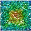
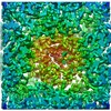
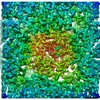
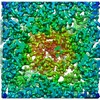
| Annotation | MicroED 2Fo-Fc map from a platinum coated, unpolished proteinase K lamella (#9) | ||||||||||||||||||||||||||||||||||||||||||||||||||||||||||||||||||||
| Projections & slices | Image control
Images are generated by Spider. generated in cubic-lattice coordinate | ||||||||||||||||||||||||||||||||||||||||||||||||||||||||||||||||||||
| Voxel size | X: 0.35208 Å / Y: 0.35208 Å / Z: 0.32666 Å | ||||||||||||||||||||||||||||||||||||||||||||||||||||||||||||||||||||
| Density |
| ||||||||||||||||||||||||||||||||||||||||||||||||||||||||||||||||||||
| Symmetry | Space group: 1 | ||||||||||||||||||||||||||||||||||||||||||||||||||||||||||||||||||||
| Details | EMDB XML:
CCP4 map header:
| ||||||||||||||||||||||||||||||||||||||||||||||||||||||||||||||||||||
 Z (Sec.)
 X (Row.)
 Y (Col.)


-Supplemental data
- Sample components
-Entire : Proteinase K
| Entire | Name: Proteinase K |
|---|---|
| Components |
|
-Supramolecule #1: Proteinase K
| Supramolecule | Name: Proteinase K / type: complex / ID: 1 / Parent: 0 / Macromolecule list: #1 |
|---|---|
| Source (natural) | Organism: Parengyodontium album (fungus) |
| Molecular weight | Theoretical: 28.93 KDa |
-Macromolecule #1: Proteinase K
| Macromolecule | Name: Proteinase K / type: protein_or_peptide / ID: 1 / Number of copies: 1 / Enantiomer: LEVO / EC number: peptidase K |
|---|---|
| Source (natural) | Organism: Parengyodontium album (fungus) |
| Molecular weight | Theoretical: 28.930783 KDa |
| Sequence | String: AAQTNAPWGL ARISSTSPGT STYYYDESAG QGSCVYVIDT GIEASHPEFE GRAQMVKTYY YSSRDGNGHG THCAGTVGSR TYGVAKKTQ LFGVKVLDDN GSGQYSTIIA GMDFVASDKN NRNCPKGVVA SLSLGGGYSS SVNSAAARLQ SSGVMVAVAA G NNNADARN ...String: AAQTNAPWGL ARISSTSPGT STYYYDESAG QGSCVYVIDT GIEASHPEFE GRAQMVKTYY YSSRDGNGHG THCAGTVGSR TYGVAKKTQ LFGVKVLDDN GSGQYSTIIA GMDFVASDKN NRNCPKGVVA SLSLGGGYSS SVNSAAARLQ SSGVMVAVAA G NNNADARN YSPASEPSVC TVGASDRYDR RSSFSNYGSV LDIFGPGTSI LSTWIGGSTR SISGTSMATP HVAGLAAYLM TL GKTTAAS ACRYIADTAN KGDLSNIPFG TVNLLAYNNY QA UniProtKB: Proteinase K |
-Macromolecule #2: water
| Macromolecule | Name: water / type: ligand / ID: 2 / Number of copies: 71 / Formula: HOH |
|---|---|
| Molecular weight | Theoretical: 18.015 Da |
| Chemical component information |  ChemComp-HOH: |
-Experimental details
-Structure determination
| Method | cryo EM |
|---|---|
 Processing Processing | electron crystallography |
| Aggregation state | 3D array |
-Sample preparation
| Concentration | 20 mg/mL |
|---|---|
| Buffer | pH: 7.5 |
| Grid | Support film - Material: CARBON / Support film - topology: HOLEY / Support film - Film thickness: 12 / Pretreatment - Type: GLOW DISCHARGE / Pretreatment - Time: 15 sec. / Details: unspecified |
| Vitrification | Cryogen name: ETHANE / Chamber humidity: 100 % / Chamber temperature: 273.0 K / Instrument: FEI VITROBOT MARK IV |
| Details | Proteinase K purchased from Sigma. |
- Electron microscopy
| Microscope | FEI TALOS ARCTICA |
|---|---|
| Temperature | Min: 77.0 K / Max: 90.0 K |
| Image recording | Film or detector model: FEI CETA (4k x 4k) / Digitization - Dimensions - Width: 4096 pixel / Digitization - Dimensions - Height: 4096 pixel / Number grids imaged: 1 / Number real images: 100 / Number diffraction images: 100 / Average exposure time: 3.0 sec. / Average electron dose: 0.03 e/Å2 Details: Continuous rotation with a rotation rate of 0.2 degrees per second and a readout every 3 seconds |
| Electron beam | Acceleration voltage: 200 kV / Electron source:  FIELD EMISSION GUN FIELD EMISSION GUN |
| Electron optics | C2 aperture diameter: 100.0 µm / Illumination mode: FLOOD BEAM / Imaging mode: DIFFRACTION / Camera length: 2055.0 mm |
| Sample stage | Specimen holder model: FEI TITAN KRIOS AUTOGRID HOLDER / Cooling holder cryogen: NITROGEN |
| Experimental equipment |  Model: Talos Arctica / Image courtesy: FEI Company |
-Image processing
| Details | This was the new CetaD |
|---|---|
| Final reconstruction | Resolution method: DIFFRACTION PATTERN/LAYERLINES / Software - Name: PHENIX (ver. 1.15.2) |
| Molecular replacement | Software - Name: PHENIX (ver. 2.8.2) |
| Crystallography statistics | Number intensities measured: 70454 / Number structure factors: 13802 / Fourier space coverage: 90.8 / R sym: 0.35 / R merge: 0.32 / Overall phase error: 27.24 / Overall phase residual: 27.24 / Phase error rejection criteria: 0 / High resolution: 2.08 Å / Shell - Shell ID: 1 / Shell - High resolution: 2.08 Å / Shell - Low resolution: 30.23 Å / Shell - Number structure factors: 13802 / Shell - Phase residual: 27.24 / Shell - Fourier space coverage: 90.8 / Shell - Multiplicity: 5.1 |
-Atomic model buiding 1
| Initial model | PDB ID: Chain - Chain ID: A / Chain - Residue range: 106-384 / Chain - Source name: PDB / Chain - Initial model type: experimental model |
|---|---|
| Refinement | Space: RECIPROCAL / Protocol: OTHER / Overall B value: 28.1 |
| Output model | PDB-6pkn: |