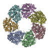
+ Open data
- Basic information
| Entry | Database: EMDB / ID: EMD-2002 | |||||||||
|---|---|---|---|---|---|---|---|---|---|---|
| Title | ATP-triggered molecular mechanics of the chaperonin GroEL | |||||||||
 Map data Map data | GroEL-ATP14 Rd2-Rd4ATP | |||||||||
 Sample Sample |
| |||||||||
 Keywords Keywords | Tetradecamer of GroEL with ATP bound in both rings | |||||||||
| Function / homology |  Function and homology information Function and homology informationGroEL-GroES complex / chaperonin ATPase / virion assembly / : / isomerase activity / ATP-dependent protein folding chaperone / response to radiation / unfolded protein binding / protein folding / response to heat ...GroEL-GroES complex / chaperonin ATPase / virion assembly / : / isomerase activity / ATP-dependent protein folding chaperone / response to radiation / unfolded protein binding / protein folding / response to heat / protein refolding / magnesium ion binding / ATP hydrolysis activity / ATP binding / identical protein binding / membrane / cytosol Similarity search - Function | |||||||||
| Biological species |  | |||||||||
| Method | single particle reconstruction / cryo EM / Resolution: 8.5 Å | |||||||||
 Authors Authors | Clare DK / Vasishtan D / Stagg S / Quispe J / Farr GW / Topf M / Horwich AL / Saibil HR | |||||||||
 Citation Citation |  Journal: Cell / Year: 2012 Journal: Cell / Year: 2012Title: ATP-triggered conformational changes delineate substrate-binding and -folding mechanics of the GroEL chaperonin. Authors: Daniel K Clare / Daven Vasishtan / Scott Stagg / Joel Quispe / George W Farr / Maya Topf / Arthur L Horwich / Helen R Saibil /  Abstract: The chaperonin GroEL assists the folding of nascent or stress-denatured polypeptides by actions of binding and encapsulation. ATP binding initiates a series of conformational changes triggering the ...The chaperonin GroEL assists the folding of nascent or stress-denatured polypeptides by actions of binding and encapsulation. ATP binding initiates a series of conformational changes triggering the association of the cochaperonin GroES, followed by further large movements that eject the substrate polypeptide from hydrophobic binding sites into a GroES-capped, hydrophilic folding chamber. We used cryo-electron microscopy, statistical analysis, and flexible fitting to resolve a set of distinct GroEL-ATP conformations that can be ordered into a trajectory of domain rotation and elevation. The initial conformations are likely to be the ones that capture polypeptide substrate. Then the binding domains extend radially to separate from each other but maintain their binding surfaces facing the cavity, potentially exerting mechanical force upon kinetically trapped, misfolded substrates. The extended conformation also provides a potential docking site for GroES, to trigger the final, 100° domain rotation constituting the "power stroke" that ejects substrate into the folding chamber. | |||||||||
| History |
|
- Structure visualization
| Movie |
 Movie viewer Movie viewer |
|---|---|
| Structure viewer | EM map:  SurfView SurfView Molmil Molmil Jmol/JSmol Jmol/JSmol |
| Supplemental images |
- Downloads & links
-EMDB archive
| Map data |  emd_2002.map.gz emd_2002.map.gz | 485.7 KB |  EMDB map data format EMDB map data format | |
|---|---|---|---|---|
| Header (meta data) |  emd-2002-v30.xml emd-2002-v30.xml emd-2002.xml emd-2002.xml | 11.1 KB 11.1 KB | Display Display |  EMDB header EMDB header |
| Images |  EMD2002.jpeg EMD2002.jpeg | 28.6 KB | ||
| Archive directory |  http://ftp.pdbj.org/pub/emdb/structures/EMD-2002 http://ftp.pdbj.org/pub/emdb/structures/EMD-2002 ftp://ftp.pdbj.org/pub/emdb/structures/EMD-2002 ftp://ftp.pdbj.org/pub/emdb/structures/EMD-2002 | HTTPS FTP |
-Related structure data
| Related structure data |  4ab2MC  1997C  1998C  1999C  2000C  2001C  2003C  4aaqC  4aarC  4aasC  4aauC  4ab3C M: atomic model generated by this map C: citing same article ( |
|---|---|
| Similar structure data |
- Links
Links
| EMDB pages |  EMDB (EBI/PDBe) / EMDB (EBI/PDBe) /  EMDataResource EMDataResource |
|---|---|
| Related items in Molecule of the Month |
- Map
Map
| File |  Download / File: emd_2002.map.gz / Format: CCP4 / Size: 26.4 MB / Type: IMAGE STORED AS FLOATING POINT NUMBER (4 BYTES) Download / File: emd_2002.map.gz / Format: CCP4 / Size: 26.4 MB / Type: IMAGE STORED AS FLOATING POINT NUMBER (4 BYTES) | ||||||||||||||||||||||||||||||||||||||||||||||||||||||||||||||||||||
|---|---|---|---|---|---|---|---|---|---|---|---|---|---|---|---|---|---|---|---|---|---|---|---|---|---|---|---|---|---|---|---|---|---|---|---|---|---|---|---|---|---|---|---|---|---|---|---|---|---|---|---|---|---|---|---|---|---|---|---|---|---|---|---|---|---|---|---|---|---|
| Annotation | GroEL-ATP14 Rd2-Rd4ATP | ||||||||||||||||||||||||||||||||||||||||||||||||||||||||||||||||||||
| Projections & slices | Image control
Images are generated by Spider. | ||||||||||||||||||||||||||||||||||||||||||||||||||||||||||||||||||||
| Voxel size | X=Y=Z: 2.02 Å | ||||||||||||||||||||||||||||||||||||||||||||||||||||||||||||||||||||
| Density |
| ||||||||||||||||||||||||||||||||||||||||||||||||||||||||||||||||||||
| Symmetry | Space group: 1 | ||||||||||||||||||||||||||||||||||||||||||||||||||||||||||||||||||||
| Details | EMDB XML:
CCP4 map header:
| ||||||||||||||||||||||||||||||||||||||||||||||||||||||||||||||||||||
-Supplemental data
- Sample components
-Entire : GroEL-ATP14 Rd2-Rd4
| Entire | Name: GroEL-ATP14 Rd2-Rd4 |
|---|---|
| Components |
|
-Supramolecule #1000: GroEL-ATP14 Rd2-Rd4
| Supramolecule | Name: GroEL-ATP14 Rd2-Rd4 / type: sample / ID: 1000 Oligomeric state: tetradecamer of GroEL with 14 ATP molecules bound Number unique components: 2 |
|---|---|
| Molecular weight | Experimental: 800 KDa / Theoretical: 800 KDa |
-Macromolecule #1: hsp60
| Macromolecule | Name: hsp60 / type: protein_or_peptide / ID: 1 / Name.synonym: GroEL / Details: ATPase Mutant, D398A / Number of copies: 14 / Oligomeric state: tetradecamer / Recombinant expression: Yes |
|---|---|
| Source (natural) | Organism:  |
| Molecular weight | Experimental: 56 KDa / Theoretical: 56 KDa |
| Recombinant expression | Organism:  |
| Sequence | GO: protein refolding / InterPro: Chaperonin Cpn60/GroEL/TCP-1 family |
-Macromolecule #2: ATP
| Macromolecule | Name: ATP / type: ligand / ID: 2 / Name.synonym: ATP / Details: ATP is bound to all 14 subunits / Recombinant expression: No |
|---|---|
| Source (natural) | Organism: synthetic construct (others) |
| Molecular weight | Experimental: 550 Da / Theoretical: 550 Da |
-Experimental details
-Structure determination
| Method | cryo EM |
|---|---|
 Processing Processing | single particle reconstruction |
| Aggregation state | particle |
- Sample preparation
| Concentration | 4 mg/mL |
|---|---|
| Buffer | pH: 7.4 Details: 50 mM Tris-HCl pH 7.4, 50 mM KCl, 10 mM MgCl2 and 200uM ATP |
| Grid | Details: cflat grids r2/2 |
| Vitrification | Cryogen name: ETHANE / Chamber humidity: 100 % / Chamber temperature: 95 K / Instrument: OTHER / Details: Vitrification instrument: Vitrobot / Timed resolved state: vitrified within 30 seconds / Method: grids were blotted for 2-3 seconds |
- Electron microscopy
| Microscope | FEI TECNAI F20 |
|---|---|
| Temperature | Min: 95 K / Max: 95 K / Average: 95 K |
| Details | The data was collected with leginon at SCRIPPS |
| Image recording | Category: CCD / Film or detector model: GATAN ULTRASCAN 4000 (4k x 4k) / Average electron dose: 15 e/Å2 |
| Electron beam | Acceleration voltage: 120 kV / Electron source:  FIELD EMISSION GUN FIELD EMISSION GUN |
| Electron optics | Calibrated magnification: 148500 / Illumination mode: FLOOD BEAM / Imaging mode: BRIGHT FIELD / Cs: 2 mm / Nominal defocus max: 3.5 µm / Nominal defocus min: 0.7 µm |
| Sample stage | Specimen holder: Eucentric / Specimen holder model: GATAN LIQUID NITROGEN |
| Experimental equipment |  Model: Tecnai F20 / Image courtesy: FEI Company |
- Image processing
| Details | The particles were automatically picked using FindEM |
|---|---|
| CTF correction | Details: each particle was phase flipped |
| Final reconstruction | Applied symmetry - Point group: C7 (7 fold cyclic) / Algorithm: OTHER / Resolution.type: BY AUTHOR / Resolution: 8.5 Å / Resolution method: FSC 0.5 CUT-OFF / Software - Name: SPIDER, IMAGIC / Details: SIRT was used to reconstruct the final map / Number images used: 6500 |
| Final angle assignment | Details: theta 80-100, phi 0-51.42 |
 Movie
Movie Controller


 Z (Sec.)
 Y (Row.)
 X (Col.)


