[English] 日本語
 Yorodumi
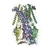
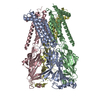
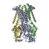
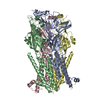
- EMDB-19420: Sub-tomogram averaging results of pentameric 5-HT3A receptor on c... -
+ Open data
- Basic information
| Entry |  | ||||||||||||||||||
|---|---|---|---|---|---|---|---|---|---|---|---|---|---|---|---|---|---|---|---|
| Title | Sub-tomogram averaging results of pentameric 5-HT3A receptor on cell-derived vesicles | ||||||||||||||||||
 Map data Map data | |||||||||||||||||||
 Sample Sample |
| ||||||||||||||||||
 Keywords Keywords | 5ht3Ar / pentamer / in-situ subtomogram-averaging / cell-derived vesicles / MEMBRANE PROTEIN | ||||||||||||||||||
| Biological species |  | ||||||||||||||||||
| Method | subtomogram averaging / cryo EM / Resolution: 19.6 Å | ||||||||||||||||||
 Authors Authors | Chu X / Kudryashev M | ||||||||||||||||||
| Funding support |  Germany, Germany,  China, 5 items China, 5 items
| ||||||||||||||||||
 Citation Citation |  Journal: EMBO J / Year: 2024 Journal: EMBO J / Year: 2024Title: Structure of tetrameric forms of the serotonin-gated 5-HT3 receptor ion channel. Authors: Bianca Introini / Wenqiang Cui / Xiaofeng Chu / Yingyi Zhang / Ana Catarina Alves / Luise Eckhardt-Strelau / Sabrina Golusik / Menno Tol / Horst Vogel / Shuguang Yuan / Mikhail Kudryashev /    Abstract: Multimeric membrane proteins are produced in the endoplasmic reticulum and transported to their target membranes which, for ion channels, is typically the plasma membrane. Despite the availability of ...Multimeric membrane proteins are produced in the endoplasmic reticulum and transported to their target membranes which, for ion channels, is typically the plasma membrane. Despite the availability of many fully assembled channel structures, our understanding of assembly intermediates, multimer assembly mechanisms, and potential functions of non-standard assemblies is limited. We demonstrate that the pentameric ligand-gated serotonin 5-HT3A receptor (5-HT3AR) can assemble to tetrameric forms and report the structures of the tetramers in plasma membranes of cell-derived microvesicles and in membrane memetics using cryo-electron microscopy and tomography. The tetrameric structures have near-symmetric transmembrane domains, and asymmetric extracellular domains, and can bind serotonin molecules. Computer simulations, based on our cryo-EM structures, were used to decipher the assembly pathway of pentameric 5-HT3R and suggest a potential functional role for the tetrameric receptors. | ||||||||||||||||||
| History |
|
- Structure visualization
| Supplemental images |
|---|
- Downloads & links
-EMDB archive
| Map data |  emd_19420.map.gz emd_19420.map.gz | 936.8 KB |  EMDB map data format EMDB map data format | |
|---|---|---|---|---|
| Header (meta data) |  emd-19420-v30.xml emd-19420-v30.xml emd-19420.xml emd-19420.xml | 14.5 KB 14.5 KB | Display Display |  EMDB header EMDB header |
| FSC (resolution estimation) |  emd_19420_fsc.xml emd_19420_fsc.xml | 2.4 KB | Display |  FSC data file FSC data file |
| Images |  emd_19420.png emd_19420.png | 28.7 KB | ||
| Masks |  emd_19420_msk_1.map emd_19420_msk_1.map | 1 MB |  Mask map Mask map | |
| Filedesc metadata |  emd-19420.cif.gz emd-19420.cif.gz | 5.2 KB | ||
| Others |  emd_19420_half_map_1.map.gz emd_19420_half_map_1.map.gz emd_19420_half_map_2.map.gz emd_19420_half_map_2.map.gz | 738.8 KB 737.9 KB | ||
| Archive directory |  http://ftp.pdbj.org/pub/emdb/structures/EMD-19420 http://ftp.pdbj.org/pub/emdb/structures/EMD-19420 ftp://ftp.pdbj.org/pub/emdb/structures/EMD-19420 ftp://ftp.pdbj.org/pub/emdb/structures/EMD-19420 | HTTPS FTP |
-Validation report
| Summary document |  emd_19420_validation.pdf.gz emd_19420_validation.pdf.gz | 773.6 KB | Display |  EMDB validaton report EMDB validaton report |
|---|---|---|---|---|
| Full document |  emd_19420_full_validation.pdf.gz emd_19420_full_validation.pdf.gz | 773.1 KB | Display | |
| Data in XML |  emd_19420_validation.xml.gz emd_19420_validation.xml.gz | 7.5 KB | Display | |
| Data in CIF |  emd_19420_validation.cif.gz emd_19420_validation.cif.gz | 9.7 KB | Display | |
| Arichive directory |  https://ftp.pdbj.org/pub/emdb/validation_reports/EMD-19420 https://ftp.pdbj.org/pub/emdb/validation_reports/EMD-19420 ftp://ftp.pdbj.org/pub/emdb/validation_reports/EMD-19420 ftp://ftp.pdbj.org/pub/emdb/validation_reports/EMD-19420 | HTTPS FTP |
-Related structure data
- Links
Links
| EMDB pages |  EMDB (EBI/PDBe) / EMDB (EBI/PDBe) /  EMDataResource EMDataResource |
|---|
- Map
Map
| File |  Download / File: emd_19420.map.gz / Format: CCP4 / Size: 1 MB / Type: IMAGE STORED AS FLOATING POINT NUMBER (4 BYTES) Download / File: emd_19420.map.gz / Format: CCP4 / Size: 1 MB / Type: IMAGE STORED AS FLOATING POINT NUMBER (4 BYTES) | ||||||||||||||||||||||||||||||||||||
|---|---|---|---|---|---|---|---|---|---|---|---|---|---|---|---|---|---|---|---|---|---|---|---|---|---|---|---|---|---|---|---|---|---|---|---|---|---|
| Projections & slices | Image control
Images are generated by Spider. | ||||||||||||||||||||||||||||||||||||
| Voxel size | X=Y=Z: 5.52 Å | ||||||||||||||||||||||||||||||||||||
| Density |
| ||||||||||||||||||||||||||||||||||||
| Symmetry | Space group: 1 | ||||||||||||||||||||||||||||||||||||
| Details | EMDB XML:
|
-Supplemental data
-Mask #1
| File |  emd_19420_msk_1.map emd_19420_msk_1.map | ||||||||||||
|---|---|---|---|---|---|---|---|---|---|---|---|---|---|
| Projections & Slices |
| ||||||||||||
| Density Histograms |
-Half map: #2
| File | emd_19420_half_map_1.map | ||||||||||||
|---|---|---|---|---|---|---|---|---|---|---|---|---|---|
| Projections & Slices |
| ||||||||||||
| Density Histograms |
-Half map: #1
| File | emd_19420_half_map_2.map | ||||||||||||
|---|---|---|---|---|---|---|---|---|---|---|---|---|---|
| Projections & Slices |
| ||||||||||||
| Density Histograms |
- Sample components
-Entire : 5-hydroxytryptamine receptor 3A pentamer
| Entire | Name: 5-hydroxytryptamine receptor 3A pentamer |
|---|---|
| Components |
|
-Supramolecule #1: 5-hydroxytryptamine receptor 3A pentamer
| Supramolecule | Name: 5-hydroxytryptamine receptor 3A pentamer / type: organelle_or_cellular_component / ID: 1 / Parent: 0 / Macromolecule list: all |
|---|---|
| Source (natural) | Organism:  |
-Macromolecule #1: 5-hydroxytryptamine receptor 3A pentamer
| Macromolecule | Name: 5-hydroxytryptamine receptor 3A pentamer / type: protein_or_peptide / ID: 1 Details: StrepII-tags, linkers, TEV protease recognition sequence, mouse 5HT3AR (with Ala insertion at position 277 (in the M2-M3 linker) as found in the human receptor) Enantiomer: LEVO |
|---|---|
| Source (natural) | Organism:  |
| Sequence | String: MWSHPQFEKG GGSGGGSGGG SWSHPQFEKG GGSGGGSGGG SWSHPQFEKG GGSGGGSGGG SWSHPQFEKE NLYFQGATQA RDTTQPALLR LSDHLLANYK KGVRPVRDWR KPTTVSIDVI MYAILNVDEK NQVLTTYIWY RQYWTDEFLQ WTPEDFDNVT KLSIPTDSIW ...String: MWSHPQFEKG GGSGGGSGGG SWSHPQFEKG GGSGGGSGGG SWSHPQFEKG GGSGGGSGGG SWSHPQFEKE NLYFQGATQA RDTTQPALLR LSDHLLANYK KGVRPVRDWR KPTTVSIDVI MYAILNVDEK NQVLTTYIWY RQYWTDEFLQ WTPEDFDNVT KLSIPTDSIW VPDILINEFV DVGKSPNIPY VYVHHRGEVQ NYKPLQLVTA CSLDIYNFPF DVQNCSLTFT SWLHTIQDIN ITLWRSPEEV RSDKSIFINQ GEWELLEVFP QFKEFSIDIS NSYAEMKFYV IIRRRPLFYA VSLLLPSIFL MVVDIVGFCL PPDSGERVSF KITLLLGYSV FLIIVSDTLP ATAIGTPLIG VYFVVCMALL VISLAETIFI VRLVHKQDLQ RPVPDWLRHL VLDRIAWILC LGEQPMAHRP PATFQANKTD DCSGSDLLPA MGNHCSHVGG PQDLEKTPRG RGSPLPPPRE ASLAVRGLLQ ELSSIRHFLE KRDEMREVAR DWLRVGYVLD RLLFRIYLLA VLAYSITLVT LWSIWHSS |
-Experimental details
-Structure determination
| Method | cryo EM |
|---|---|
 Processing Processing | subtomogram averaging |
| Aggregation state | cell |
- Sample preparation
| Buffer | pH: 7.4 |
|---|---|
| Grid | Model: Quantifoil R2/2 / Material: GOLD |
| Vitrification | Cryogen name: ETHANE |
- Electron microscopy
| Microscope | FEI TITAN KRIOS |
|---|---|
| Image recording | Film or detector model: GATAN K3 BIOQUANTUM (6k x 4k) / Average electron dose: 4.9 e/Å2 |
| Electron beam | Acceleration voltage: 300 kV / Electron source:  FIELD EMISSION GUN FIELD EMISSION GUN |
| Electron optics | C2 aperture diameter: 70.0 µm / Illumination mode: FLOOD BEAM / Imaging mode: BRIGHT FIELD / Nominal defocus max: 5.5 µm / Nominal defocus min: 3.5 µm |
| Experimental equipment |  Model: Titan Krios / Image courtesy: FEI Company |
 Movie
Movie Controller


 Z (Sec.)
 Y (Row.)
 X (Col.)


